Introduction
Gaining skills in functional endoscopic sinus surgery (FESS) undoubtedly poses a number of problems. Patients' outcomes must not be put at risk by the lack of experience of trainee surgeons, and experienced supervisors must be competent to assume responsibility for the outcome of every procedure.Reference Carter, Russell, Dunkley and Cuschieri1 Considering the three-dimensional complexity of the anatomy of the paranasal sinuses, and the region's close relationship to the orbit, optic nerve, internal carotid artery and intracranial structures, the potential for serious complications is ever-present; thus, adequate training should be undertaken before the trainee surgeon begins performing FESS procedures on patients.Reference McFerran, Grant, Ingrams and Fife2
Training on cadaveric human heads is compulsory in many resident programmes, in order to teach surgical aspects of the complex nasal and sinus anatomy and thus to ensure patient safety.Reference Kinsella, Calhoun, Bradfield, Hokanson and Bailey3–Reference Gardiner, Oluwole, Tan and White5 However, it is often difficult to obtain a satisfactory supply of human heads for training purposes, either due to strict medicolegal regulations (in some countries) or to other practical reasons (e.g. the expense of such practical courses, the complexity of storage and the use of cadavers). In our opinion, the primary goal of beginners' training is not only the study of the endoscopic surgical anatomy of the human nose and paranasal sinuses, for which supervised cadaveric dissections remain the ‘gold standard’. Equally important aims of FESS training are to gain surgical skills, to become familiar with controlled, simultaneous manipulation of two instruments within the narrow space of the nasal cavity, and to improve the trainee surgeon's orientation within the operating field.Reference Manickavasagam, Segaram and Harkness4 These aims are easily achievable by using an animal model with an appearance and dimensions as similar as possible to those in humans.
Gardiner et al. described a sequence of successfully completed endoscopic surgical manoeuvres performed on a sheep's head model, ranging from simple handling of the endoscope during examination to more complex bimanual instrumentation, including endoscopic dacryocystorhinostomy (DCR).Reference Gardiner, Oluwole, Tan and White5 Although the configuration of the sheep's head is obviously different to that of the human head, the nasal cavity appears to be quite similar and, as noted from computed tomography scans, the major sinuses (maxillary, ethmoidal and frontal) lie in approximately the same orientation as in humans. Furthermore, the model itself is cheap, easily obtainable and of good tissue quality.
Now, 14 years later, we strongly support Gardiner and colleagues' attempts to develop an animal model for FESS skills training. Furthermore, we would like to contribute our more recent knowledge gained over the past several years, during which we have studied every radiological, anatomical and surgical aspect of the use of lamb's head models for FESS skills training.
We chose lambs' heads instead of sheep's heads, since the latter's nasal cavities are too deep and thus do not correspond to the standard length of FESS instruments used for human surgery.
Gardiner et al. listed endoscopic DCR among the FESS techniques that were possible on a sheep's head model.Reference Gardiner, Oluwole, Tan and White5 The main goal of endoscopic DCR is to provide a new passage through which tears can flow directly into the nasal cavity from the lacrimal sac, instead of through the nasolacrimal duct (which is obstructed for whatever reason). Gardiner et al. described a technique for approaching the sheep lacrimal sac which was similar to that used in the human nose. The area of resection is delineated by introducing an optical fibre light source through the sheep lacrimal canaliculus into the lacrimal sac. A 0° endoscope is then introduced into the nasal cavity to visualise the agger nasi area, illuminated by the light source. After elevation of the mucoperiosteum, the bone is removed with a drill, and a sickle knife is then used to remove the medial portion of the lacrimal sac.
During our detailed study of the surgical anatomy of the lamb's head (as regards its suitability for FESS training), we did not find this kind of surgery possible in this model. The reason appeared very simple: the lacrimal sac simply does not exist in lambs.
Materials and methods
Ten fresh lamb's heads were purchased, washed under tap water and placed in a bowl of water containing three tablespoons of alcoholic vinegar. After soaking for 24 hours, the heads were left to drain for 30 minutes, wiped with a clean cloth and then frozen at −18°C until used for dissection. On the day of dissection, the heads were thawed, washed and wiped. This preparation removed excess grease and fluids and made the heads far easier to use.
Dacryocystography was performed on five lamb's heads, within the radiology department. One 20 ml ampule of Ultravist 300® contrast (Schering AG, Berlin–Wedding, Germany), the usual contrast medium for dacryocystography, was carefully administrated into one lamb's head through the upper and lower lacrimal canaliculi, under X-ray control, being mindful of the fragility of these structures.
All five lambs' heads underwent anatomical and endoscopic dissection. They were mounted on lumps of clay and positioned facing the operator. Firstly, we removed the soft tissue of the anterior part of the muzzle and the upper third of the cartilaginous nasal septum, in order to avoid greasing and fogging the endoscope. Then, we performed external dissection of the nasolacrimal drainage system, meticulously preparing the tissues layer by layer, using a blade and drill. Finally, we exposed the intact lacrimal canaliculi and nasolacrimal duct along its full length (Figure 1). However, we could not identify a lacrimal sac. Methylene blue dye was then administrated into the canaliculi in order to detect the precise point of drainage into the nasal cavity, and to attempt to identify the lacrimal sac itself.
Anatomical dissection of nasolacrimal drainage system in a lamb's head, showing a long, intact nasolacrimal duct extending all the way to the very anterior part of the nasal cavity. There is no evidence of a lacrimal sac.

Results and analysis
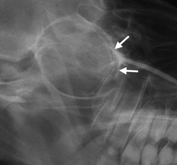
The dacryocystogram showed a rather peculiar arrangement of lacrimal passage, with the upper and lower canaliculi emptying the contrast medium directly into the nasolacrimal duct (Figure 2), without any cystic dilatation resembling a lacrimal sac. Thus, there was clear evidence of absence of a lacrimal sac. This result was obtained in all five lamb's heads tested.
Dacryocystogram of a lamb's head showing the angled junction of the upper and lower canaliculi (arrows) draining into the nasolacrimal duct, without formation of a lacrimal sac.

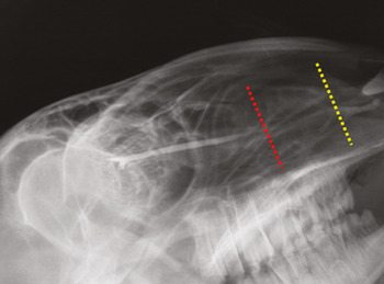
Dacryocystography also showed another variant feature of lamb anatomy. The nasolacrimal duct did not extend to the very anterior part of the nasal cavity, as it does in humans (in whom it ends with Hassner's valve, located in the ceiling of the most anterior part of the inferior meatus). Figure 3 clearly demonstrates loss of detectable contrast somewhere in the middle of the length of the muzzle, indicating that the nasolacrimal duct ends approximately halfway along the labyrinth of the inferior turbinate.
Dacryocystogram of a lamb's head; broken red line indicates the termination of detectable contrast passage, halfway along the length of the nasolacrimal duct (broken yellow line).

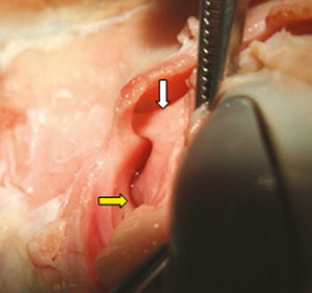
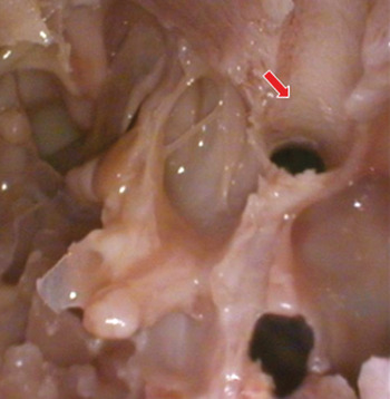
Our lamb's head dissections indicated that the upper and lower canaliculi do not form a lacrimal sac in this animal; rather, they form a sharp junction emptying straight into the nasolacrimal duct (Figure 4). After initial dissection of the maxillary sinus ostium recess and exposure of the anterior wall of the orbit, the presumed position of the bony canal containing the lacrimal sac was drilled out. However, we found nothing but the nasolacrimal duct itself, which left us unable to perform marsupialisation of the sac, as the final step in endoscopic DCR (Figure 5). For this reason, our attempts to perform endoscopic dacryocystorhinostomy were thwarted.
Posterior to anterior view of a lamb's head anatomical dissection, looking over the left eye (the bulb is pressed medially), showing the angled junction of the upper and lower canaliculi to form the nasolacrimal duct (white arrow). The beginning of the infraorbital nerve canal is also evident (yellow arrow).

Endoscopic endonasal view of a lamb's head, looking towards the left orbit. The bony nasolacrimal canal is opened (red arrow), just below the orbital floor, demonstrating the absence of a lacrimal sac.

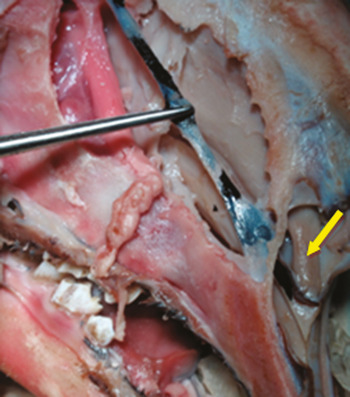
Methylene blue dye administration confirmed the dissection findings, and showed that the nasolacrimal duct actually ended under the very anterior part of the inferior turbinate (as it does in humans), not halfway along its length, as dacryocystography would suggest (Figure 6).
Lamb's head anatomical dissection showing methylene blue dye administration through the nasolacrimal canal; the dye travels all the way to the anterior pole of the inferior turbinate (yellow arrow).

Discussion
Endoscopic sinus surgery requires an extensive knowledge of both sinonasal anatomy and safe instrument usage. Many institutions around the world insist that trainee surgeons practise their FESS skills on cadaveric human heads, before they undertake their first procedure on a live patient.Reference Kinsella, Calhoun, Bradfield, Hokanson and Bailey3, Reference Montague, Kishore and McGarry6, Reference Zuckerman, Wise, Rogers, Senior, Schlosser and DelGaudio7 Functional endoscopic sinus surgery training courses and workshops provide a well structured, supervised approach to endonasal anatomy and technical training. However, such courses have the disadvantages of considerable expense and strict medicolegal requirements regarding the collection, storage and disposal of cadaveric heads, presenting great problems in a growing number of countries.
Artificial human head models have been designed and are available for training purposes, but are expensive and must be disposed of after a single use.Reference Briner, Simmen, Jones, Manestar, Manestar and Lang8
Computerised endoscopic sinus surgery simulators are another alternative FESS teaching tool. Reference Fried, Sadoughi, Gibber, Jacobs, Lebowitz and Ross9
• This study combined radiology and anatomical dissection of a lamb's head model to evaluate its usefulness for functional endoscopic sinus surgery (FESS) training
• The lamb's head is an excellent model for practising FESS techniques, with anatomy comparable to that of humans
• However, the lamb's nasolacrimal system lacks a lacrimal sac, so this animal model is unsuitable for dacryocystorhinostomy training
We believe that, for beginners, the initial goal of endoscopic endonasal surgery training is to gain surgical skills and to become familiar with the complexity of simultaneous, bimanual work in the operating field, which is distant from the field displayed on the monitor. Thus, trainees should first work with animal models, to gain skills in instrument manipulation, and then proceed to the study of human anatomy using cadaveric dissection. This process enables more efficient FESS training, giving the trainee more confidence in their surgical skills and also a better understanding of the anatomical details they will encounter. An animal model of comparable sinonasal appearance to the human head seems to be the logical choice for initial surgical skills training. Unfortunately, there are few reported attempts to develop such a model. Paying great respect to Gardiner and colleagues' introduction of a sheep's head model for FESS training, we have developed a detailed FESS skills training programme, using a lamb's head model, which combines radiology findings, three-axis frozen section analysis and meticulous research into lamb sinonasal anatomy.Reference Gardiner, Oluwole, Tan and White5, Reference Mladina10 We hope this training programme will facilitate initial FESS training for all trainees approaching this field.
Conclusion
We have developed a detailed, practical FESS skills training programme, using a lamb's head model, which combines radiology findings, three-axis frozen section analysis and meticulous research into lamb sinonasal anatomy. The lamb's head has proven to be an excellent animal model, with anatomy comparable to that of humans, which enables trainees to practise standard FESS techniques with ease, using standard FESS instruments.
However, the present study found that the lamb nasolacrimal system lacks a lacrimal sac. For this reason, endoscopic DCR training cannot be undertaken using this animal model.