Implication
Mastitis in first-calf heifers affects the profitability of dairying. Despite bacteriology being the only fully reliable way to detect mastitis, many other markers have been proposed. Owing to the relationship between the degree of infection and released nitric oxide (NOx), this study tested milk and lacteal NOx level. Infected quarters had greater NOx concentrations than non-infected quarters before (30.81 v. 22.83 μM/ml) and after (9.56 v. 5.77 μM/ml) calving. Lacteal NOx level greater than 24 μM/ml and milk NOx level greater than 6.5 μM/ml indicate pre- and postpartal mastitis status.
Introduction
Mastitis in heifers is becoming an important economic problem in dairy herds (Huijps et al., Reference Huijps, De Vliegher, Lam and Hogeeven2009), and prepartum incidence rates are high (39% to 75%; Oliver et al., Reference Oliver, Gillespie, Headrick, Lewis and Dowlen2004; Fox, Reference Fox2009) in different countries, such as the United States of America (Pankey et al., Reference Pankey, Drechsler and Wildman1991; Owens et al., Reference Owens, Nickerson, Boddie and Tomita2001), Germany (Kromker and Friedrich, Reference Kromker and Friedrich2009), Norway (Waage et al., Reference Waage, Mork, Roros, Aasland, Hunshamar and Odegaard1999), Denmark (Aarestrup and Jensen, Reference Aarestrup and Jensen1997) and Turkey (Bastan et al., Reference Bastan, Cengiz, Cengiz, Polat, Colak, Akan, Darbaz and Acar2010). There is a need for accurate, rapid and inexpensive methods to screen for potential mastitis pathogens. Staphylococcus spp., in particular, is reported to play a predominant role in the etiology of mastitis (Oliver et al., Reference Oliver, Lewis, Gillespie and Dowlen1992; Aarestrup and Jensen, Reference Aarestrup and Jensen1997; Oliver et al., Reference Oliver, Gillespie, Headrick, Lewis and Dowlen2004; Bastan et al., Reference Bastan, Cengiz, Cengiz, Polat, Colak, Akan, Darbaz and Acar2010). Coagulase-negative staphylococci (CNS) and Staphylococcus aureus (S. aureus) are the most commonly detected bacteria in heifer mastitis (Waage et al., Reference Waage, Mork, Roros, Aasland, Hunshamar and Odegaard1999; Oliver et al., Reference Oliver, Gillespie, Headrick, Lewis and Dowlen2004; Fox, Reference Fox2009). Macrophages are the major leukocytes in the secretions of uninfected mammary glands. In response to invading mastitis pathogens, chemotactic factors released by macrophages cause rapid migration of polymorphonuclear neutrophil leukocytes (PMN) into the infected area, which begin phagocytosing the invading pathogens. During the phagocytic process, the intracellular and extracellular release of toxic oxygen radicals by PMN to kill bacteria also destroy mammary secretory cells, resulting in irreversible tissue damage (Zhao and Lacasse, Reference Zhao and Lacasse2008). In addition to these radicals, the concentration of nitric oxide (NOx) increases in affected tissue. Total nitrite and nitrate is assumed to be valid as an estimate of NOx generation; nitrite oxide from nitrite and nitrate is referred to as NOx (Bouchard et al., Reference Bouchard, Blais, Desrosiers, Zhao and Lacasse1999; Komine et al., Reference Komine, Kuroishi, Komine, Watanabe, Kobayashi, Yamaguchi, Kamata and Kumagai2004). NOx is a free radical that mediates physiological and pathological changes in the body, is synthesized from l-arginine by inducible nitric oxide synthase (iNOS; Archer, Reference Archer1993) and mediated from endothelial, epithelial, neuronal and phagocytic cells (Palmer et al., Reference Palmer, Ferrige and Moncada1987; Onado and Inano, Reference Onado and Inano1998). However, NOx becomes a cytotoxic mediator when it is released in excessive amounts during Escherichia coli (E. coli) infections and in response to intramammary injection of lipopolysaccharide (LPS; Boulanger et al., Reference Boulanger, Bouchard, Zhao and Lacasse2001; Burvenich et al., Reference Burvenich, Van Merris, Mehrzad, Diez-Fraile and Duchateau2003; Komine et al., Reference Komine, Kuroishi, Komine, Watanabe, Kobayashi, Yamaguchi, Kamata and Kumagai2004).
The objective of this study was to detect NOx concentrations in mammary secretions from infected and non-infected mammary quarters of heifers before and after calving, and to evaluate the relationship of NOx with bacterial species and somatic cell count (SCC).
Material and methods
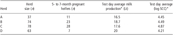
This study was carried out on four commercial dairy herds managed under similar nutritional conditions and located in the same area. Prepartum diets consisted of hay, straw and silage. The herd size ranged from 37 to 78 heifers (Table 1). Five- to seven-month pregnant Holstein heifers (n = 69) were included in the study. Heifers had no clinical signs of mastitis such as fever, pain or swelling and had not received antibiotic and anti-inflammatory treatment during the previous 30 days. Pregnancy was confirmed by rectal palpation and insemination data.
Test day average milk production and SCC in herds
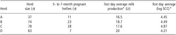
SCC = somatic cell count.
*Average milk production and log SCC results were measured for only heifers, which were used in the study.
Mammary quarters (n = 247) were sampled 80 (±15) days before expected calving. Some quarters were non-functional at the time of sampling; therefore, glandular secretions (lacteal prepartum and milk postpartum) could not be collected (data not shown). The same quarters (n = 247) were sampled 10 days (test day) after calving. The average milk production was 18.2 (±1.7) kg/day on the test day. The infectious status of the quarters (infected or healthy) was determined by diagnostic bacteriology. If a quarter was infected with a bacterium, it was considered as infected. Infected quarters with clinical symptoms were not used in the study.
Pre- and postpartum mammary secretions were collected from each mammary quarter by the same researcher according to described procedure (Kromker and Friedrich, Reference Kromker and Friedrich2009). Teat ends were cleaned and disinfected with ethanol (70%) before sampling. Light pressure was applied to the teat by using a gentle milking action and ~4 ml of secretion was collected into sterile plastic tubes. After sampling, teat ends were dipped into iodine-based (0.75%) ready-to-use solution (Dipal®, De Laval, Tumba, Sweden). Ten days after calving, milk samples were collected using the same method. After teat ends were disinfected, the first few streams of secretion were discarded, and then 10 ml was collected aseptically into sterile plastic tubes. All samples (secretions and milk) were maintained at 4°C until they reached the laboratory.
Diagnostic bacteriology was performed according to procedures described by Harmon et al (Reference Harmon, Eberhart, Langlois and Wilson1990). Ten microliters of sample was spread on MacConkey and blood agar (without esculin) containing 5% defibrinated sheep blood. The plates were incubated at 37°C for 24 and 48 h. Colonies were classified according to Gram staining, morphology and hemolysis properties in order to determine intramammary infection (IMI), contamination and mixed infections (Harmon et al., Reference Harmon, Eberhart, Langlois and Wilson1990; Blowey and Edmondson Reference Blowey and Edmondson1995; Wilson et al., Reference Wilson, Gonzalez and Das1997; Quinn et al., Reference Quinn, Carter, Markey and Carter1999; McDougall et al. Reference McDougall, Pankey, Delaney, Barlow, Murdough and Scru2002). If three or more of the same colonies were obtained on a plate, this was considered as an IMI. When three or more different colonies were obtained, this was considered as contamination. If two different types of pure colonies were detected, this was defined as mixed infection. Identification of one or more colonies of S. aureus was considered as an IMI. Colonies were re-cultured on blood agar for biochemical tests on pure colonies and the bacteria were specifically identified (Quinn et al., Reference Quinn, Carter, Markey and Carter1999). Catalase and coagulase tests were used for Gram-positive cocci. If the Gram-positive cocci had positive results for catalase and coagulase tests, they were considered to be S. aureus. If the result was coagulase negative, the bacteria were considered CNS. Catalase-negative bacteria were not detected in the study. Colony morphology was evaluated and the ‘IMViC’ (Indole – Methyl red – Voges Proskauer – Citrate) test was administrated on Gram-negative colonies for identification of E. coli. If the colonies had dry, flat, pink morphology and the test results were indole (+), methyl red (+), Voges Proskauer (−) and citrate (−), this colony was considered to be E. coli and separated from Klebsiella spp. If the culture result was negative at the first sampling or if contamination was detected, sampling was repeated 5 days later to avoid false negative and positive (due to contamination) results. The quarters were considered to be non-infected (healthy) if the culture results were negative for both sampling. The quarters that were culture negative at the first sampling but culture positive at the second sampling were allocated to the infected group. In addition, some quarters had mixed infections (two different types of pure colonies, and not considered as contaminated) before or after calving. These quarters were not taken to study in order to avoid a false correlation between NOx level and bacteria types.
Total NOx were measured in lacteal and milk samples. For the NOx assay, equal volumes of sample and potassium phosphate buffer, pH 7.5, were placed in an ultra-filter (10 000 MWCO Sartorius, Viva Science, Goettingen, Germany). The samples were centrifuged at 20°C for 45 min (5000 × g). The ultra-filtrate was used in the test. The nitrate present in the sample was reduced to nitrite with nicotinamide adenine dinucleotide phosphate (NADPH) in the presence of the enzyme nitrate reductase as previously described (Gilliam et al., Reference Gilliam, Sherman, Griscavage and Ignarro1993). Briefly, 500 μl samples were mixed with 50 μl of co-enzyme solution (1.5 mg NADPH and 0.03 mg FAD in 1 ml of potassium phosphate buffer, pH 7.5) and 20 μl of nitrate reductase solution (4 U nitrate reductase in 0.6 ml of redistilled water) of the incubated at room temperature for 30 min. Then, 150 μl of the incubated solution was mixed with an equal volume of Greiss reagent (75 μl of sulfanilamide reagent and 75 μl of N-(1-naphthyl)-ethylenediamine dihydrochloride reagent) in a microtiter plate. The reaction was allowed to proceed for 5 min and the resulting nitrite was allowed to react with sulfanilamide and N-(1-naphthyl)-ethylenediamine dihydrochloride to give a red–violet diazo dye. Total nitrite was evaluated by reading the optical density of each sample at 550 nm. The NOx concentration was determined by comparing it with a potassium nitrate standard curve (0 to 80 μM). Parallel evaluation was performed for all samples.
Quarter milk SCC was determined 10 days after calving using flow cytometry (Bentley IBCm, Bentley Instruments, Inc., Chaska, USA). If SCC was not determined within 12 h, preservative (azidiol) was added to the milk sample and stored at 4°C. Azidiol (0.018 g/100 ml) was prepared according to the reported procedure and 33 μl was added to 10 ml of milk sample (Sierra et al., Reference Sierra, Sanchez, Contreras, Luengo, Corrales, De la Fe, Guirao, Morales and Gonzalo2009). Quarter SCC was transformed to log10 base.
NOx data were subjected to two-way ANOVA (SAS Institute, 2002) using the PROC MIXED procedure. The linear model included the main effects of health status (non-infected v. infected quarters) and sampling period (prepartum v. postpartum) and their interaction, which were fixed effects and cow × quarter was the random effect. Before computation, SCC data were subjected to log transformation in order to normalize data. For the log (SCC), the model included only health status as a fixed effect and cow and quarter were random terms. For both variables, infected quarters were also subdivided into causative agents to determine differences among the bacterial species. Moreover, Pearson's correlation was performed to determine the relationship between NOx concentration and SCC. Statistical significance was declared at P < 0.05.
Results
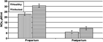
Infected quarters had a greater NOx concentration than healthy quarters in both pre- and postpartum periods (30.81 v. 22.83 μM/ml, P < 0.05 and 9.56 v. 5.77 μM/ml, P < 0.0001, respectively; Figure 1). Overall, the mean NOx levels in the prepartum period (n = 247) were greater than the NOx levels in the postpartum period (n = 247; 26.82 v. 7.67 μM/ml, P < 0.0001; Figure 1). However, it was determined that the interaction between the sampling period and health status had no effect on the NOx concentration (P < 0.16; Figure 1).
NOx concentration for healthy and infected quarters during the prepartum and postpartum periods.

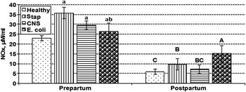
Then the bacterial species were considered. Healthy quarters (n = 147; Table 2) and quarters infected with S. aureus (n = 27), CNS (n = 61) and E. coli (n = 12) had prepartum NOx concentrations of 22.83, 35.61, 29.47 and 26.83 μM/ml in the prepartum period, respectively (P < 0.05; Figure 2). Postpartum NOx concentrations were 5.77 (n = 152), 9.77 (n = 32), 7.08 (n = 46) and 15.90 (n = 17) μM/ml for healthy quarters and those infected with the same bacteria species, respectively (P < 0.05; Figure 2). It was also determined that the interaction between sampling period and the health of the mammary gland in relation to the species of bacteria present had no effect on NOx concentrations (P < 0.08; Figure 2). S. aureus caused the greatest prepartum NOx concentration, whereas E. coli caused the greatest postpartum NOx concentration (P < 0,001; Figure 2).
The number of quarters and health status according to the sampling period

CNS = coagulase-negative staphylococci.
Prepartum and postpartum NOx concentrations in healthy quarters and in quarters infected by various bacterial species. Different superscripts (small for prepartum and capital for postpartum) significantly differ (P < 0.05).

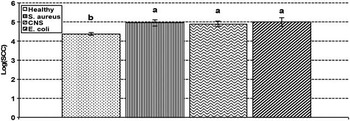
As expected, infected quarters had higher SCC than healthy quarters (mean log SCC 4.95 v. 4.39, P < 0.0001). The mean log SCC was 4.39, 4.97, 4.91 and 5.01 for healthy quarters and quarters infected with S. aureus, CNS, E. coli, respectively (P < 0.0001; Figure 3). NOx level remained unchanged as SCC increased (Figure 4).
Milk SCC for infected and uninfected mammary quarters. Different superscripts significantly differ (P < 0.0001).

Relationship between log (SCC) and NOx concentration in uninfected (y = −0.21x + 6.72, R 2 = 0.001) and infected (y = −0.56x + 11.58, R 2 = 0.006) quarters.

Discussion
Previous in vitro and in vivo studies dealing with cows suggest that NOx concentrations increase during clinical infections (Bouchard et al., Reference Bouchard, Blais, Desrosiers, Zhao and Lacasse1999; Blum et al., Reference Blum, Dosogne, Hoeben, Vangroenweghe, Hammon, Bruckmaier and Burvenich2000; Boulanger et al., Reference Boulanger, Bouchard, Zhao and Lacasse2001; Komine et al., Reference Komine, Kuroishi, Komine, Watanabe, Kobayashi, Yamaguchi, Kamata and Kumagai2004). NOx was reported to be higher for infected quarters than for healthy quarters, suggesting that NOx could be an alternative diagnostic tool for subclinical mastitis (Atakisi et al., Reference Atakisi, Oral, Atakisi, Merhan, Pancarci, Ozcan, Marasli, Polat, Colak and Kaya2010). In the current study, similar results were achieved for heifers in which average NOx concentrations were higher in infected quarters before and after calving when compared with healthy quarters (Figure 1).
In previous reports, E. coli caused significant NOx production in response to intramammary injection of LPS (Bouchard et al., Reference Bouchard, Blais, Desrosiers, Zhao and Lacasse1999; Blum et al., Reference Blum, Dosogne, Hoeben, Vangroenweghe, Hammon, Bruckmaier and Burvenich2000; Burvenich et al., Reference Burvenich, Van Merris, Mehrzad, Diez-Fraile and Duchateau2003). Although basal NOx concentration in milk was reported to be 1.6 μM/l, the concentration increased 10- to 11-fold during experimentally induced E. coli infection (Blum et al., Reference Blum, Dosogne, Hoeben, Vangroenweghe, Hammon, Bruckmaier and Burvenich2000). In this study, NOx concentrations for bacteria species were similar before calving. However, E. coli caused higher NOx (15.90, P < 0.05) production than other bacteria (S. aureus and CNS) after calving (Figure 2). This result was associated with high glandular activity, indicating that E. coli might be more destructive to the epithelial cells when the epithelial tissue become active and NOx response against E. coli could be stronger. High NOx concentrations were reported during mastitis induced with E. coli endotoxin and intramammary infections caused by Staphylococcus spp. Alveolar epithelial cells and bovine PMN have been considered the source of NOx production by some authors (Bouchard et al., Reference Bouchard, Blais, Desrosiers, Zhao and Lacasse1999; Boulanger et al., Reference Boulanger, Bouchard, Zhao and Lacasse2001; Komine et al., Reference Komine, Kuroishi, Komine, Watanabe, Kobayashi, Yamaguchi, Kamata and Kumagai2004).
NOx concentrations were greater in prepartum secretions than postpartum milk samples collected from healthy quarters (Figures 1 and 2). This result could be associated with two factors. It might be related to major development of the mammary gland during pregnancy, such that lobuloalveolar systems are formed and secretion starts by induction of elevated hormones such as estrogen and progesterone during pregnancy (Tucker, Reference Tucker1987; Connor et al., Reference Connor, Meyer, Li, Van Amburgh, Boisclair and Capuco2007). In addition, blood circulation in this stage normally increases and many nitrogenous substrates are carried to the mammary gland by red blood cells (Baumrucker, Reference Baumrucker1985). All of these changes related to physiological cellular activity might lead to higher production of NOx in glandular tissue before calving. On the other hand, two different secretions (lacteal and milk) were compared in this study and increases in milk production after calving could dilute the measured concentration while the total amount has actually increased. Although average test day milk yield was measured in this study, the amounts of mammary secretions in quarters for heifers were not measured. This parameter can be clarified in future studies.
SCC is considered to be an indicator of mammary gland health (Hallberg et al., Reference Hallberg, Dame, Chester, Miller, Fox, Pankey, Nickerson and Weaver1995; Barkema et al., Reference Barkema, Deluyker, Schukken and Lam1999; Bradley and Green, Reference Bradley and Green2005), and it is suggested that SCC status will change depending on bacterial species. There were, however, partially controversial results about the correlation between infection status and SCC in early lactation for some quarters. In this study, sampling was performed 10 days after calving and high SCC results were achieved for some of the quarters that were healthy according to bacteriologic analysis. Therefore, this result seems to support previous studies, which found that SCC was physiologically higher in early lactation and reached normal levels 2 weeks after calving, and that SCC on the day of the test used to detect of IMI status may result in higher values during early lactation (Dohoo, Reference Dohoo1993; Consentino and Luscher, Reference Consentino and Luscher1995; Quinn et al., Reference Quinn, Carter, Markey and Carter1999; De Vliegher et al., Reference De Vliegher, Barkema, Stryhn, Opsomer and De Kruif2004).
In the present study, SCC was generally higher in the infected quarters than in the healthy quarters (Figure 3) in early lactation, although SCC status did not vary among the bacterial species. Similarly, Barkema et al. (Reference Barkema, Deluyker, Schukken and Lam1999) and Bradley and Green (Reference Bradley and Green2005) also reported higher SCC for infected quarters.
Boulanger et al. (Reference Boulanger, Bouchard, Zhao and Lacasse2001) stated that bovine mammary epithelial cells and mononuclear phagocytes produce NOx under inflammatory conditions. Atakisi et al. (Reference Atakisi, Oral, Atakisi, Merhan, Pancarci, Ozcan, Marasli, Polat, Colak and Kaya2010) found a positive correlation between milk SCC and NOx production in the mammary gland. Although Bouchard et al. (Reference Bouchard, Blais, Desrosiers, Zhao and Lacasse1999) reported NOx production in somatic cells, the possibility that NO was produced by cell types other than somatic cells was discussed. Conversely, the correlation between SCC and NOx concentrations was not significant and this result was associated with NOx production from other sources such as endothelial and epithelial cells. In addition to PMN, the major type of white blood cell present during inflammation, endothelial and epithelial cells can also release NOx (Blum et al., Reference Blum, Dosogne, Hoeben, Vangroenweghe, Hammon, Bruckmaier and Burvenich2000; Boulanger et al., Reference Boulanger, Bouchard, Zhao and Lacasse2001). In addition, Boulanger et al. (Reference Boulanger, Zhao, Lauzon and Lacasse2007) reported that PMN does not produce NOx. According to the presented study, infected glandular tissue and leukocytes may be the major sources of NOx during mastitis. Unfortunately, detection of the cell type responsible for NOx production was not possible in this study, but it was clear that somatic cells had a limited effect on NOx levels.
This paper is specific to the status of first-calf heifers and compares changes in the pre- and postpartum NOx concentrations in mammary secretions. Although, it was difficult to determine a NOx cut-off point for bacteria species, significant increments were obtained for IMI. In conclusion, elevated NOx concentrations were dependent on bacterial species actually for E. coli and S. aureus, which caused heifer mastitis, but not SCC. Further studies are needed to elucidate alteration of lacteal and milk NOx level in response to structural and functional development of mammary gland during the transition period.
Acknowledgment
The authors express their sincere appreciation to Dr Armagan Hayirli for statistical analysis of data and critical reading and proofreading of this paper.