Cardiotoxicity has been reported as a rare side effect of etoposide, primarily in adults.Reference Ozben, Kurt and Oflaz1–Reference Airey, Dodwell, Joffe and Jones3 We report a child undergoing chemotherapy with etoposide, cyclosporine, and steroids for haemophagocytic lymphohistiocytosis who acutely developed left-sided cardiac failure due to myocardial ischaemia.
Case report
A 7-year-old Caucasian male child presented with fever of 104 degrees Fahrenheit, fatigue, pallor, and hepatosplenomegaly. Initial investigations showed 10.8 grams of haemoglobin per decilitre, 91,000 platelets per cubic millimetre, 2630 white blood cells per cubic millimetre, which contain 41% neutrophil and 51% lymphocyte, 0.7 milligrams of bilirubin per decilitre, 314 and 235 international units of serum aspartate transaminase and serum alanine transaminase, respectivlely, per litre, 5.1 and 2.1 grams of total protein and albumin, respectively, per decilitre. Prothrombin time and activated partial thromboplastin time were 20 and 48 seconds, respectively. Other studies include 8760 nanograms of serum ferritin per millilitre, 242 milligrams of triglycerides per decilitre, 1669 international units of lactate dehydrogenase per litre, 60 milligrams of serum fibrinogen per decilitre, and D-Dimer 2.58 milligrams per litre fibrinogen equivalent units. Infectious workup was negative, including Ebstein–Barr virus, human herpes virus 6, Hepatitis A, B, and C, adenovirus, and cytomegalovirus. His serum soluble interleukin-2 receptor was markedly elevated, that is, more than 7500 units per millilitre, and bone marrow showed evidence of haemophagocytosis. Perforin and MUNC 14-4 mutations were negative and there was no family history of haemophagocytosis-like illness. The patient was diagnosed with acquired haemophagocytic lymphohistiocytosis and started on daily dexamethasone, that is, 10 milligrams per square metre per day in two divided doses, twice daily administration of cyclosporine 1.5 milligrams per kilogram per dose, and twice weekly administration of etoposide 150 milligram per square metre per dose as per the haemophagocytic lymphohistiocytosis 2004 protocol. At the initiation of chemotherapy, his weight was 23.5 kilograms, height was 123 centimetres, and body surface area was 0.89 square metres. Initially, cyclosporine and dexamethasone were given intravenously, but were switched to an oral form after 1 week.
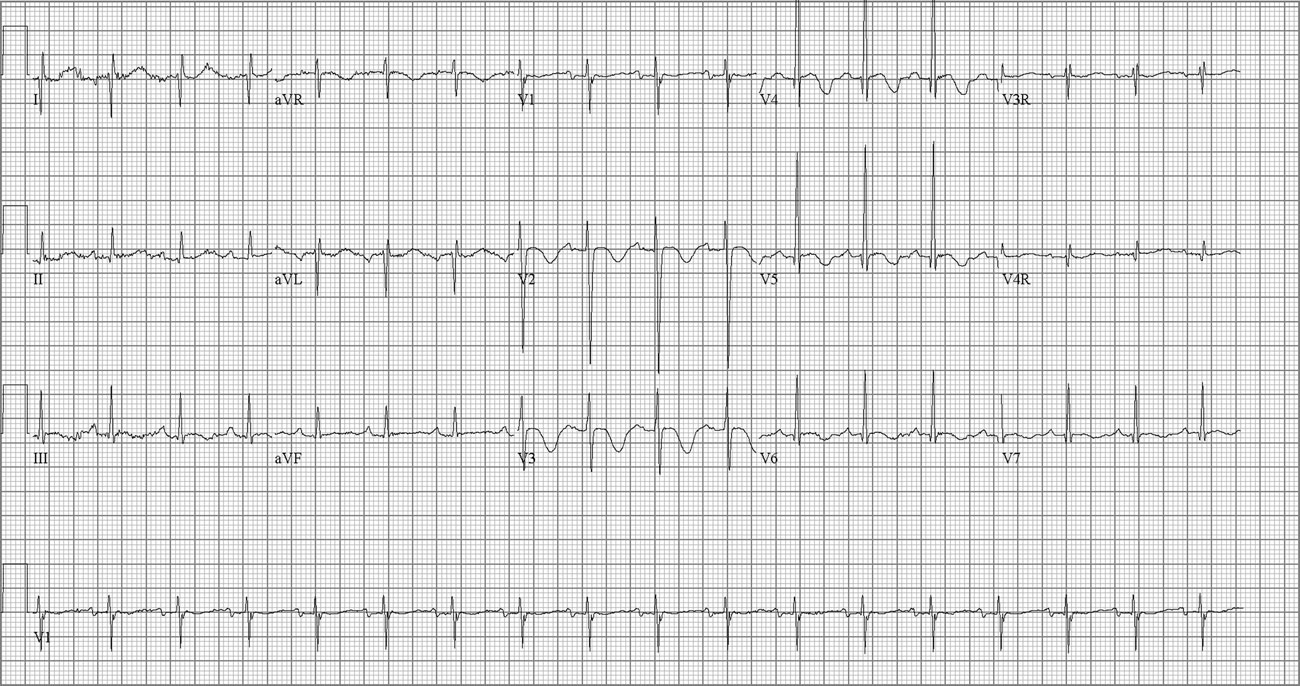
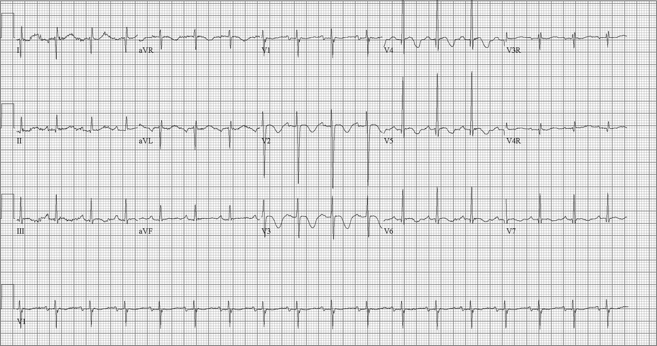
The patient was tolerating chemotherapy well and had gradual improvement of his symptoms to baseline around 10 days after starting therapy. He had a brief episode of tachypnea and oxygen requirement after the third dose of etoposide that was presumed to be due to volume overload and was resolved with a dose of furosemide. Soon after his fourth dose of etoposide, he developed cough and substernal chest pain, and pulmonary oedema was noted on chest radiograph. Broad-spectrum antibiotics and diuretic therapy were initiated. The patient then developed abdominal pain, haemoptysis, hypotension, and respiratory failure requiring vasopressors and mechanical ventilation. Laboratory studies conducted at that time showed 9.8 grams of haemoglobin per decilitre, 39,000 platelets per cubic millimetre, 490 white blood cells per cubic millimetre, 16.2 seconds of prothrombin time, and 29.6 seconds of activated partial thromboplastin time. No evidence of haemolysis is noted on peripheral blood smear and the patient did not receive erythrocyte or platelet transfusion within 4 hours of any of these etoposide administrations. Liver enzymes, such as aspartate transaminase and alanine transaminase, 50 and 75 international units per litre, respectively, 2539 nanograms of serum ferritin per millilitre, and 252 international units of lactate dehydrogenase per litre have significantly improved from diagnosis and 15 milligrams of blood urea nitrogenper decilitre, 0.4 milligrams of serum creatinine per decilitre, and 2.4 milligrams of uric acid per decilitre remained normal. Infectious workup including blood cultures and viral serology tests was negative. Echocardiogram showed a shortening fraction of 15% – pre-chemotherapy 38% – and severely depressed global left ventricular systolic function. Right ventricular function was preserved. Troponin was elevated at 0.14 nanograms per millilitre; normal level was less than 0.06. During his cardiac decompensation, his electrocardiogram showed non-specific ST-T changes. Once stabilised, the patient developed hypertension necessitating intravenous enalapril and sodium nitroprusside. Repeat echocardiogram showed improving left ventricular systolic function with shortening fraction of 22%. With fluid restriction, aggressive diuretic therapy, and enalapril, the patient’s cardiac and clinical status improved and he was extubated. Subsequently, he was started on carvedilol. An electrocardiogram performed a week after decompensation showed prominent antero-lateral Q waves consistent with anterolateral myocardial infarction (Fig 1). Subsequent echocardiograms showed improving ventricular dysfunction with ventricular septal dyskinesis.
Normal sinus rhythm, prominent Q waves in leads I and aVL consistent with antero-lateral infarct.
Chemotherapy in combination with cyclosporine, dexamethasone, and etoposide was held for 6 days due to this clinical decompensation, but stress dose hydrocortisone was continued. After his recovery, he was restarted on oral cyclosporine and steroids; however, further etoposide therapy was held after four doses. He successfully completed 8 weeks of steroids and cyclosporine. Haemophagocytic disease activity completely resolved and his left ventricular function normalised; shortening fraction was 40%.
Discussion
Myocardial ischaemia and infarction has been reported in adult patients receiving etoposide in combination with bleomycin, cisplatin, and ifosfamide.Reference Ozben, Kurt and Oflaz1–Reference Airey, Dodwell, Joffe and Jones3 In addition, myocardial infarction has been described after etoposide when used as a single agent.Reference Aisner, Van Echo, Whitacre and Wiernik4, Reference Schecter, Jones and Jackson5 Proposed mechanisms of etoposide induced myocardial infarction and ischaemia include coronary artery vasospasm, direct injury to the myocardial wall, or induction of an immune response.Reference Airey, Dodwell, Joffe and Jones3, Reference Schwarzer, Eber, Greinix and Lind6 In our patient, viral myocarditis was considered, but without global cardiac dysfunction, it was thought to be unlikely. Ampulla cardiomyopathy (Takotsubo-shaped) was another consideration, but there was no evidence of apical ballooning and hypokinesis of left ventricle to support this entity.Reference Takeoka, Nakamae and Nakamae7 In addition, ampulla cardiomyopathy has been described mainly during the active phase of haemophagocytic lymphohistiocytosis, whereas our patient’s serum markers were improved from diagnosis. Other medications were considered as a cause of myocardial ischaemia, including cyclosporine, but were felt to be less likely, and were restarted without incidence after the patient recovered from the acute ischaemic event. Hence, we presume that myocardial ischaemia in our patient is due to etoposide based on the development of ischaemia in relation to etoposide administration and his lack of other cardiac risk factors. We are unable to explain the mechanism underlying these findings, as we do not have other studies such as coronary angiography or myocardial biopsy. Etoposide-related cardiotoxicity is rarely reported, long-term outcomes and preventive screening are not in place. However, increasing awareness of this phenomenon is essential, especially in children where it is less likely to be considered.