Implications
During weaning, piglets come across stress that may result in changes on intestinal structure. These changes can impair absorptive functions of the intestine and contribute to poor growth performance. Antibiotics have been used to improve growth performance for many years, but antibiotic resistance may cause problems for animal and human health. Chito-oligosaccharide (COS) has been reported as a potential alternative to in-feed antibiotics. In this study, jejunal explants of piglets exposed to different concentrations of chito-oligosacharide induced no effect on intestinal morphology. Therefore, in piglets weaned at 21 days COS may not be efficient to replace antibiotics as growth promoters.
Introduction
Weaning stress has been reported to impair intestinal architecture and function, including intestinal morphology, immune system, and the barrier function (Hu et al., Reference Hu, Xiao and Song2013). The manipulation of gut functions and microbial habitat have been recognized as an important way to improve growth performance of weaned pigs. Antibiotics, as growth promoters and therapeutic medicine, have been used in animal production for many years (Barton, Reference Barton2000). However, consumers are becoming increasingly concerned about drug residues in meat products. In addition, it has been suggested that the continuous use of antibiotics may contribute to a reservoir of drug-resistant bacteria, which may be capable of transferring their resistance to pathogenic bacteria in both animals and humans (Van der Fels-Klerx et al., Reference Van der Fels-Klerx, Puister-Jansen, Van Asselt and Burgers2011). As a result, many countries have banned or are banning the inclusion of antibiotics in swine diets as a routine means of growth promotion. Therefore, many alternatives to the use of antibiotics have been suggested, introduced and tested (Thacker, Reference Thacker2013).
Consumption of functional oligosaccharides, known as dietary fibers, has been shown to improve growth performance and enhance the host health status (Gibson and Roberfroid, Reference Gibson and Roberfroid1995). Chito-oligosaccharide is composed by repeated units of N-acetylglucosamine β-1,4 linked, has low molecular weight (1 to 10 kDa), high solubility, and low viscosity (Chae et al., Reference Chae, Jang and Nah2005). Chito-oligosaccharide has been reported to induce different functions such as antimicrobial (Sánchez et al., Reference Sánchez, Mengíbar, Rivera-Rodríguez, Moerchbacher, Acosta and Heras2017), anti-inflammatory (Fernandes et al., Reference Fernandes, Spindola, de Sousa, Santos-Silva, Pintado and Malcata2010), and also antioxidant, antitumoral and immunostimulatory functions (Li et al., Reference Li, Xing, Liu and Li2016). Chito-oligosaccharide is obtained by chemical and enzymatic hydrolysis of chitosan – deacetylated chitin, which presents low solubility, limiting the application in animals. Chitin, considered the second most abundant polysaccharide in nature following plant cellulose, is found in the cell wall of fungi and exoskeletal area of insects and Crustacea (Muzzarelli and Peters, Reference Muzzarelli and Peters1997). As an alternative to animal extracts, COS can be produced through fermentation by microorganisms such as Bacillus spp. (Wang et al., Reference Wang, Yoo, Kim, Lee and Kim2009). This biotechnological obtaining is independent of climate and environmental factors and has the advantage to be a controlled process, which leads to standard results (Souza and Garcia-Cruz, Reference Souza and Garcia-Cruz2004).
In previous studies, the dietary COS supplementation was shown to improve growth performance in weaned piglets, which may be attributable to intestinal morphology, immunity enhancement and gut microbiota modulation (Liu et al., Reference Liu, Piao, Kim, Wang, Shen, Lee and Li2008; Chen et al., Reference Chen, Kim, Cho, Yoo, Wang, Huang, Kim and Shin2009). However, the range of COS effective doses in these studies is large (30 mg/kg ~5 g/kg) and the in vivo effects of this oligosaccharide are variables (Xiong et al., Reference Xiong, Yang, Wang, Hu, Liu, Wu, Deng, Hou, Nyachoti, Xiao and Yin2015; Wang et al., Reference Wang, Yoo, Kim, Lee and Kim2009). Considering these conflicting results and the need to broaden the knowledge about the effects of COS on the intestinal tissue of piglets, the aim of this study was to assess the effects of exposure to COS in different doses on intestinal morphology using the ex vivo model of pig intestinal explant.
Material and methods
Animals
Five piglets (Landrace×Large White×Duroc) of 24 days old were used to collect the explants. The animals were weaned at 21 days of age, and then subjected to a standard diet after weaning in separate bays. At 24 days the piglets were euthanized with an intravenous injection of sodium pentobarbital (40 mg/kg of BW). The Institutional Ethics Committee for Animal Experimentation approved all animal procedures (number 11361.2014.30).
Chito-oligosaccharide
Chito-oligosaccharide was produced in broth containing 0.7 g/l of mild chrysalis flour of the silkworm Bombyx mori and 0.3 g/l peptone, at pH 9.6, fermented by Bacillus spp. It was used 10% inoculum with incubation at room temperature, under agitation (110 rpm) during 96 h. The mixture was then centrifuged and the supernatant, as a crude extract, was sterilized to be utilized in the assays. Total solids (AOAC, 2005) as 5% and COS concentration (6.48 mg/ml) by the MBTH (3-methyl-2-benzothiazolinone hydrazine) method (Horn and Eijsink, Reference Horn and Eijsink2004) were determined. MBTH gives COS concentration using N-acetylglucosamine 0 to 100 µM as standard, with absorbance determination at 620 nm. This method reduces tenfold the protein interference, when compared with others (Anthon and Barrett, Reference Anthon and Barrett2002).
Jejunal explants technique
The explants were obtained as described elsewhere (Silva et al., Reference Silva, Gerez, Drape and Bracarense2014). Briefly, 5 cm middle jejunum segments were collected immediately after euthanasia, and washed with buffered saline and opened longitudinally. The explants were collected with the aid of a biopsy punch (8 mm) and placed in six-well plates (EasyPath, São Paulo, Brazil), (three explants/well) with 3 ml of the following treatments: control – only culture media (Dulbecco’s modified Eagle’s medium; Gibco – BRL Life Technologies, Carlsbad, CA, USA) plus penicillin/streptomycin (1.25 μl/ml; Gibco – BRL Life Technologies), gentamicin (10 μl/ml, Novafarma, Goiás, Brazil), fetal bovine serum (100 μl/ml; Invitrogen, São Paulo, Brazil) and l-glutamine (0.4 μl/ml; Sigma-Aldrich, St. Louis, MO, USA); culture media with COS in doses of 0.025 mg/ml (25COS), 0.05 mg/ml (50COS), 0.1 mg/ml (100COS) and 0.15 mg/ml (150COS). The concentrations of COS used in this experiment correspond to an ingestion of 25 mg COS/kg, 50 mg COS/kg, 100 mg COS/kg and 150 mg COS/kg of supplemented feed, respectively. The doses of COS used in the current experiment were based on the results of previous studies with weaned piglets (Liu et al., Reference Liu, Piao, Kim, Wang, Shen, Lee and Li2008; Xiong et al., Reference Xiong, Yang, Wang, Hu, Liu, Wu, Deng, Hou, Nyachoti, Xiao and Yin2015; Oliveira et al., Reference Oliveira, Silva, Castro-Gómez, Lozano, Gavioli, Frietzen, Silva, Novais, Frederico and Júnior2017; Suthongsa et al., Reference Suthongsa, Pichyangkura, Kalandakanond-Thongsong and Thongsong2017).
From each animal, six explants (replicates) were collected for each treatment. The total number of explants from each pig was 30. A total of 150 explants were exposed to different treatments, at 37°C with orbital shaking for 4 h. After the incubation, explants were fixed in 10% buffered formalin solution for morphological and morphometric assessment.
Histological and morphometric assessment
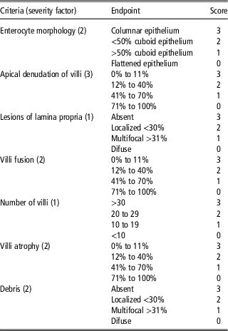
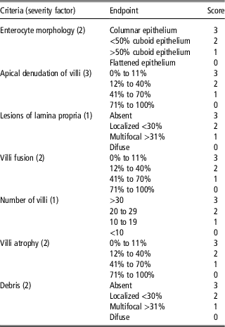
Following fixation, the explants were dehydrated in increasing alcohol solutions, diaphanized in xylene, and embedded in paraffin. Sections of 5 μm were stained with hematoxylin and eosin for histological analysis. An intestinal histological score previously described (Maidana et al., Reference Maidana, Gerez, El Khoury, Pinho, Puel, Oswald and Bracarense2016) was adapted to compare histological changes between the treatments. The maximum score (39 points) indicates the overall integrity of the intestine. Each explant was evaluated to observe the presence and intensity of the criteria disposed on Table 1. For morphometric evaluation, a minimum of 10 well-oriented, intact villi were measured from the crypt mouth to the villus tip and all measurements (villus height and crypt depth) were made with the aid of the Motic analysis system (Motic Instruments, Richmond, Canada).
Explants histological scoring: endpoints used and severity factor
Statistical analysis
The experimental design used in the present study was entirely randomized and consisted of five repetitions per treatment resulting in six replicates from each animal. Data used for statistical analysis were represented as means±SD and analyzed using the free software Rstudio Team (2016). They were statistically analyzed using normality (Shapiro–Wilk) and homogeneity (Bartlett and Levene) tests. When these two assumptions were met, the ANOVA was applied, followed by a linear regression analysis if necessary.
Results
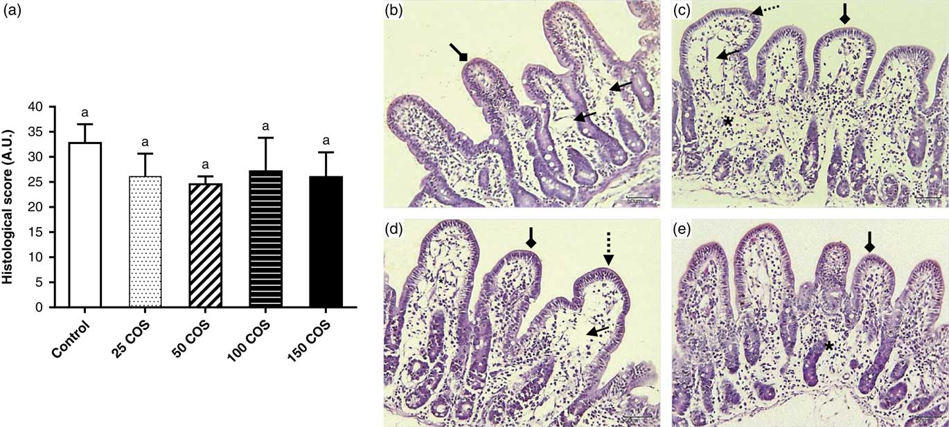
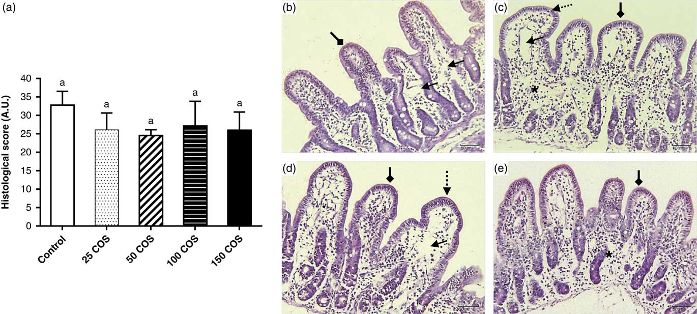
After 4 h of incubation, untreated explants (control group) presented mild dilatation of the lymphatic vessels and a mean histological score of 32.82±3.73 (Figure 1a and b). The main histological findings in explants treated with 25COS, 50COS or 100COS were mild edema of the lamina propria (Figure 1c and e) and moderate villi fusion (Figure 1c and d). The mean histological score for the explants exposed to 25COS, 50COS, 100COS and 150COS was 26.07±4.58, 24.54±1.58, 27.11±6.71 and 25.97±4.95, respectively. No statistical difference was observed in the histological score between explants exposed to COS compared with the control group (P=0.08) (Figure 1a and Supplementary Material Table S1). It is also noted that the simple columnar epithelium of the intestine was preserved in the different treatments (Figure 1b, c, d and e). Mean villi height and crypt depth was assessed in explants exposed to different concentrations of COS, and no significant difference was observed in these groups when compared with the control group, respectively (P=0.44; 0.38) (Table 2). In addition, villus : crypt ratio in the intestinal explants exposed to COS remained statistically similar to control group (P=0.17) (Table 2).
Histological scores of swine jejunal explants. (a) Explants exposed to different treatments: Control ( ) 4 h in Dulbecco’s modified Eagle’s medium (DMEM), chito-oligosaccharide (COS) in doses of 0.025 mg/ml (25COS) (
 ), 0.05 mg/ml (50COS) (
 ), 0.1 mg/ml (100COS) (
 ), 0.15 mg/ml (150COS) (
 ), respectively. Values are mean±SD. Means followed by the same letters do not differ significantly by ANOVA (P⩽0.05). (A.U. – arbitrary units). (b) Explants exposed to DMEM (Control n=30); (c) 25COS-exposed explant (n=30); (d) 50COS-exposed explant (n=30); (e) 100COS-exposed explant (n=30). The lesions are marked with different arrows: slight dilatation of the lymphatic vessels
 ; moderate fusion
 ; mild edema of the lamina propria (*) and epithelium simples columnar
 (Bar=50 µm; H&E staining).
Effects of chito-oligosaccharide (COS) on intestinal morphology of swine jejunal explants (mean±SD)

Means (n=30) within a row followed by same letters do not differ significantly by ANOVA (P⩽0.05). Explants exposed to Dulbecco’s modified Eagle’s medium (control) or chito-oligosaccharide (COS) in concentration of 25 mg/ml (25COS), 50 mg/ml (50COS), 100 mg/ml (100COS) and 150 mg/ml (150COS).
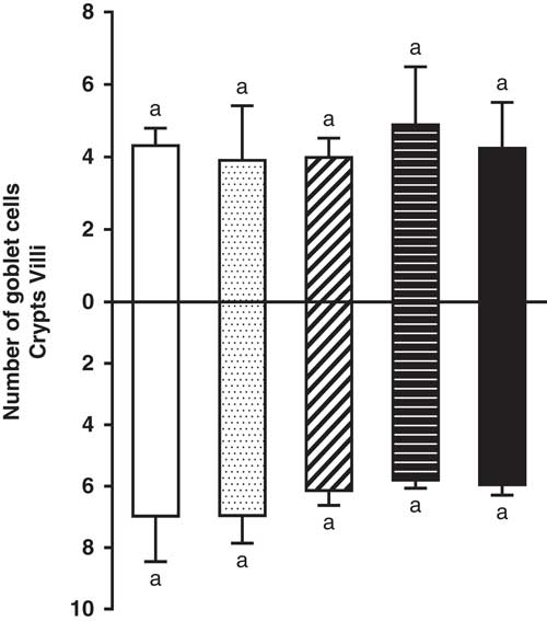
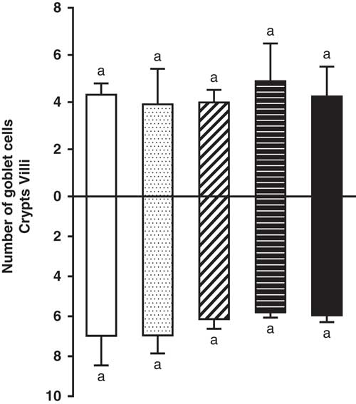
The number of villus and crypt goblet cells in explants treated with 25COS, 50COS, 100COS and 150COS were analyzed. No differences were observed in number of goblets cells among the treatments (P=0.82; 0.27) (Figure 2 and Supplementary Material Table S1).
Mean goblet cell density on villi and crypts of swine jejunal explants exposed to control treatment (), chito-oligosaccharide (COS) to 0.025 mg/ml (), COS to 0.05 mg/ml (), COS to 0.1 mg/ml (), COS to 0.15 mg/ml (). Values are mean±SD represented by vertical bars. Means followed by the same letters do not differ significantly by ANOVA (P⩽0.05).
Discussion
In the last years, there is an increasing interest in strategies focusing on the reduction or elimination of antibiotics as growth promoters. Prebiotic oligosaccharides have been investigated as an effective alternative to antibiotic addition in post-weaning diets of piglets (Xiao et al., Reference Xiao, Tang, Yin, Zhang, Hu, Feng and Wang2013). In addition, the effect of COS supplementation has been evaluated on growth performance, immunological status, gut microbiota and intestinal morphology in weaned pigs (Swiatkiewicz et al., Reference Swiatkiewicz, Swiatkiewicz, Arczewska-Wlosek and Jozefiak2015). In the present study, we have assessed the effect of COS in different concentrations on the intestinal morphology of pigs using an ex vivo approach. Regarding explant culture model, it is interesting to observe that besides reducing the number of experimental animals (3Rs principles – Russel and Burch, Reference Russel and Burch1959), relevant guiding principle for more ethical use of experimental animals, the ex vivo model allows a suitable preservation of the normal histological structure as seen in in vivo models (Lucioli et al., Reference Lucioli, Pinton, Callu, Laffitte, Grosjean, Kolf-Clauw, Oswald and Bracarense2013). To the best of our knowledge, it is the first study to analyze the effects of COS on the intestinal morphology of pigs using an ex vivo model.
Previous studies have reported positive effects of COS on the growth performance and intestinal morphology of weaned pigs (Liu et al., Reference Liu, Piao, Kim, Wang, Shen, Lee and Li2008; Zhou et al., Reference Zhou, Cho and Kim2012; Suthongsa et al., Reference Suthongsa, Pichyangkura, Kalandakanond-Thongsong and Thongsong2017). Weaned pigs fed diets containing COS at 200 mg/kg of feed for a 21-day period showed a significant increase on villus height and villus : crypt ratio in jejunum and ileum (Liu et al., Reference Liu, Piao, Kim, Wang, Shen, Lee and Li2008). In accordance, pigs supplemented with 150 mg/kg COS for 28 days exhibited an improvement in the absorption capacity which was characterized by increased villus height and villus height/crypt depth ratio in the three segments of the small intestine (Suthongsa et al., Reference Suthongsa, Pichyangkura, Kalandakanond-Thongsong and Thongsong2017). In the present study, no significant difference on histological score (villi height, crypt depth and villus : crypt ratio) was observed between explants exposed to the different concentrations of COS and the control group. Similar results were reported in weaned pigs fed diets containing increasing levels of COS (200, 400 or 600 mg/kg) for 14 days (Yang et al., Reference Yang, Ferket, Hong, Zhou, Cao, Zhou and Chen2012). In contrast, post-weaning piglets receiving diets containing 30 mg/kg of COS for 14 days showed a significant decrease in villi height and villus : crypth ratio in the ileum and a tendency to reduce villus height in the duodenum and jejunum (Xiong et al., Reference Xiong, Yang, Wang, Hu, Liu, Wu, Deng, Hou, Nyachoti, Xiao and Yin2015). In addition, it was reported that dietary supplementation with COS (100 mg/kg) obtained from fermentation of chrysalis flour by Bacillus spp. reduced significantly the villi height and villus : crypth ratio in the jejunum of weaned pigs after 18 days of treatment (Oliveira et al., Reference Oliveira, Silva, Castro-Gómez, Lozano, Gavioli, Frietzen, Silva, Novais, Frederico and Júnior2017). Therefore, the controversial effects observed in the before mentioned studies are most probably due to different dosage used and the duration of the experimental treatments. According to Xu et al. (Reference Xu, Shi, Yan, Li, Guo and Li2013) an optimal response in weight gain and histological parameters was obtained in weaned piglets receiving a 0.05% dietary level of COS. This level inclusion corresponds to 500 mg/kg, and in the present study the maximum level evaluated corresponds to 150 mg/kg.
One hypothesis for the improvement reported in the intestinal morphology in animals receiving COS is the presence of the radical N-acetyl glucosamine in the COS molecule (Kim and Rajapakse, Reference Kim and Rajapakse2005). This radical can bind to certain types of pathogenic bacteria, interfering with their adhesion capacity to the gut mucosa (Wang et al., Reference Wang, Yoo, Kim, Lee and Kim2009; Kong et al., Reference Kong, Zhou, Lian, Blachier, Liu, Etan, Nyachoti and Yin2014). Moreover, in vitro and in vivo studies indicates that COS are more rapidly and efficiently utilized by beneficial intestinal microorganisms than chitosan, increasing the proportions of Lactobacillus spp. and Bifidobacterium spp. specially in weaning pigs (Wang et al., Reference Wang, Yoo, Kim, Lee and Kim2009). In addition, COS shorts the cultivation time of probiotics, being comparable with prebiotics traditionally used, pointing it as promising prebiotic (Shang et al., Reference Shang, Jiang, Cai, Hao, Li and Yu2018). Thereby, changes in intestinal microbiota composition may provide a favorable environment for the proliferation of enterocytes. This is in agreement with a previous report where weaned piglets fed diets with increasing COS supplementation (150 and 225 mg/kg) for a 56-day period showed a significant increase on enterocyte crypt proliferation in jejunum (Suthongsa et al., Reference Suthongsa, Pichyangkura, Kalandakanond-Thongsong and Thongsong2017). Similarly, piglets exposed to 150 mg/kg of COS (with 7.5 kDa molecular weight and more than 90% degree of deacetylation (DD)) showed a significant increase in enterocyte proliferation of jejunal crypts compared with animals fed control and/or supplemented diets with 150 mg/kg COS with 65 kDa and ~80%DD and 65 kDa and ~90%DD, respectively (Thongsong et al., Reference Thongsong, Suthongsa, Pichyangkura and Kalandakanond-Thongsong2018). Therefore, short periods of treatment and structural properties of COS may influence intestinal cell recovery, responsible for an improvement in gut morphology in weaned piglets.
Goblet cells are highly polarized secretory cells whose function is to secret mucin glycoprotein, and their number in the intestinal wall reflect the intestinal potential of mucin production (Moncada et al., Reference Moncada, Kammanadiminti and Chadee2003). Mucins comprise a significant portion of mucus localized on intestinal mucosa and contribute to the intestinal barrier function (Pelaseyed et al., Reference Pelaseyed, Bergström, Gustafsson, Ermund, Birchenough, Schütte, van der Post, Svensson, Rodríguez-Piñeiro, Nyström, Wising, Johansson and Hansson2014). Therefore, changes in mucus production or in goblet cell density may facilitate pathogen invasion and cause loss of tissue homeostasis. In our study, no difference was observed on goblet cells density among the treatments, both on villus and crypt region. In accordance, jejunum of piglets exposed to diets supplemented with 30 or 100 mg/kg by 14 and 18 days, respectively, showed no effect of this prebiotic oligosaccharide on goblet cells density (Oliveira et al., Reference Oliveira, Silva, Castro-Gómez, Lozano, Gavioli, Frietzen, Silva, Novais, Frederico and Júnior2017; Xiong et al., Reference Xiong, Yang, Wang, Hu, Liu, Wu, Deng, Hou, Nyachoti, Xiao and Yin2015). Conversely, dietary chitosan at 300 mg/kg for 21 days induced a significant increase on the number of goblet cells in jejunal villi of weaned piglets (Xiao et al., Reference Xiao, Tang, Yin, Zhang, Hu, Feng and Wang2013). Probably, higher doses of COS are necessary to induce changes on goblet cells density. It is to note that data on the literature reporting the effect of COS on swine goblet cells are scarce and in the available database, no study concerning this aspect was found. Further investigation is needed to elucidate the role of COS on goblet cells function.
The present study investigated the effects of COS on intestinal tissue. In recent years, results reporting controversial effects of COS in pigs are found on database. Alternatives to the use of antibiotics as growth promoters are of increasing interest, and COS has been postulated as a possible substitute. Our results indicate that COS exposure in levels ranging from 25 to 150 mg/kg induced no effect on intestinal morphology of pig’s explants. Further studies including higher concentrations of COS and interactions with toxic substances are being conducted also using this ex vivo model to better evaluate the potential of COS as a beneficial feed additive.
Acknowledgments
A.P.F.R.L.B. was supported by fellowship from CNPq, Brazil. J.R.G. and L.Y.B. were supported by Capes/Fundação Araucária fellowship, Brazil. The authors are grateful to Raúl Jorge Hernan Castro-Gómez for having given the samples of COS; to Karina Basso and Thalita Evani Silva de Oliveira for helping in technical assistance for histological processing.
Declaration of interest
The authors have declared that no conflict of interest exists.
Ethics statement
The experimental procedures were conducted in accordance with the Institutional (Universidade Estadual de Londrina, Brazil) Ethics Committee for Animal experimentation (number 11361.2014.30).
Software and data repository resources
The authors declare that no data has been deposited in an official repository.
Supplementary Material
To view supplementary material for this article, please visit https://doi.org/10.1017/S1751731118001076