INTRODUCTION
Anthelmintic resistance to most of the commercially available anthelmintics is a worldwide problem, jeopardizing parasitic worm control in several industries. The macrocyclic lactone anthelmintics (MLs) are the most widely used and the threat to effective worm control posed by resistance already present in trichostrongyloid parasites of sheep and cattle is severe (Mejía et al. Reference Mejía, Fernandez Igartúa, Schmidt and Cabaret2003; Anziani et al. Reference Anziani, Suarez, Guglielmone, Warnke, Grande and Coles2004; Jackson et al. Reference Jackson, Rhodes, Pomroy, Leathwick, West, Waghorn and Moffat2006; Waghorn et al. Reference Waghorn, Leathwick, Rhodes, Jackson, Pomroy, West and Moffat2006; Soutello et al. Reference Soutello, Seno and Amarante2007; Suarez and Cristel, Reference Suarez and Cristel2007; Demeler et al. Reference Demeler, Van Zeveren, Kleinschmidt, Vercruysse, Hoglund, Koopmann, Cabaret, Claerebout, Areskog and von Samson-Himmelstjerna2009). There is an urgent need to understand the mode of action and mechanism of resistance in order to help develop means to delay resistance and to develop new diagnostic approaches. The two major ML groups are the avermectins typified by ivermectin (IVM) and milbemycins, typified by moxidectin (MOX). Despite research in this field over many years, the mode and site of action of the MLs are not fully understood. There is evidence that MLs inhibit larval development, motility and pharyngeal pumping (Kohler, Reference Kohler2001; Moreno et al. Reference Moreno, Nabhan, Solomon, Mackenzie and Geary2010). There is also an ongoing debate if inhibition of pharyngeal pumping or motility is the primary site of anthelmintic activity of MLs. It has been reported that the concentrations to inhibit pharyngeal pumping are lower than those required for inhibition of motility in several nematode species (Geary et al. Reference Geary, Sims, Thomas, Vanover, Davis, Winterrowd, Klein, Ho and Thompson1993; Richards et al. Reference Richards, Behnke and Duce1995; Sheriff et al. Reference Sheriff, Kotze, Sangster and Martin2002). However, observations by Gill and Lacey (Reference Gill and Lacey1998) as well as Sheriff et al. (Reference Sheriff, Kotze, Sangster and Hennessy2005) provided evidence that suggested paralysis of the somatic muscles rather than inhibition of feeding was important for action in vivo. In further support of this, a recent in vitro study by Kotze et al. (Reference Kotze, Hines and Ruffell2012) described observations of changes in adult worm motility at concentrations lower than those required for the inhibition of feeding (0·015 nm of the avermectin abamectin for motility compared with 0·16 nm for feeding).
In earlier studies we compared the effects of six MLs in two in vitro assays of free-living stages of susceptible and resistant parasite species in vitro. In the larval migration inhibition assay (LMIA) MOX showed similar potency and greater RRs than IVM (Demeler et al. Reference Demeler, Gill, von Samson-Himmelstjerna and Sangster2013) in two species of sheep nematode parasites, Haemonchus contortus and Ostertagia (Teladorsagia) circumcincta. In an extension of this work we were interested in using objective measurement to compare the effects of these drugs on muscle contraction and motility in adult worms of the same isolates and to explore mechanisms of actions and of resistance.
The force of muscle contraction in large helminths including H. contortus can be measured directly with force transducers. Sangster et al. (Reference Sangster, Davis and Collins1991) reported the development of a sensitive force transducer technique, including the ability to add drugs to the somatic neuromusculature directly via the pseudocoelomic cavity and to generate concentration-responses. This approach was used successfully to detect effects of cholinergic drugs and investigate the effect of resistance against levamisole (Sangster and Bjorn, Reference Sangster and Bjorn1995). Another instrument, the Micromotility Meter ™ (B & P Instruments, Mason, MI, USA) was reported to be sensitive and accurate in monitoring and recording the in vitro motility of helminths (Bennet and Pax, Reference Bennet and Pax1987). Folz and colleagues developed a motility assay involving this instrument to measure the effects of several anthelmintics (including IVM) in H. contortus and Trichostrongylus colubriformis larvae (Folz et al. Reference Folz, Pax, Thomas, Bennett, Lee and Conder1987a , Reference Folz, Pax, Thomas, Bennett, Lee and Conder b ). The principle of this device is the measurement of reflected light rays. A light located under the bottom of the test tube projects upwards through the tube and its content and refracts horizontally from the meniscus, where it gets measured by a photo detector. Movements of worms cause changes in reflection and therefore of voltage signals received which are amplified, converted into digital data and streamed into PicoScope Software. Higher movements of worms cause larger deviations while signals received from dead worms are almost constant. Geary et al. (Reference Geary, Sims, Thomas, Vanover, Davis, Winterrowd, Klein, Ho and Thompson1993) used the Micromotility Meter to measure the effect of IVM in female adult H. contortus after 1 h of incubation. They detected a rapid onset of inhibition of motility by IVM with concentrations at 10−8 M and above.
In the present study the Muscle Transducer, considered to measure the response of the longitudinal somatic muscles to the tested drugs was used to evaluate the effect of five different ML anthelmintics in adult H. contortus. Since MLs are known inhibitors of contractility their ability to inhibit induced contractions was measured. The Micromotility Meter was used to investigate the effects of four ML anthelmintics on whole adult worms of O. circumcincta.
MATERIALS AND METHODS
Parasites
The drug susceptible H. contortus McMaster (H.c. McM) isolate, the IVM-resistant H. contortus CAVR isolate, and the drug susceptible O. circumcincta McMaster (O.c. McM) isolate were obtained from the McMaster Laboratory, CSIRO (Armidale, Australia). The moxidectin-resistant Ostertagia isolate O. circumcincta WAMIRO (O.c. WAMIRO) was obtained from Dr Brown Besier (WA Department of Agriculture, Australia). All isolates including details of their maintenance have been previously described (Demeler et al. Reference Demeler, Gill, von Samson-Himmelstjerna and Sangster2013).
Donor sheep were infected with single isolates at least 4 weeks prior to collection of worms. Adult H. contortus were picked singly from the abomasum immediately after slaughter while adult O. circumcincta were recovered from the abomasum following migration of worms through a 250 μm aperture sieve in 0·9% saline at 37 °C. Both species were maintained separately in a RPMI-based medium (O. circumcincta. pH 6·6/H. contortus, pH 6·8) in an incubator at 37 °C in an atmosphere containing 5% CO2.
Materials
Ivermectin (IVM, 99·8% pure) was obtained from Sigma (Australia). The two components of IVM, IVM B1a, IVM B1b, as well as milbemycin A3 and A4 (Mil A3, A4; 99% pure) were supplied by the Microbial Screening Technologies (MST, Kemps Creek, Australia). Moxidectin (MOX) was supplied by Fort Dodge Animal Health (Australia) as a 95% pure preparation.
In the Muscle Transducer experiment with H. contortus, the following five MLs were evaluated: IVM, IVM B1a, IVM B1b, Mil A3 and Mil A4. MOX was not available at the time of the study. For each drug the stock solution (10−2 m in 100% Dimethyl sulphoxide (DMSO)) was diluted in Moniliformis buffer (Sangster et al. Reference Sangster, Davis and Collins1991) to achieve final concentrations between 10−5 and 10−11 m (resistant isolate) and 10−9 and 10−15 m (susceptible isolate), maintaining a concentration of 0·5% DMSO. Acetylcholine (ACh) solution at 10−5 m was prepared in buffer.
Acetylcholine (ACh) stock solutions (10−1 m in distilled water) were diluted in Moniliformis buffer and DMSO was added to a concentration of 0·5%.
In the Micromotility Meter experiment the following four MLs were evaluated: IVM, IVM B1b, MOX and Mil A4. In order to limit the number of animals in the experiment (due to ethical considerations) it was decided to restrict the use of adult worms in the Micromotility Meter to four substances. Two avermectins, the commercially available drug IVM (composed of IVM B1a and IVM B1b) and IVM B1b plus two milbemycins, the commercial ingredient MOX and Mil A4 were used. These were chosen because they were commercial products used in the field and a member of each group with the highest resistance ratios in our previous studies (Demeler et al. Reference Demeler, Gill, von Samson-Himmelstjerna and Sangster2013). Drugs were diluted in RPMI medium (pH 6·8) maintaining a concentration of 1% DMSO and giving final drug concentrations between 10−5 and 10−9 m (resistant isolate) and 10−7 and 10−11 m (susceptible isolate). For negative controls the worms were killed by heating in a water bath at 70 °C for 20 min. The readings derived from those worms were almost identical to readings obtained from worms exposed to the stock solution of IVM (10−2 m), so that the value for calculation of the curve was set to 10−2 m. For positive controls worms were kept in RPMI medium containing 1% DMSO with no drugs and the respective value for curve calculation was set to 10−16 m.
Muscle transducer
Cleaned adult female worms were maintained in RPMI medium (pH 6·8) in an incubator at 37 °C for a maximum of 3 days until used. The previously described Muscle Transducer system and cannulated injection method (Sangster et al. Reference Sangster, Davis and Collins1991, Reference Sangster, Song and Demeler2005; Sangster and Bjorn, Reference Sangster and Bjorn1995; Marks et al. Reference Marks, Sangster, Maule, Halton, Thompson, Geary and Shaw1999) were used. The transducer responds linearly up to 200 mg with sensitivity <1 mg. The responses of treated worms were recorded on a chart recorder. A single female worm was held in a bath on a thermostatically controlled hot-plate at 38 °C. Incisions were made at two points of the cuticle to open the pseudocoelomic cavity for cannulation. The cannula was used for a series of injections each of approximately 25 μL in the following sequence:
Moniliformis buffer (the tension on the worm was adjusted to zero response to buffer), ACh (10−5 M, twice), test compound, test compound+ACh-mixture, ACh (10−5 M) and KCl (50 mm). Between each injection except after the test compound, an injection of 25 μL of buffer was used to wash out the compounds (see sequence in Fig. 1). The response amplitude above background measured on the chart recorder of ACh (A), test compounds (D), the ACh-test compound-mixture (M) were measured. The values for M/A for each drug concentration were compared with the equivalent value from worms with test compound replaced by buffer (ACh only in the injection) and inhibition in percentage was calculated (complete inhibition = 0). Each data point on the graphs was derived from a minimum of 6 worms, which were collected from at least 2 different donor sheep. A full concentration-response dataset for a single drug including controls was generated on the same day. The consolidated concentration-response data for the susceptible and the resistant isolate for each drug were each fitted by non-linear regression (four parameter logistic model, variable slope) in the computer programme GraphPad Prism® 5.0 for Windows and graphed. Because the highest concentration of some of the tested substances did not inhibit muscle contraction by >50% we calculated an EC25 and their 95% confidence limits (CL) to make comparisons. In the programme, the average value of 1·2 (positive control) was set to 100% at a given concentration (TOP) and a value of 0·4 (negative control) was set as a negative baseline (BOTTOM). The P-value was calculated to compare (lack of overlap of CL) statistical differences between the means of the pairs of isolates. The resistance ratio (RR) was calculated as the mean EC25 (resistant isolate)/mean EC25 (susceptible isolate).
Response amplitudes recorded on a chart recorder over a series of injections using adult H. contortus in the Muscle Transducer system. Approximately 25 μL of each compound were injected in the following sequence: A = ACh (10−5 m), W = Moniliformis buffer (to wash out remaining ACh), D = test compound, M = test compound+ACh–Mixture and KCl (50 mm).

Micromotility meter
Individual adult O. circumcincta (males and females at random) were manually picked from the storage container with a small forceps, washed and placed in groups of 7–8 individuals into the wells of a 24-well plate (Falcon Tissue Plates). For testing the effects of drugs, each well contained in a total volume of 1·5 mL the worms, RPMI medium and a different concentration of the drug at 10-fold dilutions in different wells. The worms were incubated in these plates for 24 h in an atmosphere containing 5% CO2 at 37 °C. Following incubation, the worms from each well were placed into single glass tubes (inner diameter 6 mm) containing the incubation medium (1·5 mL) and enough fresh medium to bring the meniscus in line with the window of the light source in the Micromotility Meter as described by Folz et al. (Reference Folz, Pax, Thomas, Bennett, Lee and Conder1987a ). The tubes were maintained in a 37 °C warm water bath. For the measurements, a single tube was placed in the channel of the Micromotility Meter (also held at 37 °C). After a settling period of 10 s, motility was measured for 50–60 s. Dead helminths generated a reading comparable to those obtained from medium alone. One drug, at each concentration, was measured in triplicate (three separate tubes) on one day and the mean value generated for each drug and concentration. All experiments were performed 4–6 times (for each isolate-drug-concentration combination). A PicoScope© system (Pico Technology, USA) was used to acquire the readings (in mV) from the Micromotility Meter at a sampling interval of 20 ms. The data were streamed into Excel and a movement index was calculated by averaging 2500–3000 values derived from the absolute value of the difference in mV between consecutive electrical records. Indices for negative controls were subtracted from each index as well as from the positive controls. The movement indices of the triplicates were averaged to obtain a single value per drug concentration on one day and effects were then expressed in percentage of the positive control (set at 100%). These percentages, obtained from 4–6 repeated experiments were co-analysed to generate a concentration-response curve, where an EC50, the 95% CL and the P values were calculated (GraphPad Prism®) as described above for the Muscle Transducer experiments.
RESULTS
Muscle transducer
ACh (10−5 m) elicited significant and rapid muscle contractions and no differences in response amplitude were observed between susceptible and resistant isolates in response to ACh. A typical recording is shown in Fig. 1. It depicts three ACh-induced contractions followed by a relaxation on injection of IVM, then a reduced ACh peak from the mixture and ACh alone (Fig. 1).
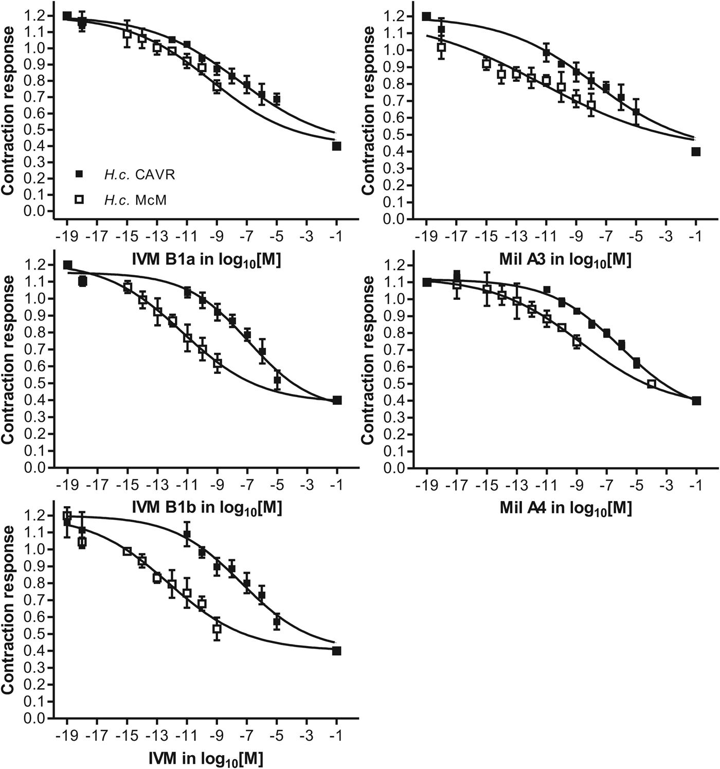
For each drug a sigmoidal concentration-dependent curve was obtained (Fig. 2). The concentration-response curves were relatively flat, running over a wide range of concentrations (Fig. 2) and inhibition was incomplete. It was also irreversible in that subsequent injections of ACh failed to elicit an increased response or a return to the initial ACh-induced contraction. Compared with drug-susceptible worms, all concentration-response curves obtained for resistant worms shifted to the right and all differences between isolates at EC25 values were statistically significant (Table 1). Due to solubility limitations the highest concentration of ML that could be used was 10−5 m. This concentration did not reliably reduce muscle contraction by >50%. While some drugs achieved a reduction of 50%, the EC25 (concentration to reduce contraction by 25%) allowed the comparison within the linear portion of the curve for all drugs. IVM was the most potent drug (EC25 = 0·004 nm) followed by IVM B1b and both milbemycins while IVM B1a was significantly less potent (EC25 2·261 nm). The milbemycins generated lower RR values and IVM B1a ranked in the middle with P values <0·05 for these three compounds. High RRs were generated by IVM (112000) and IVM B1b (13714·29).
Activity of tested drugs as inhibitors of longitudinal muscle contraction of susceptible (□) vs resistant (■) H. contortus isolates (concentrations in nM) in the Muscle Transducer experiment. Error bars show s.d. IVM, ivermectin; IVM B1b, ivermectin B1b; IVM B1a, ivermectin B1a; Mil A3, milbemycin A3; Mil A4, milbemycin A4.

EC25 with the 95% confidence interval (CL) in nM and the resistance ratios (RR) for the tested MLs in the Muscle Transducer (MT) for susceptible (H.c. McM) and resistant (H.c. CAVR) isolates of H. contortus

*P<0·0001; **P<0·05.
Micromotility Meter
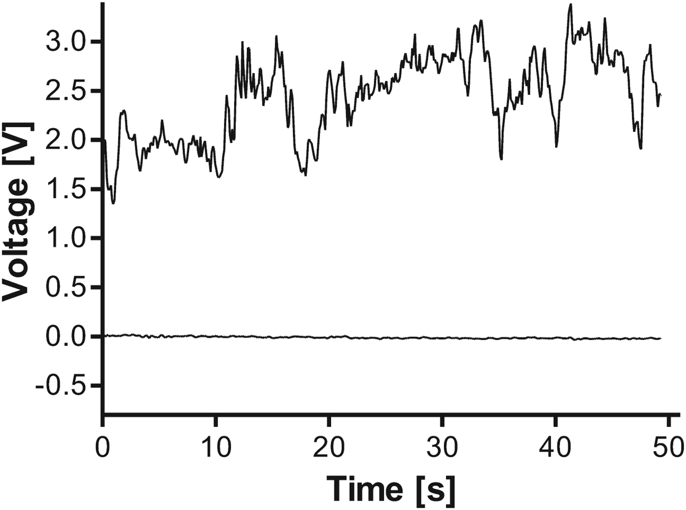
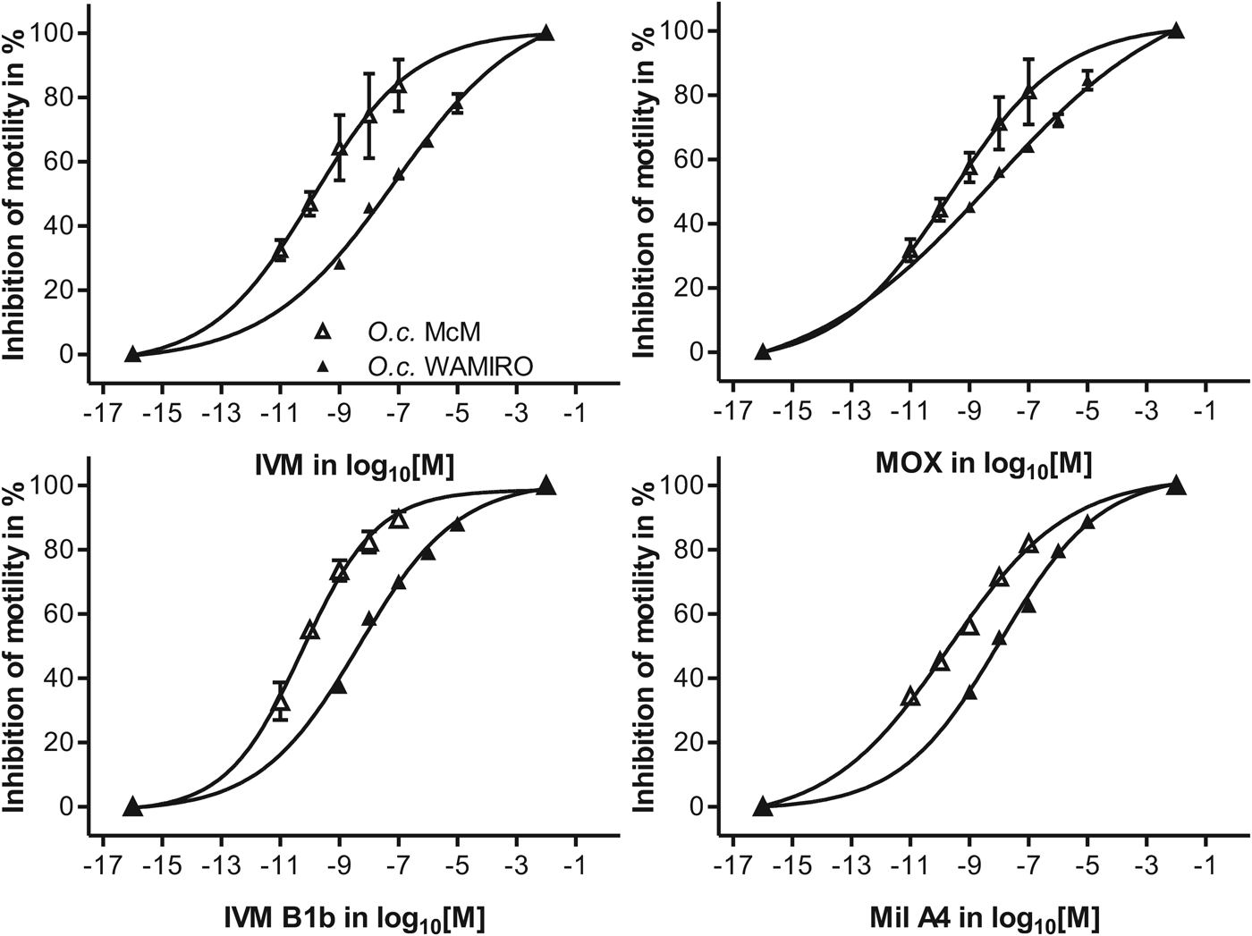
The Micromotility Meter proved to be a reliable assay giving reproducible results for each combination of isolate and drug concentration. A typical example for a read-out using PicoScope software as derived for positive and negative controls is shown in Fig. 3. A concentration-response curve was generated for every drug tested (Fig. 4). The calculation of the movement index in Excel was an efficient means of automating the collection and calculation of data.
Typical traces obtained for motility measurements of O. circumcincta from the Micromotility Meter. The upper line was derived from a negative control (no drugs), the almost straight line (lower line) represents a positive control (dead worms).

Activity of tested drugs as inhibitors of adult motility of susceptible vs resistant O. circumcincta isolates (concentrations in nm) in the Micromotility Meter experiment. Error bars show the s.e. of the mean (s.e.m.). IVM, ivermectin; IVM B1b, ivermectin B1b; MOX, moxidectin; Mil A4, milbemycin A4.

Compounds were typically effective in the nm range (Table 2). The most potent drug was IVM B1b with an EC50 of 0·054 nm for the susceptible O. circumcincta isolate, followed by IVM, Mil A4 and MOX (Table 2). The differences in the concentration-response curves between the susceptible and resistant isolates, reported as RRs, were generally small for this species but all were significantly different. An interesting difference occurred in the group of avermectins, where IVM B1b ranked higher than (but not significantly different) IVM in potency, but the resistance against IVM was found to be more than 6 times larger (RR 82·59 for IVM B1b vs. 599.84 for IVM). MOX scored the lowest RR of these drugs.
EC50 with the 95% confidence limits (CL) in nM and the resistance ratios (RR) for the tested MLs in the Micromotility Meter (MM) for susceptible (O.c. MCM) and resistant (O.c. WAMIRO) isolates of O. circumcincta

*P<0·0001; **P<0·05.
DISCUSSION
The focus of this study was to compare the effects of MLs on contraction of somatic muscles in susceptible and ML-resistant adult worms. The use of highly resistant isolates provided clear differentiation of the isolates in terms of RRs. These isolates may not be representative of other isolates present in the field. Another problem for direct comparison of in vitro and in vivo results may be the fact that efficacy data from the field are usually only available for therapeutic doses but rarely for defined overdoses of the respective host species. However, these in vitro methods provide robust tools for investigating the pharmacology of resistance.
The use of the two different methods in two different species means that direct comparisons are not possible. The transducer study can only be applied to relatively large worms such as the adult female H. contortus and requires considerable manipulative skills. The Muscle Transducer system allows injection of agents into the pseudocoelomic cavity and has been used in the past to measure excitatory as well as inhibitory responses (Marks et al. Reference Marks, Sangster, Maule, Halton, Thompson, Geary and Shaw1999). It provides a significant advantage over other assays in that nerve and muscle cells are exposed to the drugs and that responses are rapid. The Micromotility Meter has been reported as suitable for a range of sizes of worms. In our hands H. contortus became too entangled to provide reliable readings and some movement, even from partially paralysed individual males, evoked saturation of the electrical signal. The data for O. circumcincta were reproducible and allowed for relatively rapid analysis even though the machine had just a single channel. In both assays the data had relatively large CLs accentuated by relatively flat concentration-response lines. Statistical differences based on lack of overlap of CL were not commonly found between drugs, although differences between isolates, reported as significant RR values, were recorded for every comparison.
MLs have a number of putative sites of action in worms including inhibitory action on the pharynx, inhibition of body muscle contraction and effects on sensory neurons. There is evidence that their primary site of action in nematodes is by inhibition of glutamate-activated Cl− channels (GluCl) in nerves and interneuronal junctions where they cause release of Cl− ions and hyperpolarization leading to inhibition. Haemonchus contortus has a family of genes that code for the receptor subunits that form these channels (Wolstenholme and Rogers, Reference Wolstenholme and Rogers2005; McCavera et al. Reference McCavera, Walsh and Wolstenholme2007). Different GluCl subunits have been localized to neuromuscular sites including the motor neuron system of the body, especially commissures and ventral nerve cord, cells in the pharynx and sensory neurons in the head (Portillo et al. Reference Portillo, Jagannathan and Wolstenholme2003). Given the ability of the MLs to cause relaxation and paralysis, inhibitory neurons at somatic sites are likely sites of action of the MLs. Unresponsiveness of muscle cells to ACh in the Muscle Transducer also suggests that contraction is inhibited by activated GABAA receptors. The neuromuscular structure of nematodes is such that the interneuronal and neuromuscular connections are either exposed to the pseudocoelomic cavity or are located in adjacent structures such as the ventral nerve cord or commissures.
In studying the pharmacology of these receptors the transducer studies have particular advantages. The first is that because cannulation of the worm requires only two snips in the body wall, the cylindrical structure of the worm remains intact thus preserving the commissures and most of the nerve cord and neuromuscular connections. Second, because fixed concentrations of drug and mixtures are delivered to the pseudocoelomic cavity, the neuromuscular system is exposed to known concentrations of drug. For embedded target tissues the diffusion path is short. Third, direct local application of drugs avoids the potential complicating effects of ingestion of compound, diffusion through the cuticle and other possible sites of action. Fourth, following addition of sufficient concentrations of MLs relaxation was immediate suggesting a direct effect on nerves present in the preparation and innervating muscle. The 60 s period that expired between initial exposure of each worm to ML and injection of the ML/ACh mixture was required for quantitation. Further, by this time the MLs had caused their maximum inhibitory effect and this was irreversible. The ability to generate concentration-responses to a range of ML compounds in the present study strengthens the case for the somatic neuromuscular system as the major site of action of MLs. It follows that neural inhibition leads to muscle relaxation. The resistance to these compounds is also present at this site. The local nature of the effects illustrates that this response is independent of other putative sites of action in the worm and the rapidity of effect supports a neuromuscular effect in response to a ligand/receptor interaction. These data also mitigate against efflux mechanisms or metabolism as major mechanisms of resistance as the time frames are short.
In the transducer study concentrations (EC25) required to inhibit muscle contraction varied between tested substances and ranged between 0·004 nm for IVM and 2·261 nm for IVM B1a. The high potency of IVM is surprising and in contrast with that of its components. The experiments with IVM were repeated several times and the high potency confirmed. Activity of all compounds in the nM range indicates that this assay is highly sensitive. Although the potency of all substances was dramatically lower in the resistant isolate, it is interesting to compare potency. While the rank potency in the susceptible isolate was IVM>IVM B1b>Mil A3>Mil A4>IVM B1a, for the resistant isolate it was Mil A3>IVM B1b>IVM>Mil A4>IVM B1a. However, the overlap of CLs means that these rankings overlap. Another pattern that emerged was that the most potent compound had the highest RR. These data suggest that IVM B1b may be the major component contributing to IVM resistance in adult worms even though it accounts for only 20% of the composition of IVM. The importance of 25% inhibition of contraction is not known, however, the drugs alone caused relaxation and a 25% reduction of a large induced contraction would be expected to impair function. While the effects of the ML compounds have been described on intact worms, their effects on muscle preparations have been rarely described. Kass et al. (Reference Kass, Stretton and Wang1984) demonstrated that IVM had inhibitory effects on muscle preparations of Ascaris suum split longitudinally. The present study is the first to analyse the pharmacological effects of ML anthelmintics in preparations with intact body integrity. It is also the first to use such a preparation to describe the pharmacology of resistance.
The motility effects on O. circumcincta were performed on intact worms. In contrast to the transducer study the means by which drugs reach their site of action is unknown. For the MLs it is likely some drug is ingested and then diffuses from gut to the nerves, but the fact that MLs are potent inhibitors of pharyngeal pumping (Geary et al. Reference Geary, Sims, Thomas, Vanover, Davis, Winterrowd, Klein, Ho and Thompson1993) would limit the importance of that route early in in vivo treatment or in vitro incubation. Transcuticular diffusion is an alternative route of entry to sites of action. When studying intact whole worms, such as in the Micromotility Meter and in the study of Kotze et al. (Reference Kotze, Hines and Ruffell2012), long incubation times are required to achieve consistent effects. This may be to allow for equilibration of the drug throughout the tissues. It is not possible to know what concentrations are present at the nerve sites in the body or if remote sites of action such as pharynx or sensory neurons had indirect effects on motility especially over the time courses used in the studies. The irreversible nature of the inhibition makes it impossible to tease out the kinetics of the response. Nevertheless, the assay examines responses in the intact parasitic stages and in this context the comparison between IVM and MOX provides useful data.
The O.c. WAMIRO isolate is known to be highly resistant to IVM (efficacy 0%) and MOX (efficacy 5%) in vivo (Besier, personal communication). In using adults of the susceptible isolate in this study we found a rank potency (EC50) of IVM B1b>IVM>Mil A4>MOX with IVM B1b significantly more potent than both Mil A4 and MOX. The rank order in the resistant isolate was IVM B1b>MOX>Mil A4>IVM and the RRs cover the extremes from ∼600 for IVM to 27·73 for MOX. Geary et al. (Reference Geary, Sims, Thomas, Vanover, Davis, Winterrowd, Klein, Ho and Thompson1993) reported that IVM concentrations of 1–10 nm inhibited motility of adult female H. contortus in the Micromotility Meter by 50%. They were able to inhibit motility by ∼50% and up to 80% with IVM concentrations of approximately 10 and 100–1000 nm, respectively. Kotze et al. (Reference Kotze, Hines and Ruffell2012) also observed effects on motility in whole H. contortus and compared a susceptible isolate to one resistant to IVM and MOX. As in the present study where similar O. circumcincta isolates were compared after exposure to IVM, sub-nM concentrations of the IVM analogue abamectin achieved observable effects on motility (Kotze et al. Reference Kotze, Hines and Ruffell2012) and RRs were 1000-fold or more. These workers also discussed what level of motor impairment might upset the host-parasite relationship sufficiently to achieve drug efficacy. The observations of effects in the sub nM range suggest that the observed effects and those in the present study are relevant.
Considering the present work, the rank orders of potency and RRs in the transducer study are mirrored in the motility study and suggest a similarity of action and resistance (although MOX was missing from the transducer study). Potencies are in the same order of magnitude for the two studies, but RRs are far larger in the transducer study. Despite the use of different species, the similarities support the hypothesis that the motility assays used for adult worms reflect activity at the somatic site of action identified by the transducer study. The somatic site is also the most likely site of resistance in both isolates of the two species tested here and those used in other studies. Thus resistance is most likely to involve differences in receptor expressed on neurons.
Effects on motility of adult parasitic stages of H. contortus and O. circumcincta mirror those in studies on the motility of larvae of the same species (Demeler et al. Reference Demeler, Gill, von Samson-Himmelstjerna and Sangster2013). These motility/migration assays rely on the ability of MLs to inhibit body muscle contraction. The most direct comparison to the use of adult worms in the present study was the study of Demeler et al. (Reference Demeler, Gill, von Samson-Himmelstjerna and Sangster2013), where the same isolates and the same drugs were used in the LMIA. The drugs are far less potent against third-stage larvae. For example, for IVM EC50s were 266 nm in larvae and 0·125 nm in adults. Differences may be due to a range of factors but expression of receptors, the cuticular structure of the larvae, the non-feeding status, the incubation conditions and the end-point detection methods are possible sources of difference. Although the rank order of potency varied between the studies (larvae IVM>IVM B1b>MOX>Mil A4; adults IVM B1b>IVM>Mil A4>MOX) the avermectins ranked consistently above the milbemycins. Considering resistance, the patterns were different: in larvae MOX had a relatively high RR but it was lowest for adults. Potency aside, these data suggest that the site of action of MLs has similar but not identical pharmacology between the stages. Nevertheless, the RR generated by the MOX-resistant O.c. WAMIRO isolate in the Micromotility Meter (RR = 27·73) is close to the one obtained for larvae in the LMIA (RR = 22·45). RRs are good comparators because, as ratios, they compare within species and within assays. The data indicate a similarity in the pharmacology of resistance to the effects of MOX on motility at these two stages of the life cycle.
Like the study on larvae (Demeler et al. Reference Demeler, Gill, von Samson-Himmelstjerna and Sangster2013) IVM and MOX have equipotency on adult O. circumcincta in vitro (Table 2). It has been previously reported that parasite isolates resistant to IVM show decreased susceptibility to MOX in vivo but that MOX was generally more effective than IVM against resistant isolates (Conder et al. Reference Conder, Thompson and Johnson1993; Shoop, Reference Shoop1993; Gill et al. Reference Gill, Redwin, Van Wyk and Lacey1995; Sutherland et al. Reference Sutherland, Leathwick, Moen and Bisset2002; Le Jambre et al. Reference Le Jambre, Geoghegan and Lyndal-Murphy2005). In vitro studies are independent of host effects and the results suggest that the explanation for the superior efficacy of MOX in vivo is its pharmacokinetics. The longer half-life and persistence in the host is likely to improve efficacy of MOX in comparison to IVM. This means that IVM and MOX resistance is best described as ‘side resistance’. Interestingly, in both the study on larvae and the present study with adults (Fig. 4 MOX), the concentration-response lines for MOX are not parallel. This suggests that the levels of resistance are concentration dependent and that the EC50 alone is insufficient to describe resistance to MOX. The fact that the two lower concentrations are not available for the resistant isolate can possibly distort the shape of the curve. However, it does emphasize the importance of a concentration-response analysis as a means of describing the population and the characteristics of resistance and hence the value of in vitro observations.
ACKNOWLEDGEMENTS
The authors thank Michelle Power for her help with the Micromotility Meter, Ernest Lacey and Jenny Gill for the chemicals and Fort Dodge Animal Health (Sydney) for the supply of moxidectin.