Introduction
Epistaxis is an extremely common presenting complaint to primary care, emergency departments and ENT services. Although sinister causes are rare in children, early detection and specialist assessment are vital to ensure the best possible prognosis. Failure of symptoms to resolve despite treatment should always prompt consideration of alternative diagnoses and further investigation. We describe the investigation and differential diagnosis for one such patient in the context of a nasal mass. This case also highlights the challenges and potential complications encountered by the patient, family and medical team in dealing with an atypical, aggressive and locally advanced tumour in a previously well nine-year-old boy.
Case report
Case presentation
The patient presented to a local hospital with a six-month history of right-sided, intermittent epistaxis which had persisted despite multiple previous treatments with Naseptin® ointment and silver nitrate cautery. The patient denied any nasal obstruction or other systemic symptoms. There was no history of trauma and no other previous medical history of note. An ENT and systemic examination was normal, with no palpable lymphadenopathy or discernible cranial nerve palsy.
Following successive reviews by the ENT team, blood clots were suctioned from the nose, revealing a soft tissue mass high on the right side. Examination under anaesthetic confirmed the presence of a mass, which was biopsied prior to transferring the patient to a specialist paediatric unit.
Investigation
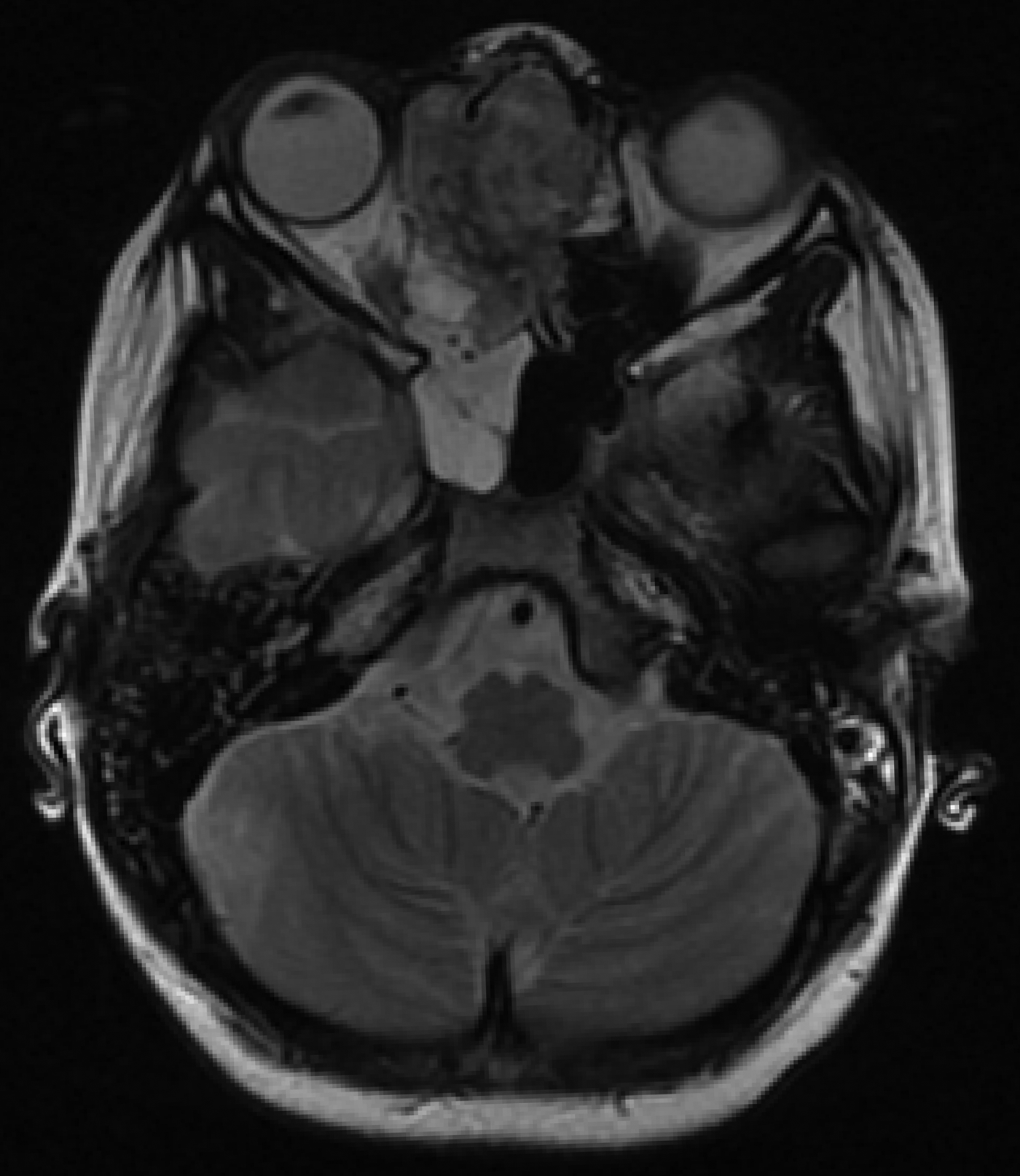
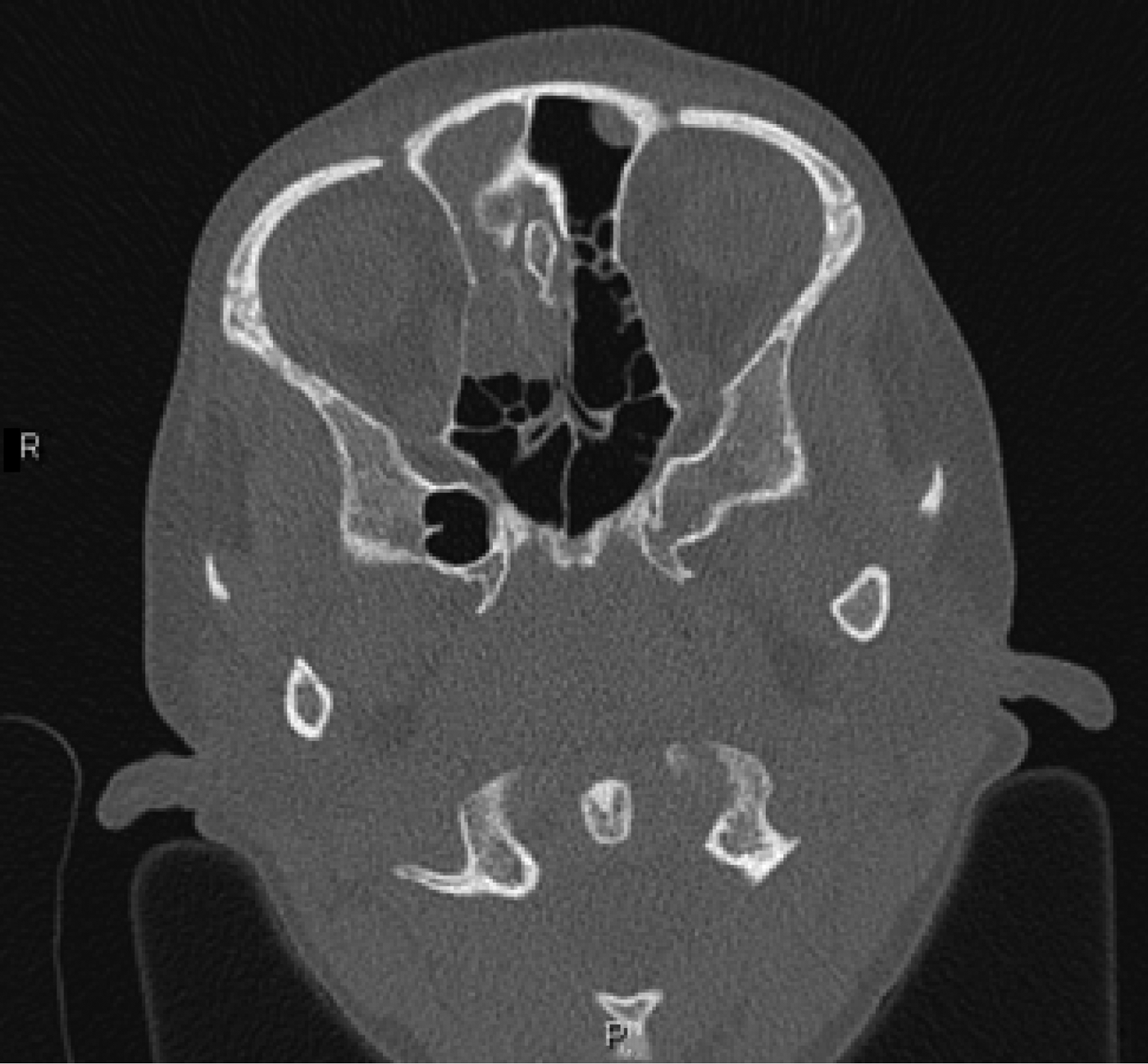
Upon the patient's admission to our centre, flexible nasendoscopy with a local anaesthetic spray was used to re-examine the mass, and head and neck magnetic resonance imaging (MRI) was arranged. The scan revealed a right-sided 3.5 cm nasoethmoid mass lesion abutting the right medial orbit and anterior skull base (see Figure 1). The lesion was seen to extend across the midline but not intracranially or intraorbitally. Prominent reactive lymph nodes were also noted in the level 2 region on the right side. Computed tomography (CT) imaging of the sinuses showed areas of bone thinning related to the cribriform plate and lamina papyracea (Figure 2). Chest and abdominal imaging showed no evidence of metastatic disease.
T2-weighted sequence magnetic resonance image showing a transverse section of the patient's head and neck. An intermediate T1–T2 signal mass is seen in the right anterior nasoethmoid region. Movement artefact is caused by the scan being performed without general anaesthesia.

Computed tomography image, showing a transverse section of the patient's sinuses, with a right-sided soft tissue mass (bone window). Movement artefact is caused by the scan being performed without general anaesthesia. R = right; P = posterior
Histological analysis of the biopsy samples confirmed a high-grade, poorly differentiated malignancy most consistent with nasopharyngeal carcinoma.
Differential diagnosis
This history initially warranted consideration of an early presentation of juvenile nasopharyngeal angiofibroma. Given the radiological findings, differential diagnoses for this patient would also include a lymphoma, rhabdomyosarcoma or olfactory neuroblastoma, thus emphasising the importance of a definitive tissue diagnosis.
Treatment
Following attendance at the regional paediatric oncology centre and rigorous multidisciplinary team discussion, a combined craniofacial resection was scheduled at a fourth hospital linked to the regional centre. In the interim, however, the tumour size increased significantly so as to protrude from the patient's nostril, and he attended an Accident and Emergency Department with worsening nasal obstruction and difficulty in breathing (see Figure 3). The patient was admitted to hospital. Whilst awaiting surgery, he experienced a major haemorrhage requiring blood transfusion and expedited surgery to debulk the tumour.
Photograph of the patient upon readmission prior to surgery, showing tumour progression. Taken by the patient's parent.
Outcome and follow up
The patient recovered well after surgery and has successfully completed adjuvant chemotherapy with cisplatin and 5-fluorouracil. Although post-operative MRI demonstrated significant progression around the skull base with dural enhancement, subsequent imaging has shown a good response to treatment, with tumour regression.
A chromosomal rearrangement, typically t(15;19), affecting the nuclear protein in testis (NUT) gene has been reported in a group of cancers described as NUT midline carcinomas.Reference French, Kutok, Faquin, Toretsky, Antonescu and Griffin1 However, analysis of a surgical specimen at a centre in the USA found it to be negative for such a mutation.
Discussion
Nasopharyngeal masses in the paediatric population may arise from benign, congenital, inflammatory or malignant lesions. Childhood malignancies in this region are rarely seen, even in specialist centres. Therefore, a methodical approach to assessing these patients, combined with an awareness of the spectrum of possible pathologies, is essential.Reference Duarte, Liu and Shapiro2 Here, we will briefly outline the diagnostic and management considerations related to this case, together with a review of this esoteric condition.
In an adolescent male presenting with epistaxis, we would suspect juvenile nasopharyngeal angiofibroma. These account for 0.05 per cent of head and neck tumours, and typically present in teenagers but can occur in younger patients.Reference Martin, Ehrlich and Abels3, Reference Yi, Fang, Lin, Lin, Xiao and Li4 Tissue biopsy is not recommended in the initial investigation of juvenile nasopharyngeal angiofibroma because of the risk of major haemorrhage. The diagnosis is usually based on cross-sectional imaging and clinical correlation. In children with a unilateral nasal mass, radiological assessment prior to biopsy may also be advantageous for detecting congenital masses connected to the cerebrospinal fluid.
The sinister findings in this case are extremely rare in children of this age in the UK, where the annual incidence of nasopharyngeal carcinoma (NPC) is reported to be 0.1 per million at age 0–9 years, rising to 0.8 per million at age 10–14 years.Reference Brennan5 However, NPC is endemic amongst the adult population in regions of China and southern Asia, where it displays a distinct geographical and racial distribution. The global disparity in NPC incidence, together with the bimodal age peaks observed in lower incidence countries such as the UK, suggests a multifactorial disease aetiology.Reference Brennan5, Reference Bray, Haugen, Moger, Tretli, Aalen and Grotmol6
Basic science and epidemiological studies have produced a large body of evidence indicating that the ubiquitous Epstein–Barr virus (EBV) is intimately related to the pathogenesis of NPC. For example, EBV gene products have been discovered in NPC precursor cells.Reference Pathmanathan, Prasad, Sadler, Flynn and Raab-Traub7 Exposure to volatile nitrosamines and tobacco have also been implicated in oncogenesis, and a number of predisposing genetic polymorphisms have been identified. However, the complex interactions amongst these factors in NPC development across different age groups are yet to be fully elucidated.
Most NPC patients present with advanced stage disease. This may be partly caused by a prolonged latent period before symptoms arise, combined with a tendency toward early metastasis. However, it is well recognised that difficulty in arriving at a diagnosis can contribute to delayed identification of NPC.Reference Leong, Fong and Low8 The clinical presentation of NPC is often subtle, and can vary considerably in the combination or absence of symptomatology. The symptoms and signs of NPC can be divided into four groups: (1) those related to a tumour in the nasopharynx (epistaxis, nasal obstruction and discharge); (2) those related to eustachian tube dysfunction caused by posterolateral extension of the tumour (serous otitis media, tinnitus and hearing loss); (3) those caused by erosion of the skull base and associated cranial nerve palsy (headache, diplopia, facial pain and numbness); and (4) neck masses, usually caused by metastatic lymph node deposits.Reference Wei and Sham9 Therefore, a high index of suspicion is required when diagnosing such patients.
Initial assessment of the nasal cavity and post-nasal space is generally considered difficult in children, but flexible laryngoscopy in the clinic (using a thin, flexible scope) is often tolerated from at least seven years of age (if not younger). In this instance, it was also possible to carry out both CT and MRI scans without the need for sedation. If flexible nasendoscopy is not possible, then cross-sectional imaging followed by examination under anaesthetic is recommended. This approach enables a biopsy to be taken at the same time once angiofibroma and encephalocele have been excluded, as described above. Ideally, anaesthesia would be administered in a specialist centre because upper airway obstruction caused by the mass prolapsing into the oropharynx may occur in the immediate post-operative period.
• Nasopharyngeal malignancy is a rare paediatric condition
• This case highlights the benefits of early flexible nasendoscopy in children with recurrent epistaxis
• Early soft tissue imaging can help delineate the cause of unexplained symptoms, including benign and malignant pathology
• Large tumours are capable of significant haemorrhage, warranting close observation and prompt intervention
Our patient was transferred amongst four different hospital trusts to establish a definitive diagnosis and for treatment. This may have significantly added to feelings of anxiety and responsibility for the family in coordinating his care. It may also have prolonged the time to treatment, and increased the risk of complications such as deep extension, skull base erosion, intracranial spread and distant metastasis.
This case illustrates that in paediatric patients with rapidly enlarging tumours, there is also a risk of acute, life-threatening haemorrhage, with consequent surgical challenges. Indications for surgical resection of NPC are limited and the mainstay of treatment is neoadjuvant chemotherapy, irradiation or adjuvant interferon β therapy, depending on staging and prognostication.Reference Brennan5, Reference Buehrlen, Zwaan, Granzen, Lassay, Deutz and Vorwek10 Although postoperative MRI suggested that surgical excision may not have been complete, In this case, surgical intervention was vital to prevent further haemorrhage. However, a good response to treatment has been reported following adjuvant chemotherapy, which we hope will be enhanced by forthcoming radiotherapy.
Acknowledgements
The authors gratefully acknowledge the contributions of Dr S Zacharoulis at the Royal Marsden NHS Trust and Mr T Odutoye of St George's Healthcare NHS Trust, as well as the invaluable assistance of the Evelina London Children's Hospital ENT secretaries.