“As any classification is necessarily incomplete and acts as a bridge between complete ignorance and total understanding in any biological system, … further modification and changes are likely to occur”
(John Goodwin, London 1982).
Historical context
Classification systems are used to aid understanding and systematic discussion through the use of logical groups and hierarchies. In clinical practice, similar principles are used to standardise disease nomenclatures. The term cardiomyopathy was first coined in 1957, to denote primary or idiopathic diseases of the myocardium, as distinct from ischaemic heart disease.Reference Brigden 1 Over the following five decades, there has been a progressive evolution in the nomenclature and classification systems used for heart muscle disorders, driven primarily by scientific advances in the understanding of disease pathogenesis and aetiology. Initial classification systems based on descriptions of clinical features in the 1960s evolved into a pathophysiological taxonomy in the 1970s and 1980s, in parallel with improvements in clinical imaging modalities, especially echocardiography and MRI. Underpinning these classification systems was an attempt to define cardiomyopathies according to their underlying aetiology, ventricular morphology, and function. This resulted in four distinct subdivisions endorsed by the World Health Organisation and the International Society and Federation of Cardiology Task Forces in 1980 and 1995: dilated, hypertrophic, restrictive, and arrhythmogenic right ventricular cardiomyopathies.Reference Oakley, Olsen and Goodwin 2 , Reference Richardson, McKenna and Bristow 3 As summarised by the 1995 Task Force, cardiomyopathies are “diseases of the myocardium associated with cardiac dysfunction” and should be “classified by the dominant pathophysiology or, if possible, by aetiological or pathogenetic factors”.Reference Richardson, McKenna and Bristow 3 A distinction was made between “primary”, or idiopathic, heart muscle disorders – cardiomyopathies – and “specific heart muscle disease”, which describe disorders with similar morphological features but distinct underlying aetiology, such as heart disease due to metabolic, inflammatory, or ischaemic processes. The genetic advances that followed in the early 21st century added a new dimension, such that the former “idiopathic” phenotypes became increasingly linked to specific gene mutations, making this term largely redundant. Unfortunately, many of the linkages exhibit highly variable or little genotype–phenotype correlation, confounded by age-dependent changes and varying penetrance, even within families. Given these difficulties, the classification of cardiomyopathies continues to be driven by clinical phenotype, with integration of causation and inheritance pattern of the disease whenever possible. A suggested “red flag” methodology for assessment of the individual patient is, therefore, described at the end of this manuscript.
In the last decade, two dominant classification schemes with distinct definitions for cardiomyopathy have evolved:
-
∙ the 2006 American Heart Association definition and classification system; and
-
∙ the 2008 European Society of Cardiology classification scheme.
Although there are some similarities between these classification schemes, there are also substantial differences, many of which are addressed in this review. A third classification system, the MOGE(S) classification, has been recently suggested, and is also briefly discussed.
The European Society of Cardiology classification system
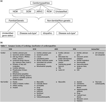
In the European Society of Cardiology classification scheme, cardiomyopathy is defined as follows: “A myocardial disorder in which the heart muscle is structurally and functionally abnormal, in the absence of coronary artery disease, hypertension, valvular disease and congenital heart disease, sufficient to cause the observed myocardial abnormality”.Reference Elliott, Andersson and Arbustini 4 The cardiomyopathies are subdivided into four distinct phenotypes – namely, hypertrophic, dilated, restrictive, and arrhythmogenic right ventricular cardiomyopathy – as well as into a fifth group of “unclassified” cardiomyopathies including left ventricular non-compaction and Takotsubo cardiomyopathy. There follows a further subdivision of these five specific morphological and functional phenotypes into familial/genetic and non-familial/non-genetic forms, with a large list of individual entities detailed in each category (Fig 1).Reference Lopes and Elliott 5
Classification system of cardiomyopathies from the European Society of Cardiology Working Group on Myocardial and Pericardial Diseases.Reference Elliott, Andersson and Arbustini 4 Panel ( a ) shows the overarching structure, whereas ( b ) details the entities that may be associated with each phenotype.Reference Lopes and Elliott 5 ARVC=arrhythmogenic right ventricular cardiomyopathy; DCM=dilated cardiomyopathy; GSD=glycogen storage disease; HCM=hypertrophic cardiomyopathy; RCM=restrictive cardiomyopathy. Reproduced with permission from Oxford University Press: Elliott P, Andersson B, Arbustini E, et al. Classification of the cardiomyopathies: a position statement from ESC. Eur Heart J 2008; 29: 271 and 273.

Hypertrophic cardiomyopathy
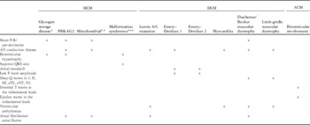
Hypertrophic cardiomyopathy is defined as increased ventricular wall thickness that is not solely explained by abnormal loading or structural heart conditions, such as valve disease, CHD, and hypertension. In adults, an end diastolic wall thickness of over 15 mm is diagnostic, whereas in children a z-score of over 2 is used. It is relatively common, affecting 1:500 people with an annual incidence of ~0.47/100,000 children.Reference Maron, Gardin, Flack, Gidding, Kurosaki and Bild 6 , Reference Wilkinson, Landy and Colan 7 The phenotypic diagnosis of hypertrophic cardiomyopathy is based on electrocardiographic and echocardiographic features, although family history, physical examination, metabolic screening, and genetic testing are used to investigate the aetiology of the disease. Typical electrocardiographic changes in children include pathological Q waves and negative T waves in more than one consecutive inferolateral lead (lead II, III, aVF, V5-V6), whereas ventricular hypertrophy according to voltage criteria is a non-specific finding.Reference Savage, Seides and Clark 8 – Reference Gregor, Widimský, Cervenka, Vísek and Hrobonová 10 Some electrocardiographic features such as short P-R interval and atrioventricular conduction disorders are typical for specific hypertrophic cardiomyopathy sub-types (Table 1). The electrocardiographic abnormalities may precede ventricular hypertrophy, as seen on echocardiography. Hypertrophic cardiomyopathy is usually associated with diminished left ventricular cavity size, while systolic function is hyperdynamic. In addition, there is often associated systolic anterior motion of the mitral valve, which may cause left ventricular outflow tract obstruction.
Typical ECG features and common arrhythmias in different cardiomyopathy sub-types.Reference Moak and Kaski 11 , Reference Rapezzi, Arbustini and Caforio 50

ACM=Arrhythmogenic cardiomyopathy; AV=atrioventricular; aVF=augmented vector foot; aVL=augmented vector left; DCM=dilated cardiomyopathy; ECG=electrocardiogram; HCM=hypertrophic cardiomyopathy; MERFF=myoclonic epilepsy with ragged red fibres
* Glycogen storage disease: Danon disease, Pompe disease, and PRKAG2 – mutations in the γ2 subunit of the adenosine monophosphate-activated protein kinase
** MELAS and Kearns–Sayre syndrome
*** Mutations in RAS-MAPK pathway: Noonan, LEOPARD, Costello, and cardiofaciocutaneous syndromes
Familial/genetic hypertrophic cardiomyopathy
Hypertrophic cardiomyopathy is most commonly hereditary and is caused by mutations in the sarcomeric protein genes in 40–60% of patients, mainly beta-myosin heavy chain (MYH7) and myosin-binding protein C (MYBPC3) mutations. The inheritance pattern is usually autosomal dominant and results predominantly in thickening of the interventricular septum causing asymmetrical hypertrophy.
Familial hypertrophic cardiomyopathy can also be associated with a huge variety of inborn errors of metabolism, malformation syndromes, and neuromuscular disorders, particularly in children where they may be seen in up to 30% of cases,Reference Moak and Kaski 11 being most common when the disease presents in infancy. In contrast to sarcomeric mutations, concentric hypertrophy of the left ventricle is the usual pattern observed. Malformation syndromes include Noonan and LEOPARD syndromes, where right ventricular involvement is frequent, whereas Friedreich’s ataxia is the classic neuromuscular disorder associated with hypertrophic cardiomyopathy. Inborn errors of metabolism account for 5% of all cardiomyopathies, and its early diagnosis is important, because disease-specific treatment may result in a better prognosis. The metabolic diseases, including glycogen storage diseases such as Pompe disease (lysosomal acid α-glucosidase deficiency) with infant-onset severe ventricular hypertrophy, skeletal myopathy, and hypotonia, as well as mucopolysaccharidoses such as Hurler syndrome, are mainly inherited in an autosomal-recessive manner. There are a few conditions including Barth syndrome (also associated with a dilated cardiomyopathy) Hunter syndrome, and Anderson–Fabry disease that have X-linked recessive inheritance pattern, whereas Danon disease has X-linked dominant inheritance. Both Anderson–Fabry and Danon disease can also affect females, usually with a later onset and a less severe phenotype.Reference Price, Stanford, Braden, Ebeid and Smith 12
Non-genetic/non-familial hypertrophic cardiomyopathy
These include infants of a diabetic mother,Reference Abu-Sulaiman and Subaih 13 heart muscle disease related to obesity,Reference Alpert 14 and, rarely, hypertrophy outside of the expected range related to athletic training.Reference Maron and Pelliccia 15
Dilated cardiomyopathy
Dilated cardiomyopathy is defined as ventricular dilation with systolic dysfunction in the absence of abnormal loading conditions, such as valve disease, CHD, and hypertension, or coronary arterial disease. Right ventricular dilation or dysfunction may also be present. Although the prevalence of dilated cardiomyopathy is unknown, its annual incidence is estimated to be 0.58 cases/100,000 children. The aetiology of dilated cardiomyopathy is known in only approximately one-third of patients at the time of diagnosis.Reference Wilkinson, Sleeper, Alvarez, Bublik and Lipshultz 16 The majority of patients present with congestive heart failure due to severely impaired left ventricular systolic function, with the pathophysiological diagnosis confirmed by echocardiography. Electrocardiographic changes can include atrioventricular conduction disorders and signs of inferolateral re-polarisation abnormalities.
Familial/genetic dilated cardiomyopathy
Familial dilated cardiomyopathy accounts for 20–25% of “idiopathic” disease, often with autosomal-dominant inheritance pattern.Reference McKenna, Codd, McCann and Sugrue 17 – Reference Hershberger and Siegfried 19 Approximately 35–40% of cases have sarcomeric gene mutations, with the majority attributed to protein-truncating mutations in the titin gene (TTN), which encodes a giant stress sensor protein. Mutations in beta-myosin heavy chain (MYH7) and myosin-binding protein C (MYBPC3) can also be associated with familial dilated cardiomyopathy, although more commonly found in familial hypertrophic cardiomyopathy. Additional genetic causes include mutations in nuclear membrane-related genes such as the lamin A/C genes, which encode intermediate filament proteins in the nuclear membrane and are associated with dilated cardiomyopathy and atrioventricular conduction disease, which can be evident before evidence of ventricular dilation, as well as Emery–Dreifuss (emerin protein mutation). Finally, mutations in the cytoskeleton can occur, such as desminopathy with associated skeletal myopathy, dilated cardiomyopathy and conduction disease.
Neuromuscular disorders associated with dilated cardiomyopathy are exemplified by dystrophin mutations. Dystrophin is a plasma membrane-associated protein, which forms a complex with proteoglycans, found both in heart and in skeletal muscles, and whose role is to protect against contraction-induced injury. Mutations in the dystrophin gene are linked to Duchenne and Becker muscular dystrophies.
Inborn errors of metabolism linked to dilated cardiomyopathy include lysosomal storage diseases such as Hurler Syndrome variants (mucopolysaccharidoses type I), carnitine deficiency, and mitochondrial myopathies.Reference McNally, Golbus and Puckelwartz 20
Non-familial/non-genetic dilated cardiomyopathy
In contrast to hypertrophic disease, acquired dilated cardiomyopathy is more common than familial forms, as exemplified by myocarditis, also called inflammatory cardiomyopathy. Myocarditis is defined as an acquired acute or chronic inflammatory process affecting the myocardium produced by a wide variety of toxins, drugs, or infectious agents. It is most often attributed to viral infections, particularly Cocksackie B enterovirus, parvovirus B19, human herpesvirus 6, and adenovirus infections, but may also be caused by non-viral agents such as Borrelia burgdorferi (Lyme disease), Corynebacterium diphtheriae, and Trypanosoma cruzi (Chagas disease).Reference Kindermann, Barth and Mahfoud 21 , Reference Drucker, Colan and Lewis 22 Cardiotoxic drugs, particularly chemotherapeutic agents such as doxorubicin, are strongly associated with dose-related left ventricular dilation and impaired function.Reference Takemura and Fujiwara 23 , Reference Yeh and Bickford 24 Immune-mediated inflammatory cardiomyopathies are characterised by the presence of autoantigens in diseases such as giant cell myocarditis and systemic lupus, as well as hypersensitivity to certain drugs such as sulphonamides. Finally, and importantly in children, fulminant Kawasaki disease can also be associated with myocarditis, seemingly independent of coronary arterial disease. Other important and reversible causes of acquired dilated cardiomyopathy include endocrine and nutritional deficiencies. Vitamin D deficiency is a more recently recognised entity, particularly in non-Caucasian children, whereas selenium and zinc deficiency have also been reported.Reference Abdullah, Bigras and McCrindle 25 – Reference Frustaci, Sabbioni and Fortaner 28 Endocrine dysfunction such as hypocalcaemia secondary to hypoparathyroidism can result in dilated cardiomyopathy and is responsive to correction of the calcium deficiency.Reference Giles, Iteld and Rives 29 , Reference Tsironi, Korovesis, Farmakis, Deftereos and Aessopos 30
Tachycardia-induced cardiomyopathies belong to the non-familial form of dilated cardiomyopathy in this classification system.
Restrictive cardiomyopathy
Restrictive cardiomyopathy is defined by the restrictive physiology of the ventricle with normal or reduced systolic and diastolic volumes and normal ventricular wall thickness. It is the least common phenotypic form, accounting for 4.5% of all cardiomyopathies. The European Society classification includes only pure forms of the disease, excluding the restrictive-hypertrophic phenotype in this category, which is regarded as a hypertrophic cardiomyopathy phenotypic variant. The clinical presentation of restrictive cardiomyopathy can be highly variable, ranging from asymptomatic to overt signs of heart failure with pulmonary hypertension.Reference Zangwill and Hamilton 31 There is a significant risk of life threatening or fatal ventricular tachyarrhythmia, such that prophylactic defibrillator implantation may be necessary, especially in patients with ischaemic symptoms, significantly prolonged P-R interval, or widened QRS complex. Early listing for transplantation is recommended given the high risk for fatal events.
Familial/genetic restrictive cardiomyopathy
These are usually inherited in an autosomal-dominant manner, and mutations in the troponin I, troponin T, α-cardiac actin, and desmin genes have all been linked to this condition.Reference Pruszczyk, Kostera-Pruszczyk and Shatunov 32 – Reference Parvatiyar, Pinto, Dweck and Potter 34
Non-familial/non-genetic forms of restrictive cardiomyopathy are attributed to autoimmune diseases such as scleroderma, hypereosinophilic syndrome, amyloidosis, radiation therapy, and anthracycline toxicity.
Arrhythmogenic right ventricular cardiomyopathy
Arrhythmogenic right ventricular cardiomyopathy is defined as the fibrofatty replacement of the right ventricular myocardium, a histopathological diagnosis in contrast to the other forms of cardiomyopathies. The estimated prevalence is 1:5000, and it may present in a similar way to dilated cardiomyopathy. Diagnostic criteria were described by the 1994 Task Force to include structural, histological, electrocardiographic, arrhythmic, and familial features of the disease. More recently, this has been expanded to include MRI findings and genetic mutations.Reference Marcus, McKenna and Sherrill 35 The left ventricle is often affected as well, or even in advance of right ventricular disease,Reference Sen-Chowdhry, Syrris, Ward, Asimaki, Sevdalis and McKenna 36 and there is a recommendation in some quarters to rename the disease as simply arrhythmogenic cardiomyopathy. The clinical manifestation of arrhythmogenic ventricular cardiomyopathy during childhood is rare, usually being diagnosed between 20 and 40 years of age, but features of the disease can develop during adolescence.Reference Turrini, Basso, Daliento, Nava and Thiene 37 Treatment options include heart failure management, antiarrhythmic medications, and defibrillator implantation as primary or secondary prevention in symptomatic patients. The management of asymptomatic patients with arrhythmogenic cardiomyopathy remains challenging.Reference Bauce, Rampazzo and Basso 38
Familial/genetic arrhythmogenic cardiomyopathy
The majority of these cases are due to a mutation in the desmosome gene with an autosomal-dominant inheritance pattern. It has been shown that almost 50% of children who carry the desmosomal gene mutation develop the disease, which fulfils the modified Task Force criteria during childhood, two-thirds of whom will have a moderate-to-severe form.Reference Sen-Chowdhry, Syrris, Ward, Asimaki, Sevdalis and McKenna 36
Unclassified cardiomyopathies
This is the remaining group in the European Society classification. The most important entity in children is left ventricular non-compaction. This is increasingly recognised in the paediatric population and is defined as prominent trabeculation with deep intertrabecular recesses of the ventricular myocardium, usually at the left ventricular apex. It is quite frequently associated with congenital heart malformations with an associated worse overall prognosis.
Takotsubo or stress-provoked cardiomyopathy is a transient cardiac syndrome related to left ventricular apical akinaesia or apical “ballooning”. It mainly affects post-menopausal women, but a few paediatric cases have been reported.Reference Brenner and Powers 39 , Reference Szardien, Möllmann, Willmer, Akashi, Hamm and Nef 40 The symptoms include abrupt angina-like chest pain with associated widespread T wave inversion and ST segment elevation, mildly elevated cardiac enzymes, and left ventricular regional wall motion abnormality, all in the absence of coronary arterial disease.
American Heart Association cardiomyopathy classification system (2006)Reference Maron, Towbin and Thiene 41
The definition of cardiomyopathy is somewhat different here to that of the European Society of Cardiology system: “Cardiomyopathies are a heterogeneous group of diseases of the myocardium associated with mechanical and/or electrical dysfunction that usually (but not invariably) exhibit inappropriate ventricular hypertrophy or dilatation and are due to a variety of causes that frequently are genetic. Cardiomyopathies either are confined to the heart or are part of generalized systemic disorders, often leading to cardiovascular death or progressive heart failure–related disability”.Reference Maron, Towbin and Thiene 41 Both classification systems exclude ventricular dysfunction secondary to congenital heart malformations, hypertension, and coronary arterial or valvar disease, given the different management approaches for these when compared with other cardiomyopathies.
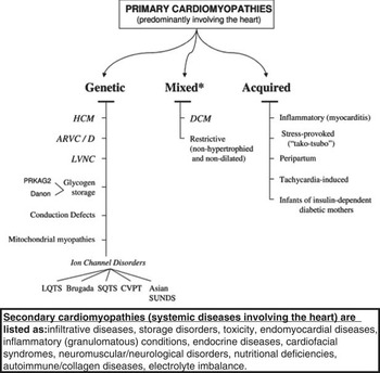
This classification system divides the cardiomyopathies into two major groups based on predominant organ involvement. Primary cardiomyopathies are those in which the heart is involved primarily, and are further subdivided into genetic, mixed (genetic and non-genetic) and acquired forms (Fig 2). Secondary cardiomyopathies are those in which cardiac involvement is part of a multi-system disorder.
Classification system of cardiomyopathies as detailed in the American Heart Association Scientific Statement.Reference Maron, Towbin and Thiene 41 ARVC/D=arrhythmogenic right ventricular cardiomyopathy/dysplasia; CVPT=catecholaminergic ventricular polymorphic tachycardia; DCM=dilated cardiomyopathy; HCM=hypertrophic cardiomyopathy; LQTS=long QT syndrome; LVNC=left ventricular non-compaction; SQTS=short QT syndrome; SUNDS=sudden unexpected nocturnal death syndrome. Reproduced with permission: Maron BJ, Towbin JA, Thiene G, et al. Contemporary definitions and classification of the cardiomyopathies. Circulation 2006; 113: 1810.

Genetic forms of primary cardiomyopathy
These are divided into different sub-groups based on the cardiomyopathy phenotype or aetiology. The category includes hypertrophic and arrhythmogenic cardiomyopathies, left ventricular non-compaction, mitochondrial myopathies, and ion channelopathies. In addition, glycogen storage diseases, which predominantly involve cardiac muscles, specifically PRKAG2 mutation and Danon disease, are included in this sub-group, whereas other glycogen storage diseases are categorised as secondary cardiomyopathies. The PRKAG2 gene encodes a subunit of adenosine monophosphate-activated protein kinase (AMPK), whose autosomal-dominant mutation is linked to hypertrophic cardiomyopathy with ventricular pre-excitation. Familial Wolff–Parkinson–White syndrome has also been associated with PRKAG2 mutation.Reference Sidhu and Roberts 42 Danon disease is caused by a mutation in the lysosome-associated membrane protein-2 (LAMP2) with X-linked dominant inheritance, with the male having the more severe phenotype and early onset. The sub-group of conduction system diseases, specifically inherited Lenègre disease, and inherited ion channelopathies such as long QT and Brugada syndromes are part of the American Heart Association classification system based on the argument that ion channel mutations alter the ion channel interfaces and architecture of cardiac muscles, although at a molecular level, and therefore should qualify as heart muscle disease. The European Society classification excludes these as the diagnosis and management regimes are also fundamentally different from other cardiomyopathies. The diagnosis of an ion channelopathy is based on typical electrocardiographic findings at rest, during exercise, or during specific pharmacological provocation tests such as Ajmaline test for Brugada syndrome and adrenaline challenge for Catecholaminergic Polymorphic Ventricular Tachycardia. As with other cardiomyopathies, genetic testing provides additional information and is used for family screening once the disease-causing mutation is found in the proband. Lenègre disease is a primary progressive cardiac conduction defect in the His-Purkinje system linked to a SCN5A mutation.Reference Kyndt, Probst and Potet 43 , Reference Probst, Allouis and Sacher 44 Most ion channelopathies are inherited in an autosomal-dominant manner, with some rarer variants having autosomal-recessive inheritance, such as long QT syndrome with congenital sensorineural deafness (Jervell and Lange–Nielsen syndrome).Reference Schwartz, Spazzolini and Crotti 45
Mixed and acquired primary cardiomyopathies
Interestingly, dilated and restrictive cardiomyopathies belong to the mixed primary sub-category, as the majority of these diseases are non-genetic. Myocarditis and tachycardia-induced cardiomyopathies belong to the acquired primary cardiomyopathies sub-group, as does Takotsubo or stress-provoked cardiomyopathy, which is a “non-classified” cardiomyopathy in the European Society system. Of note is that myocarditis is in a different sub-category than dilated cardiomyopathy, despite the overlap between these conditions, with myocarditis being the commonest cause of dilated cardiomyopathy in children.
Secondary cardiomyopathies
These include multi-systemic disorders, which are either genetic or acquired. They are not divided into further sub-categories in the American Heart Association scheme. The majority of inborn errors of metabolism, malformation syndromes, and neuromuscular disorders, as detailed in the European Society scheme, are listed here. Myotonic dystrophy, for instance, causes a progressive conduction disorder and dilated cardiomyopathy with a variable age at onset. Other aetiologies of these secondary cardiomyopathies include autoimmune diseases, endocrine problems, drug toxicity, nutritional deficiencies, and so on.
MOGE(S) classification: a phenotype–genotype nomenclature system for cardiomyopathy (2013)Reference Arbustini, Narula and Tavazzi 46
This most recent classification system for cardiomyopathies attempts to incorporate all the available information on an individual, including any genetic mutation identified and inheritance pattern, using a complex acronym-based approach. The proposed nosology is similar to the universally accepted TNM staging of tumours. The scheme uses five attributes to classify a cardiomyopathy disorder:
-
∙ M=morphofunctional notation, which describes the phenotype: hypertrophic, dilated, restrictive, non-compaction, and so on. It includes the option of documenting early disease, disease-specific clinical markers (atrioventricular block, pre-excitation) and overlapping phenotype variations such as hypertrophic with restrictive phenotype.
-
∙ O=organ involvement: cardiac, other organ systems (eye, liver, or lung) or both.
-
∙ G=genetic or familial inheritance: autosomal dominant or recessive, X-linked.
-
∙ E=aetiological annotations such as genetic condition, autoimmune disease, toxicity, myocarditis, viral agent, etc.
-
∙ S=heart failure stage (optional): NYHA class or American College of Cardiology-American Heart Association stage.
An example is MHOHGADEG-MYH7[p.R663H] , which describes that the proband has hypertrophic cardiomyopathy with only cardiac involvement, an autosomal-dominant inheritance pattern, and that an MYH7 gene mutation was identified. All members of the family can be so described, many of course being negative for all areas – M0O0GNENCSI signifying unaffected heart and organ systems with no family history, non-carrier status, and in NYHA class 1.
It is a very complex scheme on first assessment and it is currently not widely used; however, it could become a useful tool especially when documenting individual families with multiple probands. To help adoption, the authors of the MOGE(S) nomenclature have developed an internet-based application (http://moges.biomeris.com) for use in daily clinical practice.Reference Maon, Seidman and Ackerman 48 At present, the MOGE(S) system does not include ion channelopathies, conduction disease, or tachycardia-induced cardiomyopathies.
Critique of systems of classification for cardiomyopathy
The American Heart Association’s classification has been criticised for its unconventional usage of “primary” and “secondary”, which traditionally have been used in the setting of causation, secondary being equivalent to “due to”, as opposed to organ specificity as here. Furthermore, several entities do not fit well in either category with some “primary” diseases having many extra-cardiac features, such as the mitochondrial myopathies and Danon disease, whereas some “secondary” diseases are virtually entirely confined to the heart, such as endomyocardial fibrosis. On the other hand, the European Society’s category “unclassified” is not very satisfactory, leaving left ventricular non-compaction “looking for a room”, as are ion channelopathies in the American Heart Association’s view.
It can be strongly argued that we are not yet “beyond the phenotype” in clinical practice, particularly with respect to treatment regimens, which are often driven by the phenotype rather than the cause. There is great heterogeneity of genotype expression and age-related penetrance, with little genotype–phenotype correlation. Thus, mutations in the same gene can be associated with multiple phenotypes, such as sarcomeric myosin-binding protein (MYBPC3) gene mutations being associated with hypertrophic, dilated, and restrictive cardiomyopathic processes. Looking at this in reverse, hypertrophic cardiomyopathy has so far been linked to at least 15 genes and over 1400 individual mutations, whereas dilated cardiomyopathy has been linked to over 20 genes and over 40 mutations! Not surprisingly, the presence of more than one mutation is associated with an earlier age at presentation and poorer prognosis. The same phenotype can, therefore, have multiple aetiologies; hypertrophic and dilated cardiomyopathies are associated with a multitude of causes, from sarcomeric protein mutations to storage and neuromuscular diseases, as well as acquired disease such as the inflammatory cardiomyopathies. To add a further layer of complexity, there can be migration from one phenotype to another and neither classification system covers this eventuality well.
Proponents of both the European Society and American Heart Association systems have debated at length the benefits of their own system while criticising the other. This discourse, which at times has been acrimonious, is not geographically driven, as each system has advocates in both Europe and North America.
A case in point is the differing views on the high-level classification of hypertrophic cardiomyopathy. In the system of the European Society of Cardiology, hypertrophic cardiomyopathy is defined as the presence of increased left ventricular wall thickness, irrespective of cause, which is not solely explained by abnormal loading conditions.Reference Elliott, Anastasakis and Borger 47 In the structure of the American Heart Association, however, the definition is confined to those patients with isolated cardiac pathology: “unexplained left ventricular hypertrophy associated with a non-dilated ventricular chamber, in the absence of another cardiac or systemic disease, which itself would be capable of producing the magnitude of hypertrophy evident in a given patient”.Reference Maon, Seidman and Ackerman 48 The latter team argue that hypertrophic cardiomyopathy is a disease subset of left ventricular hypertrophy, independent of other disease processes, whereas the former proponents argue that all the disease entities should be classified as having cardiomyopathy, focussing on elucidating the cause of the individual patient.Reference Elliott and Mckenna 49
Red flag diagnostic approach for cardiomyopathies
“Red flags” are important features used to guide clinicians towards diagnosis or appropriate work-up for individual patients. This approach for cardiomyopathies has recently been detailed as a clinical tool in a position statement by the European Society of Cardiology Working Group on Myocardial and Pericardial Diseases.Reference Rapezzi, Arbustini and Caforio 50 The diagnostic pathway in patients with suspected cardiomyopathy includes the patient and associated family history, physical examination, electrocardiography, cardiac imaging, and laboratory tests. The family history is a crucial component in inherited cardiac conditions and a three- to four-generation family pedigree is recommended focussing on any known members with cardiomyopathy, a history of heart failure, cardiac transplantation, cardiac rhythm disturbance, need for a pacemaker/defibrillator, sudden cardiac death, stroke at a young age, or musculoskeletal disease.Reference Christiaans, Birnie and Bonsel 51 Autosomal-dominant inheritance pattern is the most frequent, whereas autosomal-recessive inheritance is least common, being suspected if the parents are not affected with the condition and/or are consanguineous. X-linked inheritance is more common in cardiomyopathies with musculoskeletal involvement. Women can be sometimes affected even with X-linked recessive disorders if there is an inactivation of the X-chromosome but usually with a later onset and milder form of the disease. The mitochondrial diseases caused by a mutation in mitochondrial DNA can be inherited only from the mother and characteristic symptoms include lactic acidosis, skeletal myopathy, hypoacusis, palpebral ptosis, encephalopathy, and retinitis pigmentosa.Reference Jacoby and McKenna 52
The age at presentation is helpful when evaluating the aetiology of the disease. Cardiomyopathy in neonates and infants are usually caused by inborn errors of metabolism – for example, Pompe disease and Danon disease – or by congenital dysmorphic syndromes such as Noonan syndrome. In contrast, sarcomeric protein disease usually manifests in adolescence or early adulthood. Cardiomyopathy related to neuromuscular disorders such as Friedrich’s ataxia and Duchenne muscular dystrophy develops mainly in adolescence. Hypertrophic cardiomyopathy can develop before the neuromuscular symptoms in Friedrich’s ataxia, and dilated cardiomyopathy might be the presenting feature in Becker’s muscular dystrophy.
Dysmorphic features are seen in malformation syndromes or mitochondrial disorders and need to be carefully assessed, particularly craniofacial abnormalities (such as hypertelorism), neck webbing in Noonan syndrome, and dermatoses – for example, café au lait spots in Noonan syndrome; angiokeratomas in Anderson–Fabry disease; and woolly hair and palmoplantar keratoderma in the Naxos variant of arrhythmogenic cardiomyopathy. Learning difficulties are also common, such as in Noonan and Costello syndromes and mitochondrial diseases, whereas visual impairment and sensorineural deafness may be a feature, such as in mitochondrial diseases and Anderson–Fabry disease.
A 12 lead electrocardiogram and usually a 24-hour recording are carried out as a part of cardiomyopathy evaluation, often giving important diagnostic markers to guide further investigations, as summarised in Table 1. Such features can precede the development of a cardiomyopathy and may be the only presenting feature of a genetic mutation, such as in lamin A/C mutations.Reference Van Rijsingen, Arbustini and Elliott 53 Ventricular pre-excitation can also be associated with left ventricular non-compaction.Reference Pignatelli, McMahon and Dreyer 54
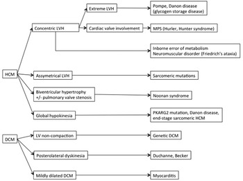
The presence of concentric or asymmetrical left ventricular hypertrophy observed using echocardiography suggests different aetiologies (Fig 3). Concentric hypertrophy is usually associated with malformation syndromes, inborn errors of metabolism, and neuromuscular disorders. Thickening of valve leaflets can be a characteristic sign of mucopolysaccharidosis. Asymmetric septal hypertrophy is commonly associated with sarcomeric protein gene mutations, whereas biventricular involvement is characteristic of malformation syndromes. Extremely severe, progressive concentric left ventricular hypertrophy in early childhood is suggestive of Pompe disease, Sengers syndrome, or Danon disease (in males).
Echocardiographic findings suggestive of the cardiomyopathy sub-types. Reproduced with modification from the position statement entitled diagnostic work-up in cardiomyopathies from the European Society of Cardiology Working Group on Myocardial and Pericardial Diseases.Reference Rapezzi, Arbustini and Caforio 50 DCM=dilated cardiomyopathy; HCM=hypertrophic cardiomyopathy; LV=left ventricular; LVH=left ventricular hypertrophy; MPS=mucopolysaccharidosis.

Armed with these findings, a direction of travel for genetic investigations and management can then be devised including treatment of the proband and family screening strategies as required.
Summary
The paediatric cardiomyopathies encompass a very heterogeneous group of disease entities. The European Society of Cardiology and American Heart Association are the two major cardiomyopathy classification systems currently being used with fundamental differences in the way they conceive the higher levels of their respective taxonomies. Both classification systems face the challenge of mixed cardiomyopathy phenotypes, with genetic heterogeneity, making logical classification challenging; however, these are the known limitations of both guidelines. Whichever high-level classification is used, the clinical reality at present remains phenotype driven. Clinical evaluation and standard diagnostic imaging dominate initial patient contact, and usually reveal diagnostic red flags, which in turn determine the direction of further specific tests and subsequent management stratagems, including family screening. Genetic testing is now fundamental and is performed at an increasingly early stage. A novel recent attempt to harmonise these competing systems of nomenclature is the MOGE(S) system; however its place in clinical practice has yet to be determined and is unproven as a fully practical solution.
Future directions for research related to nomenclature and systems of classification for cardiomyopathy include the following:
-
∙ Establishing the genetic basis for the various cardiomyopathies, in other words the links between genotype and phenotype, likely at a mutational level. This may provide the basis for a universally agreed system of classification for cardiomyopathies. Such a comprehensive system will ideally go beyond the phenotype, especially for diseases seemingly restricted to the heart.
-
∙ Working with the MOGE(S) system to produce a simpler streamlined version, which may indeed bring about a degree of harmony using a broad descriptive methodology, while allowing different levels of detail depending on the needs of the individual user (so called linearisation).