Introduction
Since the initial efforts to reconstruct ossicular discontinuity, there has been ongoing research aimed at developing improved techniques and prostheses.Reference Goode and Nishihara1 The reconstruction materials should be easy to prepare and well tolerated, with low extrusion rates.Reference Emmet2 Various materials are used, including incus autografts, Teflon®, ceramics, glass ionomer cements and metal (titanium, steel and gold wire).Reference Dornhoffer3 However, the ideal prosthesis has not yet been developed.
One of the most common middle-ear defects is loss of a portion of the incus, which is commonly reconstructed with a partial ossicular replacement prosthesis from the head of the stapes to the eardrum.Reference Brackmann, Sheehy and Luxford4 Despite the use of numerous different prostheses and surgical techniques, functional results remain unpredictable and vary widely.Reference Goode and Huttenbrink5
Ossicular prostheses are mainly used to reconstruct defects between the malleus and stapes, thus allowing stable integration with the stapes and malleus.Reference Hato, Stenfelt and Goode6 Reconstruction success is directly related to the prosthesis coupling with the remaining ossicles. Desirable properties for an ossicular prosthesis include a mass of 10–40 mg, the ability to create the correct tension between the tympanic membrane and stapes, and angulation of less than 30° with respect to the tympanic membrane.Reference Goode and Nishihara1, Reference Dornhoffer3 Most of these features are seen in the recently developed gold wire prosthesis: 15 mm of the wire prosthesis weighs 15 mg and has a diameter of 0.2 mm.Reference Katilmis, Aslan, Basoglu, Ekber, Ozturkcan and Tayfun7 The cost of middle-ear implant solutions is another important issue. For example, the hydroxyapatite partial ossicular replacement prosthesis costs over 300 Euros (equivalent to £235) in our country. However, the gold wire prosthesis discussed in this report is far less expensive than any other prosthetic material.
This study examined the hearing results and graft take rates obtained after type 2 tympanoplasty with gold wire prostheses, and compared the results with those of hydroxyapatite partial ossicular replacement prostheses in chronic otitis media patients.
Materials and methods
Study population and design
This retrospective study examined graft take rates and functional results in chronic otitis media patients who underwent type 2 tympanoplasty between March 2006 and January 2012. During this period, 1839 patients underwent tympanoplasty surgery at our institution; all operating surgeons were experienced otologists. Patients were reviewed for graft take and differences between pre- and post-operative air–bone gaps (ABGs) and pure tone averages (PTAs). Patients younger than 15 years of age who had concomitant canal wall down mastoidectomy or syndromes that can affect the middle ear (e.g. Down's syndrome, Kartagener's syndrome and cleft palate) were excluded.
Surgical procedures
All operations were performed under general anaesthesia with endotracheal intubation.
Partial ossicular replacement prosthesis surgery
The dimensions of the hydroxyapatite partial ossicular replacement prosthesis (Grace Medical, Memphis, Tennessee, USA) were a shaft inner diameter of 1.17 mm, a head diameter of 4.0 mm and a length of 5 mm. The prosthesis was prepared by simply trimming the shaft to the required length. Gelfoam® was used to stabilise the implant during packing. Optimal positioning of the prosthesis produces slight doming of the tympanic membrane or graft over the prosthesis to ensure maximum drum contact.
Gold wire surgery
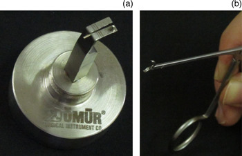
To minimise operating time, gold wire prostheses were prepared in various sizes appropriate for the left and right ears prior to surgery. For this, 0.2 mm biocompatibility-tested gold wire was trimmed to appropriate lengths. Wire hook–spiral prostheses specifically designed for this procedure were prepared in the laboratory using a Katilmis wire prosthesis preparation device (Omur Surgical Instruments, Antalya, Turkey) and sterilised (Figure 1a). In vitro cytotoxicity testing in L-929 mammalian fibroblast cells, intracutaneous and implantation testing in rabbits, and systemic injection testing in mice (test panel code number 286(i)/1314) confirmed the gold material to be biocompatible and safe for use. Biocompatibility tests were performed at the National Toxicology Centre, Pune, India, which has Food and Drug Administration, India, approval (licence number P.D-T-L-7).
Photographs showing wire hook–spiral prostheses made using the Katilmis wire prosthesis preparation device (a) and the specially designed Katilmis forceps used to fix the hook to the handle of the malleus (b).

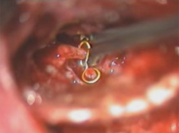
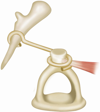
The distance between ossicles was measured. To minimise the risk of protrusion, the prosthesis was fixed at three points: one on the malleus and two on the stapes. Specially designed Katilmis forceps were used to fix the hook to the handle of the malleus (Figure 1b). The prosthesis was also fixed to the stapes by placing one and a half spirals around the stapedial capitulum. Then, within a space of approximately 1 mm × 1 mm on the medial side of the stapedial tendon, the half spiral was extended at an angle to the left for the left ear and to the right for the right ear, so that it passed under the stapedial tendon to provide further stability, thus preventing possible trauma to the facial nerve by the free tip of the prosthesis (Figures 2 and 3). Fascia was the preferred material for the eardrum graft.
Photograph showing hook fixation to the malleus after squeezing with the Katilmis forceps.

Image showing the prosthesis shape, design and position.

Outcome measures
The main outcome measures were the graft success rate and the hearing improvement level. Successful graft take was defined as complete healing of the intact graft without perforation, retraction or lateralisation for at least 12 months post-operatively. Patients were followed every month for the first three months post-operatively and then every three months for the first year. American Academy of Otolaryngology Head and Neck Surgery Committee on Hearing and Equilibrium guidelines were used to measure functional outcomes.8 Hearing results were based on the most recent audiogram available (not the best audiogram available during follow up) and were analysed by comparing pre- and post-operative air- and bone-conduction PTAs at frequencies of 0.5, 1, 2 and 3 kHz. ‘Hearing gain’ was defined as the difference between the pre- and post-operative ABGs.
Statistical analysis
Data were analysed using SPSS version 19.0 for Windows (SPSS Inc., Chicago, Illinois, USA). A normal distribution of the quantitative data was confirmed using the Shapiro–Wilk test. Parametric tests were applied to normally distributed data and non-parametric tests were applied to data with a questionable normal distribution. The independent samples t-test, Mann–Whitney U test and Wilcoxon's signed rank test were used to compare the different treatment groups. Data are expressed as the mean ± standard deviation. In all analyses, a p value of less than 0.05 was taken to indicate statistical significance.
Results
The partial ossicular replacement prosthesis group consisted of 32 patients (23 female and 9 male), and the gold wire group consisted of 26 patients (15 female and 11 male). In all cases, the indication for surgery was chronic otitis media or middle-ear exploration caused by conductive hearing loss. The average age was 31.3 ± 17.7 years (range 18–67 years) for the prosthesis group and 36.5 ± 14.8 years (range 18–67 years) for the gold wire group: there was no difference in the average age between groups (p = 0.296). All patients were followed up for at least one year after surgery. The mean follow-up period was 48.6 ± 17.2 months for the prosthesis group and 37.4 ± 13.5 months for the gold wire group (p = 0.003). The right to left ratio of the affected ears was 12:20 in the prosthesis group and 20:6 in the gold wire group.
Pre-operatively, there were no significant differences in the air- or bone-conduction thresholds between the gold wire and partial ossicular replacement prosthesis groups (Table I). The mean air-conduction threshold was 45.3 ± 13.5 dB for the prosthesis group and 50.3 ± 10.7 dB for the gold wire group (p = 0.130), while the mean bone-conduction thresholds were 9.9 ± 7.4 and 11.6 ± 6.7 dB, respectively (p = 0.371).
Pre- and post-operative pure tone averages by reconstruction material

*Data are the mean ± standard deviation (range). Pre-op = pre-operative; post-op = post-operative; PORP = partial ossicular replacement prosthesis
In the partial ossicular replacement prosthesis group, 27 patients (84.4 per cent) had subtotal perforations and 5 (15.6 per cent) had marginal tympanic membrane perforations. In the gold wire group, 13 patients (50 per cent) had subtotal perforations, 7 (26.9 per cent) had marginal tympanic membrane perforations and 6 (23.1 per cent) had intact tympanic membranes. Nine of the 32 patients in the partial ossicular replacement prosthesis group (28.1 per cent) and 12 of the 26 patients in the gold wire group (46.2 per cent) had a contralateral tympanic membrane perforation. Overall, 18.8 per cent (6/32) of patients in the partial ossicular replacement prosthesis group and 26.9 per cent (7/26) of patients in the gold wire group had undergone previous surgery on the same ear. Cholesteatomas were found in 6.3 per cent (2/32) of patients in the partial ossicular replacement prosthesis group and 26.9 per cent (7/26) of those in the gold wire group. Concomitant tympanosclerosis was seen in 34.4 per cent (11/32) of patients in the partial ossicular replacement prosthesis group and in 34.6 per cent (9/26) of those in the gold wire group. Based on the mastoid cell status, antrotomy (simple mastoidectomy) was performed in 31.3 per cent (10/32) of patients in the partial ossicular replacement prosthesis group and in 76.9 per cent (20/26) of those in the gold wire group.
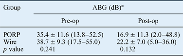
There was a significant improvement in post-operative air-conduction thresholds compared with pre-operative thresholds in both the gold wire (p < 0.001) and partial ossicular replacement prosthesis (p < 0.001) groups. However, the post-operative bone-conduction thresholds did not differ significantly from those recorded pre-operatively for either group (p = 0.780 and p = 0.464, respectively). Furthermore, there was significant improvement in ABGs compared with pre-operative thresholds in both the gold wire and partial ossicular replacement prosthesis groups (p < 0.001 for both). There were no significant differences between the pre- and post-operative ABGs for the two reconstruction materials (Table II).
Pre- and post-operative average air–bone gaps by reconstruction materials

*Data are the mean ± standard deviation (range). ABG = air–bone gap; pre-op = pre-operative; post-op = post-operative; PORP = partial ossicular replacement prosthesis
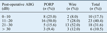
Post-operatively, the mean air-conduction threshold was 26.6 ± 12.4 dB for the partial ossicular replacement prosthesis group and 32.6 ± 10.5 dB for the gold wire group (p = 0.027), while the mean bone-conduction thresholds were 9.7 ± 7.0 and 10.4 ± 6.4 dB, respectively (p = 0.687; Table I). Hearing results in all ears according to reconstruction material are presented in Table III. Overall, the graft take rate was 91.4 per cent (53/58), with values of 90.6 per cent (29/32) for the partial ossicular replacement prosthesis group and 92.3 per cent (24/26) for the gold wire group (p = 0.848). No prosthesis extrusion or displacement was noted for any patient in either group.
Post-operative air–bone gaps by reconstruction material

ABG = air–bone gap; PORP = partial ossicular replacement prosthesis
The mean post-operative hearing gain was 18.5 ± 14.0 dB for the partial ossicular replacement prosthesis group and 16.5 ± 10.6 dB for the gold wire prosthesis group (Table IV): the difference between graft materials was not significant (p = 0.555).
Post-operative hearing gain by reconstruction material

*Data are the mean ± standard deviation (range). PORP = partial ossicular replacement prosthesis
Discussion
Chronic otitis media is a common disorder and patients may benefit from tympanoplasty to obtain a dry, ventilated middle ear, intact graft and acceptable hearing. Ossicular chain reconstruction is the most important feature for successful surgery.Reference Pinar, Sadullahoglu, Calli and Oncel9 The reconstruction material should be reasonably priced, well tolerated, biocompatible with a low extrusion rate and sufficiently flexible to provide optimal incudostapedial rebridging.Reference Martin and Harner10, Reference Nikolaou, Bourikas, Maltas and Aidonis11
To achieve good long-term results, the connection between the implant and ossicles should be sufficiently firm and tight to provide good sound transmission. The connection must also be stable because even minor dislocations can lead to a significant reduction in acoustic energy transfer. Although these problems are well known, prosthesis dislocation continues to be a common complication and major cause of long-term unsatisfactory post-operative hearing results.Reference Pinar, Sadullahoglu, Calli and Oncel9 The reported protrusion risk for hydroxyapatite prostheses is 2–16 per cent.Reference Nikolaou, Bourikas, Maltas and Aidonis11 Gold wire prostheses provide fixation at three points – one on the malleus and two on the stapes – as described previously.Reference Katilmis, Aslan, Basoglu, Ekber, Ozturkcan and Tayfun7 Gold is a biocompatible material that is easy to shape and stabilise, and is well tolerated. Schneider and Hagen reported similar results with their gold and titanium prostheses, and determined that the extrusion rate is lower with gold than with titanium.Reference Schneider and Hagen12 Gjuric and Schageri observed no implant rejection in their gold wire prosthesis patients and concluded that gold is highly biocompatible.Reference Gjuric and Schageri13
Tympanoplasty results may be influenced by many variables, such as revision surgery, variable perforation size and location, perforation drainage during surgery, eustachian dysfunction (bilateral perforation), the presence of cholesteatoma, and accompanying canal wall down mastoidectomy.Reference Songu, Aslan, Unlu and Celik14–Reference Demirpehlivan, Onal, Arslanoglu, Songu, Ciger and Can17 We attempted to include a homogeneous patient group to facilitate comparison by excluding patients younger than 15 years of age and those who had undergone concomitant canal wall down mastoidectomy. Nevertheless, we could not exclude all accompanying pathologies that may have skewed the functional results. Of the two treatment groups, the gold wire group had significantly more patients with bilateral perforations (46.2 per cent vs 28.1 per cent), previous surgery (26.9 per cent vs 18.8 per cent), cholesteatoma (26.9 per cent vs 6.3 per cent) and accompanying mastoidectomy (76.9 per cent vs 31.3 per cent). Although the post-operative hearing results for air-conduction thresholds were significantly better with the partial ossicular replacement prosthesis than with the gold wire prosthesis, there was no significant difference in post-operative hearing gain between the two reconstruction materials. In addition, the graft take rate did not differ significantly between groups. The main reason for ossiculoplasty failure is thought to relate to middle-ear pathology rather than surgical technique or prosthesis design. In atelectasis, the eardrum may collapse onto the prosthesis, causing extrusion. Our use of fascia instead of cartilage for grafting the eardrum makes this even more likely.
Another important issue for middle-ear implant solutions is that cost calculations must also be considered.Reference Gates18 While the hydroxyapatite partial ossicular replacement prosthesis costs over 300 Euros in our country, the gold wire prosthesis is far less expensive (less than 3 Euros) than this and any other prosthetic material. The main disadvantage of the wire technique is the time required to trim the wire into the correct shape and size (approximately five minutes per prosthesis). However, the time needed for this process reduced as the number of procedures increased.
• No ideal prosthesis has been developed to reconstruct ossicular discontinuities
• Ossicular chain reconstruction is the most important feature for successful surgery
• Functional outcome remains unpredictable and varies widely
• Reconstruction material should be reasonably priced, well tolerated and biocompatible with a low extrusion rate
• It should be sufficiently flexible to allow optimal incudostapedial rebridging
• Gold wire prosthesis provides comparable success and complication rates to the hydroxyapatite partial ossicular replacement prosthesis
This study has one main limitation: no selection criteria were used when choosing the prosthesis type. This decision was based on the surgeon's preference at the time of operation. Obviously, this may have left our design open to possible bias. As randomisation was not an available option, a retrospective control was run ahead of the statistical analysis to check that the pre-operative conditions of both patient groups were statistically homogeneous. Furthermore, the present study was underpowered and is subject to type 2 error. Another limitation of the study is possible observer bias. The new surgical method reviewed in the present manuscript was first described by the authors; therefore, in theory, there could have been observer bias in the assessment. Finally, it must be emphasised that the two groups are not fully comparable. The partial ossicular replacement prosthesis group has a much higher percentage of patients with a subtotal perforation.
Conclusion
The gold wire prosthesis seemed to provide comparable outcome success and complication rates to hydroxyapatite partial ossicular replacement prosthesis in the present study.