Introduction
Co-introduction of parasites (i.e.: introduction along with their non-native host into a novel locality outside of their natural range) is common, particularly among freshwater fish and their helminth parasites (Lymbery et al., Reference Lymbery, Morine, Kanani, Beatty and Morgan2014). Survival of these non-native parasites depends on a range of conditions or factors related to the environment and host(s) (MacLeod et al., Reference MacLeod, Paterson, Tompkins and Duncan2010). The establishment of parasites in novel habitats is more likely to occur for species with simple, direct life cycles that do not require intermediate hosts. A recent review by Lymbery et al. (Reference Lymbery, Morine, Kanani, Beatty and Morgan2014) showed a considerable proportion of these parasites is composed of fish monogeneans. Within monogeneans, Gyrodactylus is one of the most species-rich genera, with over 400 described species (Shinn et al., Reference Shinn, Harris, Cable, Bakke, Paladini and Bron2010). Members of the Monogenea have a direct life cycle and a relatively high level of host-specificity. Species of the genus Gyrodactylus parasitize many wild, farmed or ornament fish, and because of their mode of reproduction, which includes both viviparity and progenesis, they are often co-introduced along their fish hosts to the new areas and considered among the most invasive fish parasites (Kennedy, Reference Kennedy, Pike and Lewis1994).
Bullhead catfish species of the genus Ameiurus, including A. nebulosus, A. melas and A. natalis, were introduced from the North America to Europe as pond and aquarium fish since late 19th century (Spillmann, Reference Spillman1961). Brown bullhead, A. nebulosus (LeSueur, 1819) is the most widespread; only a few decades after introduction, the species spread to most European countries(Rutkayová et al., Reference Rutkayová, Biskup, Harant, Šlechta and Koščo2013). Black bullhead, A. melas (Rafinesque, 1820) has expanded its European range in recent decades (Béres et al., Reference Béres, Kánainé, Muller, Staszny, Farkas, Baks, Orbán, Urbányi and Kovács2017), while yellow bullhead, A. natalis (LeSueur, 1819) appears limited to Italy (Rutkayová et al., Reference Rutkayová, Biskup, Harant, Šlechta and Koščo2013). The occurrence of A. nebulosus in the Czech Republic was first documented in 1890 (Hanel and Lusk, Reference Hanel and Lusk2005) with its maximum population density occurring predominantly in the Elbe River basin during the mid-20th century. The current distribution of this species is rather patchy (Lusk et al., Reference Lusk, Lusková and Hanel2011). An accidental introduction of A. melas into the Czech Republic occurred in the Elbe River basin in 2005 (Musil et al., Reference Musil, Jurajda, Adámek, Horký and Slavík2010) and currently this species is only encountered occasionally. Species identifications based on potentially ambiguous characters, for both A. melas and A. nebulosus, has resulted in uncertainty regarding the occurrence of these species in some European countries (Rutkayová et al., Reference Rutkayová, Biskup, Harant, Šlechta and Koščo2013). Recently, molecular methods have contributed to the rapid identification of both species and their potential hybrids (Walter et al., Reference Walter, Gnyra, Söderberg and Heath2014; Béres et al., Reference Béres, Kánainé, Muller, Staszny, Farkas, Baks, Orbán, Urbányi and Kovács2017).
In their native range, A. melas and A. nebulosus are hosts of at least six monogenean species, including Gyrodactylus fairporti, G. nebulosus (A. n.), G. elegans (A. m.), Ligictaluridus floridanus, L. mirabilis (A. m.), L. monticelli (A. n.), L. pricei and Tetracleidus longus (Muzzal and Whelan, Reference Muzzal and Whelan2011). Among them, four monogenean species have been co-introduced with Ameiurus spp. to Europe. The dactylogyrid Ligictaluridus pricei (Mueller, 1936), known to infect the gills of North American catfishes, is the most frequently encountered non-native parasite in European populations of bullhead catfish (summarized in Nitta and Nagasawa, Reference Nitta and Nagasawa2015). Additionally, other reports include another dactylogyrid that infects the nasal cavity, Ligictaluridus monticelli as well as the gyrodactylid Gyrodactylus fairporti Van Cleave 1921 (Prost, Reference Prost1973; Galli et al., Reference Galli, Pugachev and Kritsky2010). Furthermore, the presence of G. nebulosus Kritsky and Mizelle, Reference Kritsky and Mizelle1968 was recently documented from Bulgarian A. melas (Vancheva et al., Reference Vancheva, Bobeva, Pehlivanov, Stefanov and Georgiev2020). Due to the specificity of the aforementioned parasite species to the North-American catfishes, the distribution of the parasites remains limited to the distribution of their non-native hosts.
During the parasite survey of wild non-native fish species in the Czech Republic (Central Europe), gyrodactylid parasites were collected from the two bullhead catfish species, A. nebulosus and A. melas. As previously noted, two gyrodactylid species, G. fairporti and G. nebulosus, are recently known as co-introduced into Europe, although the measurements of haptoral hard parts for both species slightly differed in comparison with the original description (Prost, Reference Prost1973; Vancheva et al., Reference Vancheva, Bobeva, Pehlivanov, Stefanov and Georgiev2020). Interestingly, while G. nebulosus primarily infects A. nebulosus in its native range, in Europe the species was recently reported from A. melas (Vancheva et al., Reference Vancheva, Bobeva, Pehlivanov, Stefanov and Georgiev2020). In North America, A. melas is frequently parasitised by G. fairporti (Hoffman, Reference Hoffman1999), but in Europe, this parasite has been previously reported from A. nebulosus (Prost, Reference Prost1973; Galli et al., Reference Galli, Pugachev and Kritsky2010). Our investigation used a combination of morphological and genetic analyses, an approach that is becoming a common practice in monogenean species identification (e.g. Bueno-Silva et al., Reference Bueno-Silva, Boeger and Pie2011). Specimens collected in this study displayed similar morphology, but showed variation of haptoral hard parts associated with the host species and season. Genetic analysis based on widely used markers for monogenean parasites, including sequences of partial 18S rDNA, ITS1-5.8S rDNA-ITS2 and cytochrome c oxidase subunit I (COI) (e.g. Ziętara and Lumme, Reference Ziętara and Lumme2002; Paladini et al., Reference Paladini, Huyse and Shinn2011; Vanhove et al., Reference Vanhove, Tessens, Shoelinck, Jondelius, Littlewood, Artois and Huyse2013; Kvach et al., Reference Kvach, Ondračková, Seifertová and Hulak2019), indicated the presence of two genotypes that could be separated according to host species. These results supported the description of a new gyrodactylid species infecting A. melas.
Material and methods
Fish sampling
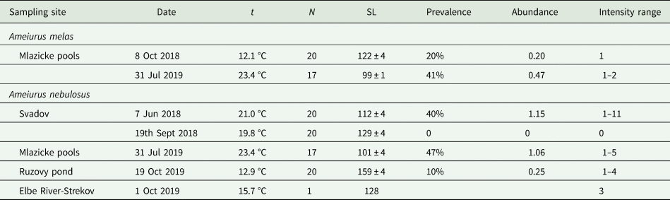
The fish were sampled at four sites in the Elbe River basin in the Czech Republic: (1) Mlazicke pools (50°22′50.0″N, 14°27′16.3″E), Elbe river oxbows near the town of Melnik; (2) Svadov (50°39′49.8″N, 14°05′51.3″E), a side arm of the Elbe River near the city of Usti nad Labem and (3) Ruzovy pond (50°37′29.6″N, 13°45′10.2″E), angling pond near the town of Jenikov, situated approximately 20 km by the Elbe River. Additionally, one fish was captured directly in the (4) Elbe River, near Strekov, Usti nad Labem city (50°38′17.7″N, 14°02′53.2″E). Fish were sampled in June and September 2018 as well as July and October 2019 using electrofishing gear (see Table 1). In total, 115 individuals of Ameiurus spp. were collected and transported alive in aerated cans to the laboratory, where they were dissected for monogenean parasites within a maximum of 2 days after capture following Kvach et al. (Reference Kvach, Ondračková, Janáč and Jurajda2016). Prior to dissection, each fish was measured (standard length, to the nearest 1 mm, Table 1).
List of sampling sites for A. melas and A. nebulosus collected due to the examination for Gyrodactylus species during 2018 and 2019, showing number of collected fish (N), mean standard length (SL, in mm) ± s.d., gyrodactylid prevalence in %, mean abundance and range of intensity (min–max) of parasite infection

Parasite collection and morphometrical analysis
The fins, skin and gills were examined for gyrodactylid parasites using an Olympus SZX10 stereomicroscope (Olympus Optical co., Hamburg, Germany). After removing the parasites, the caudal fin of each fish was preserved in 96% ethanol for later molecular analysis. Collected parasites were mounted in glycerine-ammonium-picrate (Malmberg, Reference Malmberg1970) to allow for morphological and morphometric analyses. Gyrodactylid specimens collected from A. nebulosus (G. nebulosus; acc. No. IPCAS M-744) and A. melas (G. melas n. sp.; acc. No. IPCAS M-745), dehydrated in ethanol and mounted in Canada Balsam, were deposited in the helminthological collection at the Institute of Parasitology, Academy of Sciences of the Czech Republic, České Budějovice. A subsample of gyrodactylid parasites was transferred into a drop of water on a microscope slide. The haptor was excised and preserved in a mixture of ammonium picrate and glycerine as described above. The remaining part of the body was stored in 96% ethanol at 4 °C prior to molecular analyses.
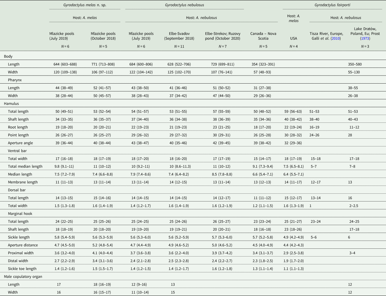
Parasite individuals were characterized according to the shape and size of the haptoral hard parts [anchors, connective bars and marginal hooks (MH)] using a light microscope (Olympus BX51) equipped with a phase-contrast and differential interference contrast. Drawings of haptoral hard components were made with the aid of a drawing attachment and phase-contrast optics. Measurements were obtained using the digital image analysis package MicroImage 4.0 for Windows (Olympus Optical co., Hamburg, Germany). Nine morphological characters of the anchors, ventral and dorsal bars, along with seven characters of MH were measured according to Shinn et al. (Reference Shinn, Hansen, Olstad, Bachmann and Bakke2004), supplemented by length and width of the whole body, pharynx and male copulatory organ (MCO) (Table 1). The MCO was observed in eight specimens, obtained from both host species. For comparative analysis, five specimens of native North-American G. nebulosus collected from A. nebulosus in Nova Scotia, Canada (Leis et al., Reference Leis, Leblanc, Easy, Dort and Cone2020) and four specimens of G. fairporti collected from A. melas in the Black River, Wisconsin, USA (Leis et al., Reference Leis, Easy and Cone2018) were subjected to the same morphometrical study.
The parasitological indices follow Bush et al. (Reference Bush, Lafferty, Lotz and Shostak1997): prevalence (P, %), mean intensity (MI), intensity range (IR, as minimum–maximum), mean abundance (A). Standard deviation (s.d.) was calculated for means. Principal component analysis (PCA) based on standardized data was used to visualize the position of G. fairporti (native), G. nebulosus (native and introduced) and G. melas n. sp. (introduced) specimens in morphological space according to Dávidová et al. (Reference Dávidová, Jarkovský, Matějusová and Gelnar2005), applying measurements of 12 morphological characters. Due to high inter-individual variation, the anchor angle, total length of MH, ventral bar membrane and dorsal bar length and width were excluded from the PCA analysis. First and second factorial axis were adopted for subsequent analysis of differences among samples based on a single parasite characteristic using Kruskal–Wallis ANOVA followed by Mann–Whitney U post-hoc tests for multiple comparisons. Statistical analyses were performed using Statistica 13.1 (StatSoft, Inc).
Molecular determination of host species
The caudal fin clips of all sampled fish were preserved in 96% ethanol. DNA was extracted with an Invisorb® Spin Tissue Mini Kit (Stratec). Two molecular methods were used to confirm the species identity as well as the potential presence of hybrids of A. nebulosus and A. melas. Following Béres et al. (Reference Béres, Kánainé, Muller, Staszny, Farkas, Baks, Orbán, Urbányi and Kovács2017), Cytochrome b gene was amplified in the volume of 12 μL reaction using PCR Multiplex kit (Qiagen) with primers CytB_L_14724F (5´-GACTTGAAAACCCACCGTTG-3´) and Cytbasa_R (5´-GAAGGCGGTCATCATAACTAG-3´). Cycling conditions for PCR were 95 °C at 15 min, then 35 cycles of 95 °C at 30 s, 56 °C at 20 s, 72 °C at 60 s. All PCR products were sequenced with specific primers (Eurofins, Germany), sequence data were analysed using Geneious® 9.0.5. and compared with sequences from GenBank (A. nebulosus-AY184271 and A. melas-AY184263). Specific multiplex PCR reactions composed of 16S rDNA partial gene (P5 5´-CCGAACTTAAGTTATAGCTGGTTGC-3´, 16sarL 59 5´-CGCCTGTTTATCAAAAACAT-3´) and specific NEB region (Neb_F 5´-CTGCTACATGCTAAGGCTAACG-3´, Neb_R 5´-GGATTATTGTGGCGATTGGT-3´) were performed using PCR Multiplex kit (Qiagen) in avolume of 12 μL. Cycling conditions for PCR were 95 °C at 15 min, then 35 cycles of 95 °C at 30 s, 56 °C at 60 s, 72 °C at 60 s. All PCR products were separated and analysed on 1.5% agarose gel. The PCR amplifies a control 16S rDNA (1175 bp) from all samples, a species-specific NEB fragment from the Ameiurus nebulosus (591 bp) and Ameiurus melas (457 bp).
Molecular characterization of gyrodactylid parasites
The anterior parts of four gyrodactylid specimens collected from A. nebulosus (two from Mlazicke pools, one from Ruzovy pond and one from Elbe-Strekov) and three from A. melas (Mlazicke pools) were placed individually in a 1.5 mL Eppendorf tube with 96% ethanol for genomic DNA extraction. The total genomic DNA of each individual was extracted using the DNeasy Blood & Tissue Kit (Qiagen, Hilden, Germany) following the protocol for purification of total DNA from animal tissues. Three widely used markers in monogeneans: (i) partial 18S rDNA, (ii) ITS1-5.8SrDNA-ITS2 and (iii) mitochondrial cytochrome c oxidase subunit I (COI) were amplified and sequenced to allow for comparison with other Gyrodactylus species.
The primers PBS18SF (forward, 5´-CGCGCAACTTACCCACTCTC-3´) and PBS18SR (reverse, 5´-ATTCCATGCAAGACTTTTCAGGC-3´) (Gilmore et al., Reference Gilmore, Cone, Lowe, King, Jones and Abbott2012) were used to amplify a variable region of 18S rDNA approximately 450 bp in length. Each amplification reaction contained 5 μL of DNA, 1 × PCR Buffer (Fermentas), 2.5 mm MgCl2, 200 μ m dNTPs, 0.5 μ m of each primer and 1 U Taq Polymerase (Fermentas) in a total volume of 30 μL. The PCRs were performed in the Mastercycler ep gradient S (Eppendorf) under the following conditions: initial denaturation for 3 min at 95 °C, 35 cycles of 1 min at 94 °C, 45 s at 54 °C, 90 s at 72 °C; and final extension of 7 min at 72 °C.
Sequence data for ITS1-5.8S-ITS2 was generated using forward primer ITS1F (5´-GTTTCCGTAGGTGAACCT-3´) (Rokicka et al., Reference Rokicka, Lumme and Ziętara2007) and reverse primers ITS2 (5´-TCCTCCGCTTAGTGATA-3´) (Cunningham, Reference Cunningham1997) or ITSR3A (5´-GAGCCGAGTGATCCACC-3´) (Matějusová et al., Reference Matějusová, Gelnar, McBeath, Collins and Cunningham2001). All PCR reactions were performed in a final volume of 25 μL, including 1 μL of DNA, 1 × PCR Buffer (Fermentas), 2.5 mm MgCl2, 200 μ m dNTPs, 0.5 μ m of each primer and 1 U Taq Polymerase (Fermentas). Cycling conditions were as follows: initial denaturation at 96 °C for 3 min, 35 cycles of 50 s at 95 °C, 50 s at 50 °C, 50 s at 72 °C; and final extension of 72 °C for 7 min.
The fragment of the COI gene was amplified with the primers Asmit1 (forward, 5′-TTTTTTGGGCATCCTGAGGTTTAT-3′) (Littlewood et al., Reference Littlewood, Rohde and Clough1997) and COI_Mono_int3 (reverse, 5′-ACATAATGAAARTGAGC-3′) (Plaisance et al., Reference Plaisance, Rousset, Morand and Littlewood2008) using following PCR protocol: 95 °C for 5 min, 35× (94 °C for 1 min, 50 °C for 1 min, 72 °C for 1 min), 72 °C for 10 min. PCR reaction was performed in 25 μL, containing 1 μL DNA-extract, 1× PCR Buffer (Fermentas), 1.5 mm MgCl2, 200 μ m dNTPs, 0.5 μ m of each primer and 1 U Taq Polymerase (Fermentas).
All PCR products were electrophoresed on 1.5% agarose gels stained with GoodView (SBS Genetech, Bratislava, Slovakia) and then purified using ExoSAP-IT™ (Affymetrix Inc., Santa Clara, USA), following the manufacturer's protocol. The purified PCR products were sequenced directly in both directions using the same primers as in the amplification reaction. For ITS sequencing, an additional internal primer ITSR3A (5′-GAGCCGAGTGATCCACC-3′) (Matějusová et al., Reference Matějusová, Gelnar, McBeath, Collins and Cunningham2001), complementary to the sequence at the 5′ end of 5.8S gene, was used. Sequencing was carried out using BigDye® Terminator v3.1 Cycle Sequencing Kit (Applied Biosystem by Thermo Fisher Scientific, Prague, Czech Republic) and an Applied Biosystems 3130 Genetic Analyzer (Applied Biosystems). The DNA sequences were assembled and edited using Sequencer software (Gene Codes Corp., Ann Arbor, MI, USA). The sequences were subjected to a BLAST search (Altschul et al., Reference Altschul, Madden, Schäffer, Zhang, Zhang, Miller and Lipman1997) in GenBank for species identification. The uncorrected p-distances were computed in MEGA X (Kumar et al., Reference Kumar, Stecher, Li, Knyaz and Tamura2018). The newly obtained sequences of Gyrodactylus species were deposited in GenBank.
Results
Host species identification
All fish from Svadov, Elbe River-Strekov and Ruzovy pond were determined to be A. nebulosus. Co-occurrence of both A. nebulosus and A. melas was confirmed in Mlazicke pools (Table 1). Among the 51 fish from Mlazicke pools, no hybrids were detected. Both cyt b and PCR multiplex analysis provided identical results.
Infection parameters
Gyrodactylid parasites were found primarily on the fins and body surface of the fish. Gill infections were observed less frequently (5% of parasites) and only on A. melas. All specimens represented a morphologically similar species with characteristics corresponding generally to Gyrodactylus nebulosus, with subtle differences in metrics and morphology of haptoral hard parts apparent between specimens obtained from both Ameirus species. The intensity of infection was low. Prevalence in A. nebulosus ranged from 0 to 47%, with the highest mean abundance in summer samples at Svadov (1.15) and Mlazicke pools (1.06). Maximum intensity of infection reached 11 specimens at Svadov in the summer. Gyrodactylid prevalence in A. melas ranged from 20 to 41%, with mean abundance between 0.20 and 0.47 in autumn and summer, respectively, and maximum intensity of infection of 2 (Table 1). At a location with the sympatric occurrence of both Ameiurus fish species (Mlazicke pools), parasite infections did not differ between the host species (t-test, t = 1.4; P = 0.18 for abundance, χ test, χ = 0.12; P = 0.73 for prevalence). In Svadov, infection parameters were significantly higher in June than September (MW-U test, Z = 3.1; P = 0.003 for abundance, χ test, χ = 10.0; P = 0.003 for prevalence).
Molecular characterization of gyrodactylids
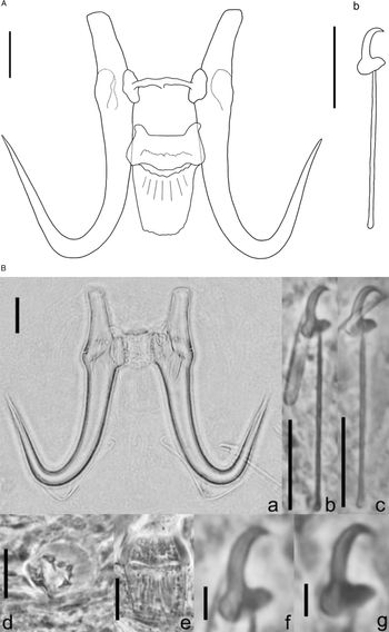
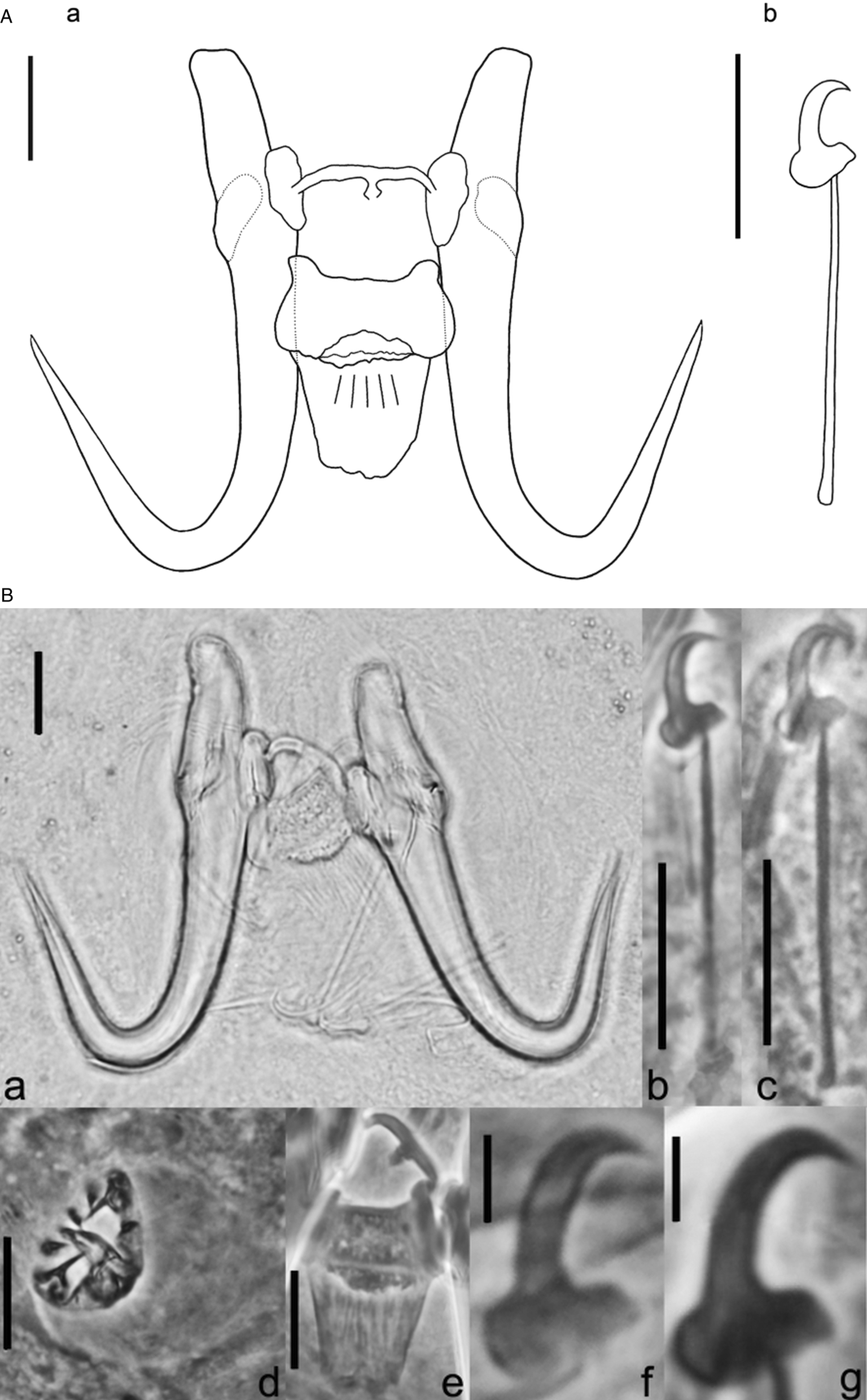
Molecular analyses showed consistent results in all three markers, with the differences correlating to the host species, i.e. A. nebulosus and A. melas. Thus, G. nebulosus consisted of two host-dependent genotypes: A. nebulosus was infected with G. nebulosus (Fig. 1A and B) and A. melas with genotype hereafter named as Gyrodactylus melas n. sp. (Fig. 2A and B).
(A) Drawings of Gyrodactylus nebulosus ex. Ameiurus nebulosus from the Svadov oxbow, Czech Republic. (a) Opisthaptoral central hook complex; (b) marginal hook. Scale bar: 10 μm. (B) Light photomicrograph of haptoral hard parts and male copulatory organ of Gyrodactylus nebulosus collected from A. nebulosus in Svadov oxbow, Czech Republic. (a) Opisthaptoral central hook complex; (b,c) marginal hooks; (d) MCO; (e) ventral bar (scale bar: 10 μm); (f,g) marginal hook sickle (scale bar: 2 μm).

(A) Drawings of Gyrodactylus melas n. sp. ex Ameiurus melas from the Mlazicke pools, Czech Republic. (a) Opisthaptoral central hook complex; (b) marginal hook. Scale bar: 10 μm. (B) Light photomicrograph of haptoral hard parts and male copulatory organ of Gyrodactylus melas n. sp. collected from Ameiurus melas in Mlazicke pools, Czech Republic. (a) Opisthaptoral central hook complex; (b,c) marginal hooks; (d) MCO; (e) ventral bar (scale bar: 10 μm); (f,g) marginal hook sickle (scale bar: 2 μm).

The partial sequences of 18S rDNA (432 bp) obtained from specimens collected from the same host species were identical and deposited in GenBank under the following accession numbers: MT501811 (G. melas n. sp.) and MT501812 (G. nebulosus). The two sequences were almost identical, differing in one base in position 217 (T-C). A BLAST search through the NCBI database revealed that our G. nebulosus was most similar to partial 18S sequences of G. nebulosus obtained from A. nebulosus in Canada (99.5%; MK863255) and from A. melas in Bulgaria (99.8%; MN556336). Gyrodactylus melas n. sp. was identical to a Gyrodactylus sp. obtained from A. nebulosus in Canada (100%; JF836106) as well as with specimens identified as G. nebulosus from A. melas in Bulgaria (100%; MN556336). Similarities with G. fairporti (MG825655) were 97.3% for G. nebulosus and 97.5% for G. melas n. sp.
The ITS1-5.8S-ITS2 sequences did not differ among specimens from the same host species, and the consensus sequences were deposited in Genebank under accession numbers MT501865 (G. melas n. sp.; 1133 bp) and MT501866 (G. nebulosus; 1131 bp). A comparison of these sequences showed 34 base-pair differences, representing a genetic variation of 3.01% (P-distance). Results from BLAST searches revealed that the ITS1-5.8S-ITS2 of G. melas n. sp. was identical to a sequence from specimens identified as G. nebulosus (MN556337) collected from A. melas in Bulgaria. The ITS sequence of G. nebulosus showed the closest hit (97%) to G. nebulosus collected from A. melas (MN556337).
The partial COI sequences (393 bp) were identical among specimens collected from the same host species; the consensus sequences were deposited in GenBank under accession numbers MT502383 (G. melas n. sp.) and MT502384 (G. nebulosus). Genetic diversity between these newly identified COI sequences was 4.83% (P-distance). A BLAST search of COI sequences showed ~85% similarity with several Gyrodactylus species (G. nyanzae – NC038214; G. turnbulli – EF446767; G. gurleyi – NC041379).
Morphometric analysis
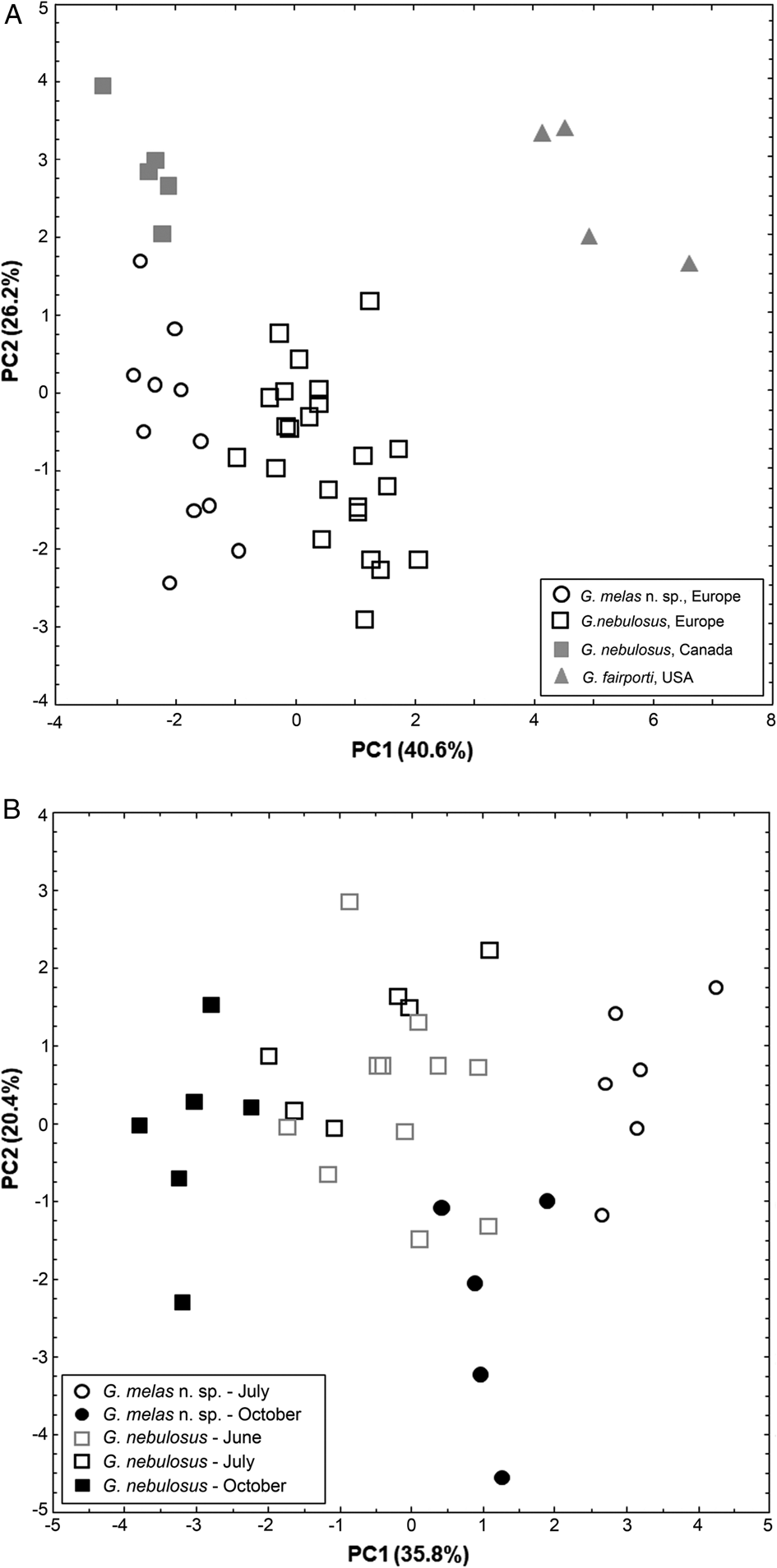
Specimens of G. nebulosus and G. melas n. sp. showed similar morphology to the original description of G. nebulosus, regarding the overall shape of the anchors, ventral bar, MH and MCO featuring one large spine and 4–6 smaller spines. However, the haptoral hard parts differed in some metrical parameters related to origin (native or introduced), host species (A. nebulosus and A. melas) and season (water temperature), as shown in Table 2 and on the plot of PCA based on the size of haptoral hard parts (Fig. 3A).
(A) Plot of principal component analysis based on 12 metrical characters of the haptoral hard parts. The plot shows the distribution of native Gyrodactylus fairporti (full grey triangles) obtained from A. melas, native G. nebulosus obtained from native (full grey squares) and non-native (empty black squares) A. nebulosus, and G. melas n. sp. parasitizing non-native A. melas (empty black circles). (B) Plot of principal component analysis based on 12 metrical characters of the haptoral hard parts. The plot shows the distribution of non-native G. nebulosus obtained from A. nebulosus (squares) and G. melas n. sp. obtained from A. melas (circles) during different months: June (empty grey squares), July (empty black marks) and October (full marks).

Morphometric parameters (mean, range; in μm) of Gyrodactylus melas n. sp., Gyrodactylus nebulosus and G. fairporti collected in their native (North America) and non-native (Europe) range

Specimens of both G. nebulosus and G. melas n. sp. differed in morphology and metrical parameters from native G. fairporti obtained from A. melas in the Black River, Wisconsin, USA (Table 2, Fig. 2A), with larger, narrower anchors (Fig. 4) and shorter MH sickles (Fig. 5) present with G. fairporti. In addition to a cluster of specimens clearly identified as G. fairporti from the Black River, WI USA, another three groups including native and non-native G. nebulosus, as well as G. melas n. sp. showed separate clusters. These related particularly to the PC1 axis (40.6% of variability), distinguishing all groups [Kruskal–Wallis ANOVA, H(3,43) = 35.5, P < 0.0001; Mann–Whitney U post-hoc tests, P ⩽ 0.002 for all comparisons] except for North American G. nebulosus and G. melas n. sp. PC1 was associated to the measurements of anchors [total length (contribution by 17.5%), shaft (16.2%), root (11.4%), point (12.7%)] and length of MH shaft (15.8%; Spearman rank correlation, all r s > 0.77 and P < 0.0001). PC2 axis (26.2%) was associated with the measurements of MH sickle [sickle toe length (contribution by 20.5%), proximal width (16.0%), aperture distance (13.3%), all r s > 0.69 and P < 0.0001] and ventral bar width (13.4%, r s = −0.52, P < 0.001), and mainly distinguished gyrodactylids from their native (USA) and non-native (Europe) range [H(3,43) = 21.1, P = 0.0001]. The measurements obtained from the slides of North American G. fairporti and G. nebulosus generally agreed with those reported in the supplemental diagnoses of the species (Leis et al., Reference Leis, Easy and Cone2018, Reference Leis, Leblanc, Easy, Dort and Cone2020).
Anchors of Gyrodactylus species parasitizing Ameiurus catfish species: (a) Gyrodactylus melas n. sp. from the fins of A. melas, Mlazicke pools; (b) Gyrodactylus nebulosus from the fins of A. nebulosus, Svadov; (c) Gyrodactylus fairporti from A. melas, Wisconsin, USA.

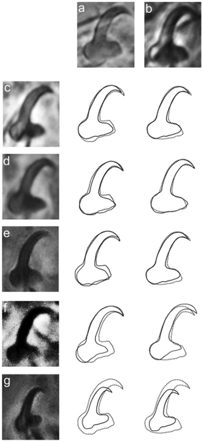
Overlay of marginal hook sickles of Gyrodactylus melas n. sp., representing the two morphotypes collected in July (a) and October (b) at Mlazicke pools (hatched line), with Gyrodactylus nebulosus collected from Svadov oxbow during June (c) and from Ruzovy pond in October (d), from Nova Scotia, Canada (e), Gyrodactylus nebulosus holotype (USNM 1366501; photo courtesy of Dr David Cone) (f) and with Gyrodactylus fairporti from Black River, USA (g); (full line).

Differences in the measurements of haptoral hard parts were associated with sampling season (water temperature, Table 1) in the non-native gyrodactylid populations (Fig. 3B). Anchor measurements were larger in the cold months (October) compared to summer (June, July), consistently for both G. nebulosus and G. melas n. sp. Individual groups clustered mainly along the first axis (35.8%). Significant differences of PC1 values between seasonal samples were found both for G. melas n. sp. (Mann–Whitney U test, Z = 2.65, P = 0.008) and G. nebulosus [Kruskal–Wallis ANOVA, F(4,34) = 27.0, P < 0.0001], except for gyrodactylids collected in June and July. The second axis (20.4%) differentiated seasonal samples of G. melas n. sp. (Z = 2.28, P = 0.022), and did not differ among samples of G. nebulosus. Both axes were associated with the same features as mentioned above.
New species description
Family: Gyrodactylidae Cobbold, 1864
Genus: Gyrodactylus von Nordmann, 1832
Gyrodactylus melas Ondračková, Seifertová, Leis n. sp. (Fig. 2A and B)
Type host: Ameiurus melas (Rafinesque, 1820)
Type locality: Mlazicke pools, Elbe River basin, Czech Republic (50°22′50.0″N, 14°27′16.3″E)
Water temperature and date of collection: 23.4 °C, 31 July 2019 (6 spec.); additional sample on 8 October 2018 (5 spec.), 12.1 °C; Table 1
Site on the host: fins, gills
Type specimens: Holotype and two paratypes (acc. No. IPCAS M-745) are deposited in the helminthological collection at the Institute of Parasitology, Academy of Sciences of the Czech Republic, České Budějovice.
Material examined: 11 flattened specimens (morphology), three ethanol-preserved specimens (DNA analysis)
DNA reference sequences:
The partial 18S sequence (432 bp), sequence of ITS1-5.8S-ITS2 region (1133 bp) and partial COI (393 bp) sequence are deposited in GenBank under accession Nos. MT501811, MT501865 and MT502383, respectively.
Etymology: The specific epithet refers to the host, A. melas.
Morphological description
General morphology based on 11 specimens collected in cold (12.1 °C) and warm (23.4 °C) water conditions: Body small, elongate, 603–808 μm long, with two distinct cephalic lobes. Haptor circular, anchors medium-stout, 49–54 long, uniformly wide along its length with proportionately short straight roots (18–21). Shaft length 33–37, point length 25–27, anchor aperture angle 36–44. Ventral bar 9–12 long, median length 6.6–8.8, sub-rectangular with slightly tapered posterior membrane (11–14); triangular strengthening of posterior median section. Anterolateral processes present as minute bumps on each end of the ventral bar. Dorsal bar typical with medial posterior projection, 13–16 long, 1.3–1.9 wide. MH total length 22–26, shaft straight, 18–20 long and slightly enlarged basally (Fig. 1). MH sickle slender, 5.3–5.9 long, toe short (1.2–1.7) and square; angling acutely from the base of the blade, 2.2–3.6 wide distally, 3.2–4.4 wide proximally, aperture 4.5–5.4; base with distinct heel, marked by a slightly flat, downward sloping dorsal edge which then curves smoothly to the point where the shaft articulates with the sickle. The MCO slightly spherical, 16–19 long, 15–17 wide, featuring one large spine and 5–6 smaller spines in a single row, observed in four specimens.
Remarks
Anchors of G. melas n. sp. are larger than those of G. ictaluri and shorter than G. fairporti, other gyrodactylids known to infect bullhead catfishes, and of similar length to G. nebulosus in general. However, anchor length was slightly shorter compared to G. nebulosus collected from the same region and season (Table 2). The terminal portions of the anchor roots were more rounded in G. melas n. sp. compared to the more angled in G. nebulosus (Figs 1, 2 and 4). Ventral bar posterior membrane more distinctly tapered in G. melas n. sp. than in G. nebulosus. MH sickles were larger than G. ictaluri and G. fairporti, and comparable in size to G. nebulosus. The most distinct difference between G. melas n. sp. and G. nebulosus is the shape of marginal sickle toe, which angles acutely from the base of the blade and is squarely truncated in G. melas n. sp., compared to a slimmer, more trapezoidal-shaped toe in G. nebulosus (Figs 1, 2 and 5).
Discussion
In this study, we provide the first confirmation that G. nebulosus, native to the North America, has been co-introduced with A. nebulosus to the Elbe River basin, Europe. The parasite was observed at four sampling sites along the Elbe River, with relatively low infection parameters. Despite being among the first reports of this parasite species in the European continent (see Vancheva et al., Reference Vancheva, Bobeva, Pehlivanov, Stefanov and Georgiev2020 for the first report), we believe this species was previously observed on A. nebulosus in Lake Dratów in Poland (Prost, Reference Prost1973) as well as the Tisza River (Galli et al., Reference Galli, Pugachev and Kritsky2010), although identified incorrectly as G. fairporti. Results of the PCA highlighted the lengths of both anchor and MH as important features for distinguishing between G. fairporti and G. nebulosus. Anchor lengths of gyrodactylids collected in this study, as well as that of parasites collected in Lake Dratów (Prost, Reference Prost1973), Tisza River (Galli et al., Reference Galli, Pugachev and Kritsky2010) and Srebarna Lake (Vancheva et al., Reference Vancheva, Bobeva, Pehlivanov, Stefanov and Georgiev2020), were slightly larger than those reported from native G. nebulosus, but still shorter than G. fairporti (Kritsky and Mizelle, Reference Kritsky and Mizelle1968; Leis et al., Reference Leis, Easy and Cone2018; present study). Nevertheless, the whole range of anchor lengths in European specimens overlapped both North-American species (i.e. G. fairporti and G. nebulosus, see Table 2). In addition, the MH measurements corresponded more closely to G. nebulosus than G. fairporti, further supporting that G. nebulosus was the species co-introduced to Europe and now occurs in several Ameiurus populations. Furthermore, the MCO reported in Prost (Reference Prost1973) certainly appears more similar to G. nebulosus.
The number and size of spines of the MCO also appeared to be a good feature for distinguishing G. nebulosus and G. fairporti (Leis et al., Reference Leis, Easy and Cone2018, Reference Leis, Leblanc, Easy, Dort and Cone2020). Unfortunately, the MCO was observable in only a small number of parasites collected in the Elbe River basin, and was often not clearly visible (see Fig. 1A). Nevertheless, the number of spines (i.e. one large and 4–6 small spines in a single row) corresponded to native G. nebulosus (Leis et al., Reference Leis, Leblanc, Easy, Dort and Cone2020), in contrast to G. fairporti, for which the MCO has a single large spine covering the pore and eight small spines that do not appear in a single row (Leis et al., Reference Leis, Easy and Cone2018). Morphologically identified specimens collected from the Elbe River basin were further confirmed through genetic results. A BLASTn search of the partial 18S and ITS1-5.8S-ITS2 sequences showed the closest hits with G. nebulosus (99–100% identity), compared to 97.34% similarity of partial 18S rDNA with native G. fairporti (MG825655; Leis et al., Reference Leis, Easy and Cone2018).
Specimens of G. nebulosus infecting A. nebulosus were morphologically and genetically consistent. Interestingly, a distinctly different genotype was identified from morphologically similar specimens infecting A. melas in the Elbe River basin. Herein, we describe this genotype as G. melas n. sp., representing a cryptic species sister to G. nebulosus. Due to the lack of unambiguous morphological characters for species discrimination, the genus Gyrodactylus is believed to contain numerous cryptic species (Hansen et al., Reference Hansen, Bakke and Bachmann2007). According to Ziętara and Lumme (Reference Ziętara and Lumme2003), sympatric cryptic species should show consistent differences in their DNA, demonstrate consistent morphometrical differences in the haptoral hard parts and be highly host specific. Parasite individuals of G. nebulosus and G. melas n. sp. sequenced in this study showed consistent differences in the partial 18S rDNA, ITS and COI sequences, associated with host species, i.e. A. nebulosus and A. melas, living in sympatry at Mlazicke pools for approximately 10 years (Musil et al., Reference Musil, Jurajda, Adámek, Horký and Slavík2010). There was one base (1 transition, 0.2%) difference at the partial 18S, 3.0% divergence in the ITS1-5.8S-ITS2 and 4.8% divergence in COI between G. nebulosus and G. melas n. sp., indicating the presence of two distinct species. Although the genetic divergence was relatively low, the 3.0% divergence in the ITS region was similar to the differences recently found with other cryptic gyrodactylid species infecting closely related host species, for example: marine gobies of the genus Pomatoschistus (1.5–1.8%; Huyse and Volckaert, Reference Huyse and Volckaert2002), cichlids (5.2%, García-Vásquez et al., Reference García-Vásquez, Hansen, Christison, Bron and Shinn2011) or poecilid fish at different localities in Mexico (1.9 or 2.3%; García-Vásquez et al., Reference García-Vásquez, Razo-Mendivil and Rubio-Godoy2015). Interspecies differences identified with mitochondrial markers have been used for gyrodactylid species discrimination (e.g. Kuusela et al., Reference Kuusela, Ziętara and Lumme2008; Xavier et al., Reference Xavier, Faria, Pladini, van Oosterhout, Johnson and Cable2015), although the genetic divergence observed between G. nebulosus and G. melas n. sp. (4.8%) was smaller than, for example, between cryptic species of poecilid fish (5.7–17.2%; Xavier et al., Reference Xavier, Faria, Pladini, van Oosterhout, Johnson and Cable2015), indicating an early stage of speciation. Partial data of the highly conserved 18S rDNA showed the lowest divergence (only 0.2%) between the two Gyrodactyus species in this study, although this difference was consistent.
Both species shared morphologically similar haptoral hard parts, though measurements were slightly smaller in G. melas n. sp. compared to G. nebulosus (Table 2), despite a comparable host age/size, intensity of infection and external conditions (in the case of sympatric occurrence of both species in Mlazicke pools). Multivariate analysis assigned anchor length as the most important feature that separates these two species in their non-native range. Conversely, the size of haptoral hard parts of North American G. nebulosus corresponded most closely to the newly described G. melas n. sp. and differed slightly from non-native G. nebulosus. However, the size ranges of the haptoral hard parts overlapped between the two species, making anchor size an insufficient feature for their morphological identification. The MH sickles, representing an important character for gyrodactylid species identification, were consistently the same size, but displayed a slightly different toe morphology that varied according to season (see Fig. 5). MH toe shape, along with host species identity may assist in the delineation of these two cryptic species.
Intraspecific variability was not found for either species in any of the genetic markers, indicating an apparent tendency for host specificity. However, it is important to note that not all parasite specimens were used for both the morphometrical and genetic analyses. Sequence data for both the partial 18S and ITS of G. melas n. sp. corresponded to the sequences of non-native G. nebulosus obtained from A. melas in the Srebarna Lake, Bulgaria (Vancheva et al., Reference Vancheva, Bobeva, Pehlivanov, Stefanov and Georgiev2020) with nearly 100% similarity (MN556336, MN556337). Such similarity indicates that the Gyrodactylus species collected from this distant locality actually represents the newly described species G. melas n. sp., and further supports our observations regarding host specificity. On the other hand, the occurrence of an identifcal genotype found on A. nebulosus in North America (Gilmore et al., Reference Gilmore, Cone, Lowe, King, Jones and Abbott2012) indicates that the host specificity may not necessarily be strict.
Gyrodactylus nebulosus was originally described from A. nebulosus collected in Strawberry Lake in North Dakota, USA (Kritsky and Mizelle, Reference Kritsky and Mizelle1968), and in its native range, the species is reported to predominantly infect its primary host A. nebulosus (Hoffman, Reference Hoffman1999; Leis et al., Reference Leis, Leblanc, Easy, Dort and Cone2020). Interestingly, Kritsky and Mizelle (Reference Kritsky and Mizelle1968) noted the presence of one G. nebulosus specimen among G. fairporti in the type slide. However, given the similarities among the cryptic species outlined in this report and that the samples were collected from A. melas, could this specimen have been G. melas n. sp., infecting rarely A. melas in its native range? Perhaps the absence of G. fairporti, the predominant gyrodactylid of this host species in its native range (Leis et al., Reference Leis, Easy and Cone2018), may have provided a free niche for G. melas n. sp. in non-native populations of A. melas in the Czech Republic.
Since the fish were collected during different months, we observed significant metrical differences among the gyrodactylid haptoral hard parts related to the season (i.e. water temperature, ranging from 12.1 to 23.4 °C), making delineation of the two gyrodactylid species based on haptoral measurements more difficult. Seasonal variation of the haptoral hard parts in monogeneans has previously been noted, with the general trend being a negative correlation between water temperature and size (Mo, Reference Mo1991, Reference Mo1993; Dávidová et al., Reference Dávidová, Jarkovský, Matějusová and Gelnar2005; Razo-Mendivil et al., Reference Razo-Mendivil, García-Vásquez and Rubio-Godoy2016). Accordingly, we observed an increase in the size of haptoral hard parts during the cold-water months (October) compared to warm-water months (June, July) for both G. nebulosus and G. melas n. sp. (see Table 2). Anchors of G. nebulosus collected in October (Ruzovy pond, Elbe-Strekov) reached a similar size (up to 59 μm) to that of G. fairporti (55–60 μm, Kritsky and Mizelle, Reference Kritsky and Mizelle1968; 58.2 ± 1.2, Leis et al., Reference Leis, Easy and Cone2018), indicating the need for thorough morphological examination, including observing the spines of the MCO, as well as consideration for using molecular methods when identifying species. Furthermore, the importance of proper identification is emphasized particularly in non-native species, as potential differences related to host preference or other biological attributes may affect their successful establishment in their new habitats.
Acknowledgements
We thank Professor Petr Ráb and representatives of the Czech Angler Association in Ústí nad Labem for an opportunity to sample the fish and members of the ichthyological and parasitological teams from the Institute of Vertebrate Biology ASCR for help with fish and parasite sampling. We would also like to thank Dr David Cone for kindly providing photomicrographs of the holotype for Gyrodactylus nebulosus. Usage of trade names does not imply endorsement by the US Government. The findings and conclusions in this article are those of the authors and do not necessarily represent the views of the US Fish and Wildlife Service.
Financial support
This study received financial support through ROZE, the research programme Life Diversity and Ecosystem Health in the frame of the Strategy AV21 and through the project PROFISH CZ.02.1.01/0.0/0.0/16_019/0000869 funded by the European Regional Development Fund in the operational programme VVV MŠMT.
Conflict of interest
We declare no conflict of interests.
Ethical standards
The research was undertaken in line with the ethical requirements of the Czech Republic, and has been approved by an appropriate ethics committee. The sampling, transportation, maintenance and care of experimental fish, as well as the method of fish killing complied with legal requirements in the Czech Republic (§ 7 law No. 114/1992 about the protection of nature and landscape and § 6, 7, 9 and 10 regulation No. 419/2012 about the care, breeding and using experimental animals). Researchers involved in this study (MO and PJ) are certified according to Czech legal requirements (§15, Law No. 246/1992 on Animal Welfare) to work with experimental animals.