Introduction to ultrafast laser fabrication of biomimetic structures
The study and simulation of biological systems is popularly known as biomimetics. Nature offers a diverse wealth of functional surfaces that are unmatched by today’s artificial materials. Based on the ideas, concepts, and underlying principles developed by nature, a highly interdisciplinary field has emerged to design, synthesize, and fabricate biomimetic structures. Reference Stratakis and Zorba1 As a result, several methodologies have been developed to form bioinspired constructs, exhibiting hierarchical structuring at length scales ranging from hundreds of nanometers to several micrometers. The two main fabrication routes are based on top-down and bottom-up processing schemes, respectively. In the top-down approach, a material is produced in bulk and then shaped into a finished part through a variety of processes. In contrast, bottom-up techniques construct the desired features from fundamental building blocks, without the need for patterning.
Laser processing is a maskless top-down approach that allows localized modifications with a large degree of control over the shape and size of the features that are formed. Reference Stratakis2 It is generally less expensive than e-beam texturing and more flexible, since it does not require complex vacuum facilities. While the minimal achievable structure size is limited by diffraction to the order of wavelength (microscale) in the far-optical field, the optimal interplay between the laser and material parameters may allow the fabrication of features with dimensions beyond this diffraction limit (nanoscale). This has been accomplished via the application of femtosecond (fs) lasers in materials processing, which has increasingly proved to be a powerful approach to overcome the diffraction limit of longer pulse and continuous laser processes and has improved the lateral resolution in the fabricated structures. Reference Sugioka and Cheng3 The most useful property of fs laser-induced modification is the limited size of the affected volume. Material structuring with laser pulses is induced by optical breakdown, which generates a plasma at the focal point of the laser. Ablation of the substrate is initially confined to a small volume due to set-in of shock wave propagation and cavitation, arising from plasma recombination before thermal diffusion. Although the intensity required to initiate breakdown is fairly high, the short pulse duration allows achieving the threshold intensity with a modest fluence. The combination of localized excitation and low-threshold fluence can greatly reduce the extent of collateral damage to surrounding areas, such that the size of the affected material can be below the diffraction-limited focusing volume. Another key advantage of using fs lasers is that nonlinear absorption effects can be driven due to the high peak powers attained. This opens new channels for direct nanowriting of transparent materials, while three-dimensional (3D) biomimetic structuring becomes feasible. In this context, fs lasers provide a unique toolset toward complex and unusual biomimetic structures, which is important for a plethora of biomimetic applications.
This article reviews the current state of the art of laser processing methodologies that have been used for the fabrication and engineering of biomimetic structures for microfluidic and tissue-engineering applications. Besides presenting the potential and significance of the laser-based biomimetic structures for such applications, the article will also delineate existing limitations and discuss emerging possibilities and future prospects.
3D biomimetic structure fabrication via two-photon polymerization
Cells are inherently responsive to their environment and are receptive to micro-/nanoscale features and chemical patterns. Reference Jeon, Schmidt, Barton, Hwang, Gamble, Castner, Grigoropoulos and Healy4–Reference Jahed, Molladavoodi, Seo, Gorbet, Tsui and Mofrad8 Controlling the surface energy is one of the important requirements for biomimetic tissue scaffold fabrication. Furthermore, certain compositions or properties similar to those of the extracellular matrix (ECM) are required, with structure features on various length scales to maintain cell phenotype and behavior. Hence, ECM components must be integrated within the scaffold through covalent conjugation.
As one kind of rapid prototyping technique, a laser-based fabrication system facilitates control over macroscopic (e.g., shape of scaffold) and microscopic (e.g., internal architecture) properties. The advantages of laser structuring include the capability of high-precision features, fast fabrication rates, noncontact interactions, reproducibility, and applicability to diverse types of materials. Direct laser writing can be used to fabricate 3D nanostructures with arbitrary geometry of diverse biomimetic materials. Reference Spagnolo, Brunetti, Leménager, De Luca, Sileo, Pellegrino, Pompa, De Vittorio and Pisanello9,Reference Terzaki, Kissamitaki, Skarmoutsou, Fotakis, Charitidis, Farsari, Vamvakaki and Chatzinikolaidou10 Moreover, this offers the creation of chemically and physically (e.g., same order of magnitude) well-defined cellular environments, which can be designed to fit the requirement of applications.
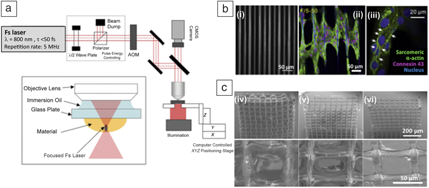
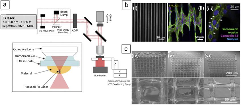
To fabricate a 3D cellular microenvironment, photo-induced polymerization using fs laser direct writing has been receiving increasing interest for precise fabrication with nanoscale resolution Reference Zhang and Chen11–Reference Paz, Emons, Obata, Ovsianikov, Peterhänsel, Frenner, Reinhardt, Chichkov, Morgner and Osten14 ( Figure 1a). During processing, a tightly focused laser beam forms an ultrahigh intensity at the center of focus using a high numerical aperture objective lens with immersion oil for index matching. For this multiphoton polymerization, diverse photosensitive materials can be used such as epoxy oligomer-containing commercial photoresist (SU-8), Reference Juodkazis, Mizeikis, Seet, Miwa and Misawa15 acrylate-based polymers, Reference Rekstyte, Zukauskas, Purlys, Gordienko and Malinauskas16 organically modified biocompatible materials Reference Kufelt, El-Tamer, Sehring, Meißner, Schlie-Wolter and Chichkov17,Reference Ovsianikov, Deiwick, Van Vlierberghe, Dubruel, Möller, Dräger and Chichkov18 (Figure 1c), titanium-containing sol-gel composite materials, Reference Psycharakis, Tosca, Melissinaki, Giakoumaki and Ranella19 and zirconium oxide-based hybrid materials. Reference Ovsianikov, Gruene, Pflaum, Koch, Maiorana, Wilhelmi, Haverich and Chichkov20–Reference Farsari, Vamvakaki and Chichkov23
Three-dimensional biomimetic scaffold fabrication using multiphoton polymerization. (a) Schematic of a multiphoton polymerization setup. Reproduced with permission from Reference Reference Paz, Emons, Obata, Ovsianikov, Peterhänsel, Frenner, Reinhardt, Chichkov, Morgner and Osten14. © 2012 AIP Publishing. (b) (i) Scanning electron microscope (SEM) images of a fabricated F/10–50 (Fiber/diameter–spacing [μm]) and (ii–iii) confocal images of cardiomyocytes aligned on the fiber showed the sarcomere structure stained by sarcomeric α -actinin and intercellular gap junctions stained by connexin 43 on the F/5–50, and white arrows pointing to connexin 43 staining. Reproduced with permission from Reference Reference Ma, Koo, Finnegan, Loskill, Huebsch, Marks, Conklin, Grigoropoulos and Healy22. © 2014 Elsevier. (c) SEM image of gelatin scaffolds produced by multiphoton polymerization; (iv) before 100 CDU/mL collagenase solution treatment, (v) after 1 h and (vi) 2 h. Top-row images are low magnification and bottom row is high magnification of the top row. Reproduced with permission from Reference Reference Ovsianikov, Deiwick, Van Vlierberghe, Dubruel, Möller, Dräger and Chichkov18. © 2011 ACS Publications. Note: λ, wavelength; τ, pulse duration; AOM, acoustic-optic modulator; CMOS, complementary metal-oxide semiconductor; fs, femtosecond.
A focused laser beam irradiated into these photocurable materials initiates the polymerization process. However, the transparency of photocurable materials is critical for 3D structure fabrication, as it allows the laser to pass through the entire material without attenuation. However, due to the ultrahigh intensity at the center of the focal point, nonlinear and localized multiphoton absorption occurs, and this reaction can only take place the vicinity of the small volume (voxel) of the focal point, where the laser intensity exceeds the threshold of multiphoton absorption. These nonlinear optical properties are utilized for selective photoinduced polymerization or machining of materials.
The achievable maximum resolution depends on the operating photoresist and wavelength. Due to the unavoidable elongation of the diffraction-limited focal spot, unequal dimensions are seen in the lateral and vertical voxel axes, resulting in a lower-resolution structure. The diffraction-limited focal spot is elongated to a greater extent in higher-index materials than in lower-index materials. Due to the increasing demand for higher resolution, a new method has been developed for further improving the resolution beyond the diffraction limit, based on stimulated emission depletion. Reference Klar, Wollhofen and Jacak24–Reference Sun, Dong, Chen, Nakanishi, Duan and Kawata26 In this process, two different types of laser beams are used. The first laser beam is focused as for standard multiphoton polymerization and the second beam inhibits this photoinduced polymerization, either via stimulated emission depletion or photo inhibition. By adjusting the position and shape of the two laser focal spots on top of each other, the resulting voxel volume can be significantly decreased.
Ultrafast laser fabrication for fluidic applications
Biomimetic surfaces with extreme wetting and stimuli-responsive properties
Direct laser micro-/nanostructuring is a material-independent and versatile technique that presents some key benefits over other more established microfabrication techniques for tailoring wetting properties and manufacturing functional surfaces with extreme wettability for a vast range of applications, including microfluidics, lab-on-a-chip devices, self-cleaning surfaces, textiles, drag reduction coatings for automotive, aerospace, and marine vehicles. Reference Stratakis, Ranella and Fotakis27 In nature, extreme wetting properties are excellently demonstrated through the superhydrophobicity and water repellence of the lotus leaf and the superhydrophilicity of carnivorous plants.
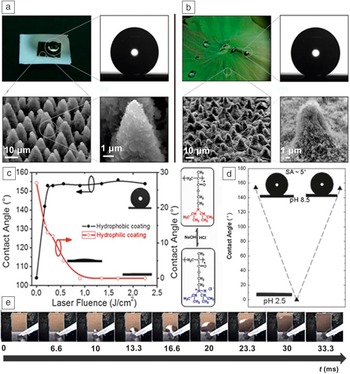
Hierarchical surfaces provide the appropriate template for observations of extreme wetting characteristics. Zorba et al. conducted studies of ultrafast laser processing of Si in an SF6 atmosphere, followed by the deposition of chloroalkylsilane monolayers; this led to the formation of double-length scale lotus leaf-like structures Reference Zorba, Stratakis, Barberoglou, Spanakis, Tzanetakis, Anastasiadis and Fotakis28 ( Figure 2a). Apart from mimicking the surface morphology of the lotus leaf, these artificial structures exhibited comparable wetting behavior to the natural surface. Accordingly, the laser-structured surfaces were superhydrophobic and water repellent, with remarkable self-cleaning ability. This structuring technique led to the production of superhydrophobic and water-repellent artificial surfaces. Reference Zorba, Stratakis, Barberoglou, Spanakis, Tzanetakis, Anastasiadis and Fotakis28
Wetting and water transport properties of femtosecond (fs) laser-fabricated structures and responsive surfaces. (a) Top left: Picture of a water droplet on a fs laser structured silicon surface (dark area of 5 × 5 mm2); (top right) Contact angle is 154° ± 10°; (bottom left) scanning electron microscope (SEM) image of the artificial surface comprising protrusions with conical asperities. (bottom right) High magnification SEM image of a single protrusion. The surface was structured in the presence of 667 mbar SF6 at a laser fluence of 2.47 J/cm2 with an average of 500 pulses. Reproduced with permission from Reference Reference Zorba, Stratakis, Barberoglou, Spanakis, Tzanetakis, Anastasiadis and Fotakis28. © 2008 Wiley. (b) Top left: picture of a lotus leaf; (top right) contact angle is 153° ± 10°; bottom left: SEM image of the leaf surface; bottom right: high magnification SEM image of a single papillose. Reproduced with permission from Reference Reference Zorba, Stratakis, Barberoglou, Spanakis, Tzanetakis, Anastasiadis and Fotakis28. © 2008 Wiley. (c) Contact angle measurements of a water drop on structured Si surfaces coated with a hydrophobic (●) and hydrophilic (○) coating, respectively, plotted as a function of laser fluence. Representative pictures of water droplets are presented as insets. The wetting response of the structured surfaces can be switched from hydrophobic to hydrophilic depending on the chemical nature of the coating. (d) Characteristic images of water droplets residing on poly(2-diethylaminoethyl methacrylate) (PDEAEMA) functionalized artificially structured surface following immersion at pH 8.5, pH 2.5 (complete wetting), and again at pH 8.5. The scheme shows the protonation/deprotonation process (the tertiary amine group becomes charged/neutral) of PDEAEMA. Note: SA, corresponding sliding angle of the respective surface. Reproduced with permission from Reference Reference Stratakis, Mateescu, Barberoglou, Vamvakaki, Fotakis and Anastasiadis33. © 2010 RSC Publishing. (e) Selective snapshots of a 4-μl water droplet spreading uphill as a function of time (t) on a vertically positioned laser structured substrate. The irradiated area, colored in light brown, is of 8 mm × 10 mm size. Reproduced with permission from Reference Reference Paradisanos, Fotakis, Anastasiadis and Stratakis30. © 2015 American Institute of Physics.

More importantly, the wetting response of the dual rough surfaces obtained can be switched to superhydrophilic (Figure 2c) upon coating of the fabricated structures with a hydrophilic layer. Reference Ranella, Barberoglou, Bakogianni, Fotakis and Stratakis29 It can be concluded that ultrafast laser texturing can provide a unique tool to control the wetting response from superhydrophobic to superhydrophilic. Moreover, it is a universal, material-independent technique to obtain multiscale surfaces with extreme wettability.
The design and fabrication of smart, fluid controllable interfaces are of great interest in novel microfluidic devices. Wettability gradients can induce net anisotropic wetting and spontaneous motion of liquids, owing to the action of the surface-tension gradient. Paradisanos et al. reported fs laser fabrication of gradient-wettable micro-/nanopatterns on Si surfaces that exhibit remarkable liquid spreading velocities, even in vertically positioned surfaces. Reference Paradisanos, Fotakis, Anastasiadis and Stratakis30 The gradual alteration of the surface roughness in the micrometer and nanometer scales gives rise to a gradient surface tension that provides the driving mechanism for liquid motion. It was shown that water droplets on the patterned surfaces spread at a maximum speed of 505 mm/s, which is the highest velocity demonstrated so far for liquid spreading on a surface tension gradient in ambient conditions (Figure 2e). Reference Paradisanos, Fotakis, Anastasiadis and Stratakis30
Finally, fs laser textured hierarchical micro- and nanostructured surfaces can be used as a platform for realizing surfaces that can be reversibly switched between the two extreme wetting states under the action of different external stimuli such as light, electric field, and pH. This is achieved via proper functionalization of fs laser-fabricated structures. Depending on the functional coating deposited onto those surfaces, different functionalities were attained, including photo-, electro-, and pH responsiveness. Reference Papadopoulou, Barberoglou, Zorba, Manousaki, Pagkozidis, Stratakis and Fotakis31–Reference Stratakis, Mateescu, Barberoglou, Vamvakaki, Fotakis and Anastasiadis33 In all cases, it was shown that the principal effect of hierarchical roughness is to cause an amplification of the response to the external stimulus. A prominent example is the fabrication of polymer functionalized pH-responsive surfaces, which can reversibly switch between superhydrophilic at low pH and superhydrophobic and water repellent at high pH. Reference Stratakis, Mateescu, Barberoglou, Vamvakaki, Fotakis and Anastasiadis33 The surfaces were developed by “grafting from” a pH-sensitive polymer brush onto fs laser-fabricated micro- and nanostructured substrates, which mimic the hierarchical morphology of natural water-repellent surfaces (Figure 2d).
Laser-fabricated 3D microfluidic channels
Recently, demand has increased for a reduction in reagent consumption for chemical/biological reactions, reduced analysis time, and lower labor costs. Reference McDonald and Whitesides34 To achieve these goals, microscale fluidic systems (e.g., lab-on-a-chip and optofluidic systems) are good candidates. However, to achieve high efficiency, accuracy, and sensitivity of analysis, increasingly complex microchips need to be fabricated that are integrated with electrical, mechanical, and optical components in these systems. Generally, poly(dimethylsiloxane) (PDMS) is used as a cost-effective patterning template. Reference Aoun, Weiss, Laborde, Ducommun, Lobjois and Vieu35,Reference Tanaka, Morishima, Shimizu, Kikuchi, Yamato, Okano and Kitamori36 Fabrication of a 3D microfluidic channel requires multiple steps such as alignment, stacking, and bonding. A direct laser writing method with glass in place of PDMS can skip complicated steps by employing fs-laser-assisted wet chemical etching (FLICE) or liquid-assisted fs laser drilling. Glass is superior to PDMS because of its stability and high transparency over a wide wavelength range.
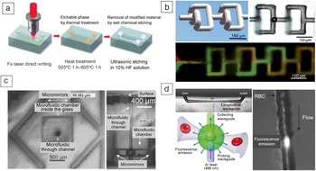
The FLICE method consists of successive multiple steps: (1) Direct laser writing (DLW)—scribing a pattern in the photosensitive glass by a tightly focused fs laser beam; (2) Thermal treatment—transformation of scribed pattern to an etchable phase; and (3) Wet etching—removal of the modified region through steps (1) and (2) with 5∼10% hydrofluoric acid solution in an ultrasonic bath Reference Sugioka and Cheng13,Reference Liao, Cheng, Liu, Song, He, Shen, Chen, Xu, Fan, Wei, Sugioka and Midorikawa37,Reference Sugioka and Cheng38 ( Figure 3a). For the DLW process, photosensitive glass (e.g., Foturan glass) and fused silica are commonly used. Foturan glass is lithium aluminosilicate glass doped with trace amounts of silver and cerium, and it can be modified with a lower intensity of laser, resulting in higher scanning speeds and shorter fabrication times.
Three-dimensional (3D) microfluidic channel using femtosecond (fs) laser direct writing. (a) Schematic of the procedures for 3D microfluidic channel fabrication using fs laser direct writing. Reproduced with permission from Reference Reference Sugioka and Cheng38. © 2012 RSC Publishing. (b) (top left) Schematic of 3D microfluidic mixer, (top right) top-view optical micrograph of two mixing units, and (bottom) result of two fluid mixing (red and green fluid). Reproduced with permission from Reference Reference Liao, Song, Li, Luo, Shen, Chen, Cheng, Xu, Sugioka and Midorikawa44. © 2012 RSC Publishing. (c) (left) Top view of the microfluidic laser and (right) side view of the microfluidic chamber and through channel. Reproduced with permission from Reference Reference Cheng, Sugioka and Midorikawa48. © 2004 OSA Publishing. (d) (top left) Optical transmission image of a fabricated microchannel, and (bottom left) schematic and (right) result of cell detection using fluorescence emission. Reproduced with permission from Reference Reference Kim, Hwang, Jeon, Hiromatsu and Grigoropoulos49. © 2009 RSC Publishing. Note: RBC, red blood cell.

During the DLW process, the nonlinear optical property (e.g., multiphoton absorption) of the fs laser is used, which makes it possible to alter the internal structure of glass in a spatially selective manner. Effective modification of the glass substrate can therefore occur in the restricted area, resulting in high-resolution microscale channel fabrication Reference Sugioka and Cheng13,Reference He, Liao, Lin, Song, Qiao, Cheng and Sugioka39–Reference Liao, Song, Li, Luo, Shen, Chen, Cheng, Xu, Sugioka and Midorikawa44 (Figure 3b).
Another approach for the 3D microfluidic channel fabrication is liquid-assisted fs laser drilling. Reference Sugioka and Cheng45 This technique is easier to implement and more environmentally friendly than FLICE. As this technique does not rely on the etching sensitivity of the material, it can be applied to any material that is transparent to the laser wavelength. From the near surface of the glass substrate, the fs laser direct writing process is applied in distilled water, which can help remove the debris generated during the drilling process and enhance the drilling length relative to the drilling process in ambient air. Channels of several hundred micrometers Reference An, Li, Dou, Yang and Gong46 or nanometers Reference Ke, Hasselbrink and Hunt47 can be fabricated using low laser energy.
The difficulties of conventional lithographic methods for microfluidic system fabrication lie in the lack of flexibility and challenges of 3D micro-optical element incorporation. One solution is simultaneous 3D channel and micro-optical component fabrication, such as mirrors Reference Cheng, Sugioka and Midorikawa48 (Figure 3c), optical waveguides Reference Kim, Hwang, Jeon, Hiromatsu and Grigoropoulos49 (Figure 3d), lenses, attenuators, and integrated structures of these components (e.g., waveguide and photonic crystals) Reference Haque, Zacharia, Ho and Herman50 in a single substrate. Fs laser-based microfabrication has been a good candidate, with demonstrated capability to fulfill these needs, opening up new avenues for fabricating various optofluidic microchips for advanced analysis systems. Reference Yang, Nava, Minzioni, Veglione, Bragheri, Lelii, Vazquez, Osellame and Cristiani51
Ultrafast laser structuring for biological applications
Ultrafast laser fabrication of biomimetic cell culture substrates
The surface morphology, surface chemistry, and thus surface energy of a biomaterial are important factors affecting cell response. Precise texturing of surfaces can have a profound impact on the growth, adhesion, and proliferation of cells on biomedical substrates for applications, including biosensors, structural implants, or tissue-engineering scaffolds. In such applications, it is not just the overall surface roughness that plays a critical role, but the detailed features on all length scales that affect cell–biomaterial interactions. Recent studies of fs laser structuring of biocompatible materials and their biological responses show the great potential of the fs laser nano-/microtexturing approach in the creation of novel culture scaffolds with better functionalities and improved performance. Reference Stratakis, Ranella, Fotakis, Schmidt and Belegratis52 The unique laser-induced structures can precisely modify the morphology and local chemistry of the surface and enable tuning of cell response, depending on the morphological features at various size scales. At the same time, it could be beneficial for the systematic exploration of the role of micro-/nanomorphology and surface energy on cell adhesion and growth.
Laser-based techniques have been employed for the precise fabrication of cell culture substrates at multiple length scales toward manipulating specific cellular functionalities, including cell growth, adhesion, proliferation, orientation, and differentiation. Ultrafast laser-fabricated arrays of biomimetic micro-/nanorough microcones (MCs) on silicon have been realized and used as cell culture platforms. Tailoring of the morphological features of such textured substrates can be achieved by tuning the laser structuring parameters, resulting in MCs of different micro- and nanoscale roughness and surface energy. Depending on the MC morphological characteristics, it is possible to preferentially tune fibroblast cells Reference Zorba, Stratakis, Barberoglou, Spanakis, Tzanetakis, Anastasiadis and Fotakis28 (cells that synthesize the ECM and collagen) and primary neuron cells adhesion, Reference Papadopoulou, Samara, Barberoglou, Manousaki, Pagakis, Anastasiadou, Fotakis and Stratakis53 growth, and proliferation. Such scaffolds enable primary neurons to grow in 3D, without the use of synthetic ECM coatings or other growth factors. Reference Barberoglou, Zorba, Pagozidis, Fotakis and Stratakis32 Simitzi et al. reported that different topographical characteristics of MCs remarkably influence the differentiation fate of PC12 cells into a neuronal phenotype. Reference Simitzi, Stratakis, Fotakis, Athanassakis and Ranella54
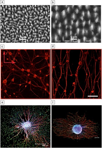
Finally, the effects of anisotropic discontinuous geometry of MCs ( Figure 4a) on neuroglia (types of cells that support neurons) and neuronal cell orientation and alignment have been investigated. Reference Simitzi, Efstathopoulos, Kourgiantaki, Ranella, Charalampopoulos, Fotakis, Athanassakis, Stratakis and Gravanis55 Topographic guidance of neurite outgrowth and axonal guidance in response to topographical cues could shed light on the mechanisms of axon initiation, axonal branching, and neuronal network formation. It was shown for the first time that even a discontinuous topographical pattern could promote sympathetic neuron and Schwann cell (cells that produce the myelin sheath around neuronal axons) alignment, provided it displays anisotropic features in a parallel orientation with interfeature distance on the micrometer scale (Figure 4a–b). In particular, parallel-oriented MCs with elliptical cross sections promoted oriented sympathetic neuron and Schwann cell outgrowth along the major axis of the ellipse. On the contrary, cells on MCs exhibiting a discontinuous but random pattern showed an isotropic orientation, resembling the growth pattern of cells grown on nontextured surfaces (Figure 4c–d). This guidance effect was also observed for the complex model of dorsal root ganglia explants (clusters of nerve cell bodies) Reference Simitzi, Efstathopoulos, Kourgiantaki, Ranella, Charalampopoulos, Fotakis, Athanassakis, Stratakis and Gravanis55 (Figure 4e–f).
(a–b) Top-view scanning electron microscope images of micropatterned Si substrates of (a) low and (b) high roughness; confocal microscope images of neurofilament positive sympathetic neurons grown on (c) low and (d) high roughness micropatterned Si substrates for six days. Scale bar: 150 μm. Confocal microscopy images of whole dorsal root ganglia explants (colored with violet) on (e) low and (f) high roughness micropatterned Si substrates. S100 positive Schwann cells and neurofilament positive sympathetic neurons are shown in green and red, respectively. Scale bar: 300 μm. Reproduced with permission from Reference Reference Simitzi, Efstathopoulos, Kourgiantaki, Ranella, Charalampopoulos, Fotakis, Athanassakis, Stratakis and Gravanis55. © 2015 Elsevier.

Ultrafast laser surface texturing of biomaterials
Biomaterials are widely used in various biomedical devices such as dental implants, orthopedic implants, artificial organs, and vascular stents. Biomedical devices that are inserted into the body are used to substitute for damaged organs or tissues. The device must be able to reproduce the functions and features of the original organ and not adversely affect the surrounding normal tissue. Metal- and ceramic-based materials have been used extensively for biomedical devices (orthopedic and dental implants) associated with bone, such as joint replacements, bone plates, bone screws, and bone cements. In addition, metal alloys such as NiTi, CoCr, and stainless steel are primarily utilized for vascular stents. Polymers are also widely used for artificial vessels, intraocular lenses, contact lenses, artificial breasts, and sutures. When the biomaterial is implanted in the body, it initially interacts with surrounding cells or the ECM. Therefore, biocompatibility of the material is the most important consideration, which can have a profound impact on whether the implant succeeds.
The goal of early-stage biomaterials was to replace the tissue with an alternative that provides suitable physical properties without toxicity. Today, repair or regenerative functions of the biomaterials are considered by controlling cell behavior at implant interfaces, Reference Hench and Polak56 where cell fate can be controlled by adjusting cell–material interactions at the material interface via surface chemistry, Reference Jeon, Schmidt, Barton, Hwang, Gamble, Castner, Grigoropoulos and Healy4,Reference Keselowsky, Collard and Garcia57,Reference Chen, Mrksich, Huang, Whitesides and Ingber58 topography, Reference Dalby, Gadegaard, Tare, Andar, Riehle, Herzyk, Wilkinson and Oreffo59–Reference Jeon, Koo, Reese, Loskill, Grigoropoulos and Healy61 and stiffness. Reference Discher, Janmey and Wang62,Reference Engler, Sen, Sweeney and Discher63 The typical size of a cell is approximately 10∼20 µm, and the size of its focal adhesion cluster, which is a large macromolecular assembly serving mechanical linkages to the ECM and transmitting regulatory signals, is approximately 50∼200 nm. Therefore, it is possible to control cell behavior by introducing micro- and nanoscale patterns on biomaterial surfaces. Among various patterning technologies, ultrafast laser ablation using far-field optics in a tight focusing configuration offers substantial advantages for the direct, maskless, and arbitrary patterning of various materials and curved surfaces, while maintaining high feature resolution. Reference Jeon, Koo, Reese, Loskill, Grigoropoulos and Healy61
Ablative direct laser writing can functionalize the surface of biomedical devices, including cell culture plates or medical implants. Reference Jeon, Koo, Reese, Loskill, Grigoropoulos and Healy61 Patterned surfaces with nanoscale craters can control cell ability to adhere to surfaces. This, in turn, results in directing cell migration, which is dependent on surface protein density or surface rigidity.
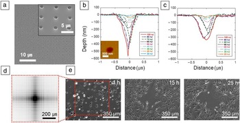
Jeon et al. suggested patterned nanocrater arrays with isometric and spatial gradient density distribution as a new, facile, and scalable method to control cell attachment and migration that are both related to cell growth and differentiation. They used a fs pulsed laser to etch precisely spaced nanometer-scale craters on quartz and polystyrene surfaces ( Figure 5a–c). Cultured fibroblast cells on patterned surfaces appear to first attach, but then move toward smoother surfaces, where nanocraters are separated by large spacing (Figure 5d). By changing the shape and arrangement of the ablated patterns, new possibilities emerge for applying this simpler and cheaper platform in cellular and medical research. For example, the method is applicable to diverse experiments as an effective tool for mechanotransduction studies in the context of regenerative medicine. Different microenvironmental stimuli can be implemented to alter intracellular structures and can contribute to stem cell differentiation and fate. Reference Jeon, Koo, Reese, Loskill, Grigoropoulos and Healy61
Nanocrater-patterned surface created by femtosecond laser direct-write lithography to control cell adhesion and migration. (a) Scanning electron microscope image of the patterned craters. The inset shows an enlarged patterned surface. (b) Cross-sectional profiles at the center line of the ablated nanocraters depending on pulse energies using 100× objective. Inset image is an atomic force microscope top view of a crater. (c) Cross-sectional profiles of the nanocraters obtained using a 50× objective. By using high numerical aperture objectives, submicrometer-scale features can be fabricated. (d) Schematic diagram representing pattern design. (e) Time-lapse phase contrast images of fibroblast cells cultured on the patterned surface. Reproduced with permission from Reference Reference Jeon, Koo, Reese, Loskill, Grigoropoulos and Healy61. © 2015 Nature Publishing Group.

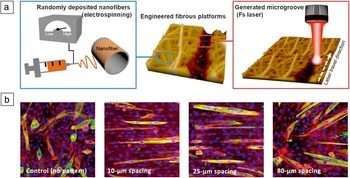
The advantage of multiphoton lithography is that surfaces of biomedical materials can be directly processed. Jun et al. used the fs laser-patterning process to create hierarchical topographies on electrospun nanofiber scaffolds. Reference Jun, Chung, Heo, Han, Park, Jeong, Lee, Lee, Kim, Seok, Shin and Jeon64 These scaffolds have been widely used for tissue-engineering applications due to their similarity in geometrical structure to native ECM and high contact surface areas that are good for cell attachment. Depending on the electrospinning process parameters such as voltage, flow rate, and mandrel rotating speeds and base materials, it is possible to control fiber diameter and alignment. Reference Murugan and Ramakrishna65 However, the low porosity of the scaffolds is a significant concern for the conventional electrospinning process, and unitary patterned fibrous platforms limit complex structures mimicking native tissues. Jun et al. suggested multiphoton laser lithography to generate hierarchical topographies that have both nano- and microscale structures on biodegradable polymer fibrous scaffolds, which regulate cell morphology and alignment with enhanced infiltration ( Figure 6 ). Reference Jun, Chung, Heo, Han, Park, Jeong, Lee, Lee, Kim, Seok, Shin and Jeon64
Hierarchical topographies on electrospun nanofiber scaffolds by femtosecond laser direct-write lithography. (a) Schematic illustration of the (left) electrospinning process, (right) laser-ablated microscale groove patterns, and (middle) engineered fibrous platform. (b) Immunofluorescence images of sarcomeric myosin heavy chain-positive C2C12 myoblasts on patterned fibrous platforms after five days of culturing (green, myosin heavy chain; red, F-actin; blue, nuclei). Well-ordered formation of myotubes are observed on the 10- and 20-μm spacing patterned surfaces, but scattered myotubes are observed on the control and 80-μm spacing patterns. Reproduced with permission from Reference Reference Jun, Chung, Heo, Han, Park, Jeong, Lee, Lee, Kim, Seok, Shin and Jeon64. © 2016 ACS Publications. Note: fs, femtosecond.

Direct-write laser texturing can be applied to metal as well as glass or polymer surfaces. It is well known that ordered micro- and nanoscale topographies are useful to control cell behavior and fate. However, ordered micro- and nanotopographical interfaces on metallic devices have not been extensively studied, mainly due to the limitation of fabrication processes, even though metal is the most commonly used material for implantable devices. In addition, the opacity of metals hinders in situ monitoring of cell behavior on patterned substrates.
Recently, a metal-based cell culture platform (MCP) using nickel titanium (nitinol) was introduced to study cell behaviors on microgrooves mimicking the vascular stent surface. Reference Jun, Chung, Park, Han, Park, Kim, Lee, Kim, Han, Kim, Seok, Kim and Jeon66 In this study, patterned PDMS substrates were fabricated with defined microscale topographies and deposited with nitinol thin film at nanoscale thickness (∼20 nm), which provides transparency (15% transmittance) of the patterned metallic surface for cell imaging. The selective effect of microscale grooves on vascular cell behavior was observed, depending on cell types, using human aortic smooth muscle cells (SMC) and human umbilical veil endothelial cells. When vascular stents were inserted, abnormal overgrowth of SMC caused recurrence of narrowing of the damaged arteries. It has been reported that topographical patterns can slow the restenosis and promote revascularization. Reference Sprague, Tio, Ahmed, Granada and Bailey67 Jun et al. elucidated the vascular cell response to the topographies by using a MCP and demonstrated that the topographical patterns can be directly fabricated on nitinol by fs laser direct-write texturing.
Biomimetic scaffold fabrication via two-photon polymerization
Hydrogels are biomaterials consisting of hydrophilic polymer networks and are widely used as 3D matrices for cell culture, owing to the similarity of their mechanical and chemical properties to real tissues. However, the aggressive conditions required for thermal polymerization and cross-linking hydrogels hinder their usage in applications such as encapsulation of cells and growth factor for tissue engineering. Photocross-linkable biomaterials offer significant advantages such as (1) rapid and mild cross-linking conditions; (2) the potential to pattern structures on thin films through photoinduced polymerization; and (3) the ability to precisely control the cross-linking and fine tuning of physical, mechanical, and chemical properties.
However, conventional lithography methods are not able to fabricate the 3D hydrogel structure with submicrometer-scale resolution. Multiphoton polymerization by fs laser could be a good candidate for 3D biomaterial fabrication with high precision, with its ability to be focused down to a submicrometer spot size. Moreover, secondary polymerization induced by the polychromic effect can be eliminated due to temporal coherence. Reference Zhang and Chen11,Reference Qin, Torgersen, Saf, Mühleder, Pucher, Ligon, Holnthoner, Redl, Ovsianikov, Stampfl and Liska68–Reference Ovsianikov, Deiwick, Van Vlierberghe, Pflaum, Wilhelmi, Dubruel and Chichkov70
Utilization of hydrogels for multiphoton polymerization will facilitate the development of live-cell encapsulated systems within 3D scaffolds in a spatially defined manner. The development and maintenance of the tissue involves complex and dynamic regulations (e.g., interaction between cell and other factors [substrate, growth factor, and ECM]), which, in turn, influence the cell’s fate by factors such as adhesion, migration, apoptosis (programmed cell death), proliferation, differentiation, and organization. By incorporating this 3D multiscale structure with ECM, a real tissue-mimicked structure with high resolution can be fabricated. Raimondi et al. measured cell viability, migration, and proliferation of primary rat mesenchymal stem cells (MSCs) on a 3D structure by multiphoton polymerization, and they observed that the 3D mechanical niche (e.g., external wall, internal volume for cell trapping) plays an important role in MSC colonization and differentiation. Reference Raimondi, Eaton, Laganà, Aprile, Nava, Cerullo and Osellame71
For a bone-tissue-engineering application, seeding MG63 osteoblast-like cells on 3D structures composed of microscale vertical tubes and a triangular lattice revealed that the physical geometry affected the alignment of the nuclei of the cells and the potential of osteogenesis (e.g., mineralization level of the osteoblast). Reference Mihailescu, Paun, Zamfirescu, Luculescu, Acasandrei and Dinescu72 Marino et al. seeded SaOS-2 osteoblasts on a trabecular-like structure (structural support to the spongey bone found at the ends of bones; osteoblasts are responsible for making the bone tissue on this structure) by multiphoton polymerization and observed that osteogenic differentiation and hydroxyapatite production were enhanced. Reference Marino, Filippeschi, Genchi, Mattoli, Mazzolai and Ciofani73
Three-dimensional scaffolds with a well-predefined structure can be used for in vitro platforms for tissue engineering and regenerative medicine. Ma and Koo et al. fabricated synthetic 3D filamentous scaffolds using multiphoton polymerization and used human in vitro cardiac tissue models for the study of cardiovascular diseases (e.g., cardiac arrhythmias) Reference Ma, Koo, Finnegan, Loskill, Huebsch, Marks, Conklin, Grigoropoulos and Healy22 (Figure 1b). The native myocardial tissues are organized in a 3D complex structure, which is critical to the electromechanical function of the heart. To mimic the human ventricular myocardium, fibers with different stiffness and pitch were fabricated, and healthy cardiomyocytes (CMs) and long QT syndrome type 3 (LQT3) CMs from human-induced pluripotent stem cells were cultured in this filamentous scaffold. Using this artificial cardiac model, they studied contractility malfunctions associated with electrophysiological consequences LQT3 and their response to drugs. Reference Ma, Koo, Finnegan, Loskill, Huebsch, Marks, Conklin, Grigoropoulos and Healy22 On this 3D fibrous scaffold, a high frequency of contractility malfunctions was observed. This suggests that there might be false-negative readouts during toxicity testing when using only two-dimensional toxicity testing systems.
Summary
Fs laser micro-/nanoprocessing offers unique capabilities for precise 3D, material independent structuring at the micro- and nanoscales. This article reviewed the current state of the art of laser processing methodologies that have been used for the fabrication and engineering of structures relevant to biomimetic, fluidic, and biological applications. Besides presenting the potential and significance of the laser-based biomimetic structures for such applications, the article also discusses emerging possibilities and future prospects.
Acknowledgments
H.J. acknowledges support from the KIST Project (2V04910). S.K. acknowledges support from the National Science Foundation Scalable Nano Manufacturing Award (1449305). E.S. acknowledges support from the European Commission-funded project LiNaBioFluid (GA 665337).
Emmanuel Stratakis is a researcher at the Institute of Electronic Structure and Laser (IESL) of the Foundation for Research and Technology–Hellas (FORTH), Greece. He received his PhD degree in 2001 in physics from the University of Crete, Greece. He then joined IESL-FORTH as a visiting researcher and the Department of Materials Science and Technology, University of Crete, as an adjunct professor. In 2007, he was elected researcher at IESL-FORTH where he is leading the “Ultrafast Laser Micro- and Nano-Processing” Laboratory. Since 2015, he has been leading the European Nanoscience Facility of IESL-FORTH, which is currently part of the NFFA-EUROPE EU Infrastructure. His current research interests include biomimetic micro-/nanostructuring of materials for fluidics, organic electronics, and tissue engineering. He has authored more than 120 publications and 10 book chapters. Stratakis can be reached by phone at +30-2810-391274 or by email at stratak@iesl.forth.gr.

Hojeong Jeon joined the Biomedical Research Institute at the Korea Institute of Science and Technology (KIST), South Korea, in 2012 as a senior research scientist. He received his BS degree in 2001 and MSc degree in 2004, both in mechanical engineering from Seoul National University, South Korea. He earned a PhD degree in 2011 in mechanical engineering from the University of California, Berkeley. His current research interests include micro-/nanosurface modification for functional biomaterials, microfabrication for tissue engineering, and biomedical devices. He has published approximately 30 papers, and received young scientist awards from the Korean Institute of Surface Engineering and the Korean Institute of Metals and Materials in 2016. Jeon can be reached by phone at +82-2-958-5140 or by email at jeonhj@kist.re.kr.

Sangmo Koo is a postdoctoral researcher in the Department of Mechanical Engineering at the University of California, Berkeley. He received his BS degree in 2005 and MSc degree in 2007 in Mechanical Engineering from Seoul National University, South Korea. He joined as an instructor and naval officer at the Korea Naval Academy from 2007 to 2010, and was awarded Best Professor Award in 2010. He received his PhD degree in 2016 from the University of California, Berkeley. His current research interests include biomaterial fabrication using femtosecond laser direct writing for manipulation of cellular mechanics and fabrication of biomimetic 3D tissue scaffold for high-throughput drug screening. Koo can be reached by phone at 510-599-1662 or by email at sangmo.koo@berkeley.edu.