INTRODUCTION
Lymphatic filariasis is a disease caused by the filarial worms Wuchereria bancrofti, Brugia malayi and B. timori. Approximately 120 million people worldwide are infected with one of these worms. Almost 90% of the infections are due to W. bancrofti, and the remainder is due mostly to B. malayi. An estimated 40 million infected individuals are seriously debilitated and disfigured by the disease (Melrose, Reference Melrose2002).
Unlike W. bancrofti, which has a global geographical distribution, B. malayi occurs only in regions of Asia such as India, Sri Lanka, Myanmar, Indonesia, Malaysia, Thailand, Vietnam and South China. Furthermore, B. malayi can also naturally infect mammals such as monkeys and cats (Edeson and Wilson, 1964). Brugia malayi is one of the targeted species in the WHO Global Programme to Eliminate Lymphatic Filariasis (GPELF), and much research has been done on the biology, drug response, immunology and diagnosis of infection. However, very little is known about the genetic variation in this species, particularly in endemic regions. In an effort to determine intraspecies variation within B. malayi, Areekit et al. (Reference Areekit, Singhaphan, Khuchareontaworn, Kanjanavas, Sriyaphai, Pakpitchareon, Khawsak and Chansiri2009b) analysed the ITS nucleotide sequences of B. malayi isolated from naturally infected cats in Thailand. The ITS (1 and 2) regions, which are found in the nuclear rDNA gene cassette, are used widely in phylogenetic and genetic diversity studies at species level due to their high rates of nucleotide change (Hillis and Dixon, Reference Hillis and Dixon1991; Weider et al. Reference Weider, Elser, Crease, Mateos, Cotner and Markow2005). In their study, Areekit et al. (Reference Areekit, Singhaphan, Khuchareontaworn, Kanjanavas, Sriyaphai, Pakpitchareon, Khawsak and Chansiri2009b) noted that the B. malayi isolates could be separated into several subgroups in the resultant phylogenetic trees. It was further observed that the ITS1 region was more informative than ITS2 in revealing intraspecies variation of Brugia spp.
In our present study, we determined the ITS1 nucleotide sequences of B. malayi isolated from microfilaria-positive human blood in Sabah, a Malaysian state in Northeast Borneo Island. Comparative analysis was performed not only among the ITS1 sequences of these isolates, but also with the sequences of B. malayi from Thailand. The latter comparison provided valuable information on genetic variation which exists between B. malayi from mainland Asia and Borneo Island.
MATERIALS AND METHODS
Detection of B. malayi microfilariae in human blood samples
The study subjects comprised 7 individuals (BM20, BM22, BM27, BM28, BM29, BM32, BM34) with B. malayi microfilaraemia from subdistrict Tangkarason, Beluran district (latitude 5°54′0N; longitude 117°32′60E) in the state of Sabah in Malaysia (Northeast Borneo Island). This is a rural area located about 400 km from Kota Kinabalu, the capital city of Sabah, and known to be endemic for the nocturnal subperiodic strain of B. malayi (Barclay, Reference Barclay1969). The microfilariae of this strain can be found in the peripheral blood at all hours but the density increases during the night.
The study protocol complied with the Declaration of Helsinki and was approved by the Committee on the Ethics of Human Research of Universiti Sains Malaysia. Written informed consent was obtained from each individual. The individuals were identified by house-to-house survey and screening by finger-prick blood sampling during night-time, from 8 pm to 11 pm. A minimum blood volume of 170 μl was obtained from each individual in EDTA-coated microcontainer tubes. Brugia malayi infection in the individuals was determined using traditional thick blood smears to detect the presence of microfilariae, real-time PCR, and by the commercially available immunochromatography IgG4 rapid test, Brugia Rapid (Reszon Diagnostic International Sdn. Bhd., Malaysia). The rapid test uses BmR1 recombinant antigen that identifies specific anti-filarial IgG4 antibodies in individuals infected with B. malayi and B. timori (Rahmah et al. Reference Rahmah, Shenoy, Nutman, Weiss, Gilmour, Maizels, Yazdanbakhsh and Sartono2003).
Thick blood smears (60 μl) were made on glass microscope slides, Giemsa-stained and examined in the field laboratory. Similarly, Brugia Rapid was performed in the field laboratory, using 35 μl of the blood sample. Meanwhile, 10 μl of blood were spotted on each of 4 protrusions of filter paper discs (TropBio, Townsville, Australia) and allowed to dry.
Extraction of total genomic DNA
Total genomic DNA was extracted from the dried blood spots using the QIAamp DNA mini kit (Qiagen, Hilden, Germany), according to the manufacturer's instructions. The DNA was used in real-time PCR using a previously described procedure (Rao et al. Reference Rao, Weil, Fischer, Supali and Fisher2006), and in the PCR amplification of the ITS1 region.
PCR, cloning and sequencing of the B. malayi ITS1 sequence
Primers (forward ITS1F 5′-GGTGAACCTGCGGAAGGATC-3′; reverse ITS2F 5′-AGC GGGTAATCACGACTG-3′) for the PCR amplification of B. malayi ITS were based on the sequences of Areekit et al. (Reference Areekit, Singhaphan, Khuchareontaworn, Kanjanavas, Sriyaphai, Pakpitchareon, Khawsak and Chansiri2009b). PCR was carried out in a 25 ml reaction mixture containing 10 mM Tris–HCl (pH 8·3), 2 mM MgCl2, 50 mM KCl, 0·01% gelatin, 200 mM of each deoxynucleoside triphosphate, 20 pmol of each primer, 1 U of Taq polymerase (Fermentas Life Sciences, Canada). The PCR mixture was pre-heated at 95 °C for 10 min for initial denaturation before 30 cycles of amplification, which consisted of denaturation at 94 °C for 1 min, annealing at 54 °C for 1 min, and elongation at 72 °C for 2 min. Final extension of the reaction was carried out at 72 °C for 10 min. The PCR fragment was cloned into plasmid pGEM-T, according to the manufacturer's instructions (Promega Corporation, Madison, USA). Recombinant pGEM-T plasmids harbouring the cloned fragment were sent to a commercial laboratory for DNA sequencing. Each recombinant plasmid was sequenced twice to ensure consistency of sequence data.
Reference filarial worm ITS1 sequences
Filarial worm ITS1 sequences used for multiple sequence alignment and phylogenetic analyses in this study were obtained from the GenBank database. The ITS1 sequences were as follows: B. malayi C12Cat1 (EU373601), C4Cat2 (EU373606), EU373612 (C7Cat3), C17Cat4 (EU373616), C22Cat5 (EU373621), cBm-1 (AY621466), cBm-2 (AY621467), cBm-3 (AY621468), hBm-1 (AY499507), hBm-2 (AY621465), hBm-3 (AY621464); B. pahangi Bp-1 (AY621469), Bp-2 (AY621470), Bp-3 (AY621471), Bp-4 (AY621472), C6Cat7 (EU373655), C27Cat6 (EU373640); and W. bancrofti isolate Guntur (AY843438) and Wb-M3 (AY621475).
Sequence data analysis and construction of phylogenetic trees
Nucleotide sequences were aligned using CLUSTAL-W, which is available online (http://www.ebi.ac.uk/Tools/msa/clustalw2). Pairwise nucleotide differences and genetic distance (defined as p-distance) between the sequences were calculated using MEGA4 (Tamura et al. Reference Tamura, Dudley, Nei and Kumar2007). Phylogenetic trees were constructed using the Neighbour-Joining (NJ) and Maximum Parsimony (MP) methods, also described in MEGA4. In the construction of the phylogenetic trees, bootstrap replicates of 1000 were used to test the robustness of the trees generated.
RESULTS
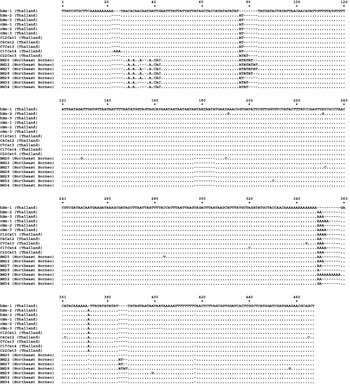
The 7 ITS1 sequences of B. malayi from Northeast Borneo (GenBank Accession numbers JQ327143-JQ327149) were aligned together with those of cat and human isolates from Thailand (Fig. 1). Comparative analysis of the alignment showed an ITS1 length variation of 431–441 bp within the Northeast Borneo isolates. These lengths were relatively shorter than those of the Thailand isolates, which had a length range of 441–450 bp. Close inspection of the alignment showed that the length variation between the North Borneo and Thailand isolates was attributed mainly to a region at positions 177–185. In this region, there was complete absence of 9 bases in the sequence of the Northeast Borneo isolates. Other regions of significant length variation were observed at positions 76–83 and 349–358.
Clustal-W multiple sequence alignment of Brugia malayi ITS1 from Northeast Borneo and Thailand. Dots indicate nucleotides identical to those of hBm-1 (Thailand).

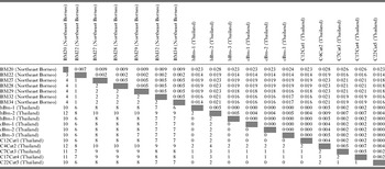
Apart from length differences, variation in nucleotide composition was also noted in the alignment. Table 1 summarizes the number of pairwise nucleotide differences and genetic distance (p-distance) among the sequences. The number of nucleotide differences (gaps not included) within the Northeast Borneo isolates ranged from 1 to 4, which corresponded to genetic distance range of 0·002 to 0·009. Nucleotide difference and genetic distance within the Thailand isolates were 0 to 4 and 0·000 to 0·009, respectively. However, when the Northeast Borneo isolates were compared with isolates from Thailand, the number of nucleotide differences was significantly larger, ranging from 6 to 12, corresponding to a higher genetic distance range of 0·014 to 0·0028.
Pairwise comparisons of nucleotide differences and genetic differences (p-distance) among Brugia malayi isolates from Northeast Borneo and Thailand

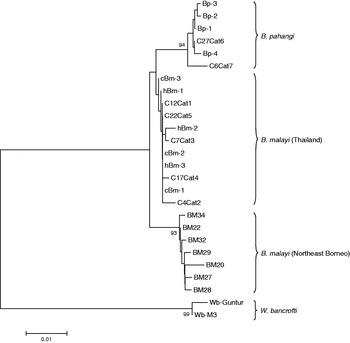
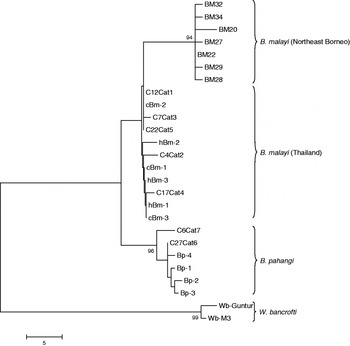
Genetic relationships of the B. malayi isolates can be better inferred if presented in the form of a phylogenetic tree. In our study, the trees were constructed using Neighbour-Joining (NJ) and Maximum Parsimony (MP) methods. In the tree analysis, closely related filarial worms such as B. pahangi and W. bancrofti were also included, with the latter serving as outgroup. In both of the phylogenetic trees (Figs 2 and 3), Brugia spp and W. bancrofti were distinctly separated into 2 main clusters. The Brugia spp. cluster could be divided into 3 subclusters, 1 consisting of B. pahangi and the remaining 2 made up of B. malayi. Interestingly, the 2 B. malayi subclusters were clearly divided based on the geographical origin of the isolates. One subcluster contained isolates from Thailand, and the other subcluster was composed of isolates from Northeast Borneo. Host was not a factor in the clustering of B. malayi as human and cat isolates from Thailand were grouped together.
Phylogenetic tree based on ITS1 sequences of Brugia malayi, B. pahangi and Wuchereria bancrofti. The tree is constructed using the Neighbour-Joining method. The percentage (>90%) of replicate trees in which the associated isolates cluster together in the bootstrap test (1000 replicates) are shown next to the branches. The phylogenetic analysis was conducted using MEGA4 (Tamura et al. Reference Tamura, Dudley, Nei and Kumar2007).

Phylogenetic tree based on ITS1 sequences of Brugia malayi, B. pahangi and Wuchereria bancrofti. The tree is constructed using the Maximum Parsimony method. The percentage (>90%) of replicate trees in which the associated isolates cluster together in the bootstrap test (1000 replicates) are shown next to the branches. The phylogenetic analysis was conducted using MEGA4 (Tamura et al. Reference Tamura, Dudley, Nei and Kumar2007).

DISCUSSION
Molecular biological approaches that produce variable profiles of DNA and nucleotide sequences from members of a species have allowed precise construction of phylogenies. The spatial relationship, evolutionary origin and history of a species population can be determined if the phylogenies are analysed in a geographical context (Hickerson et al. Reference Hickerson, Carstens, Cavender-Bares, Crandall, Graham, Johnson, Rissler, Victoriano and Yoder2010). Studies have shown that geographical isolation of populations results in reproductive isolation. This can lead to genetic variation and subsequently to divergence of the species.
A number of molecular markers or genes have been the targets for genetic variation or diversity studies in filarial worm species. During the early years of molecular phylogenetic studies of filarial worms, markers such as the HhaI repeat sequences (Xie et al. Reference Xie, Bain and Williams1994a) and 5S rRNA (Xie et al. Reference Xie, Bain and Williams1994b) have been used but primarily to distinguish species of filarial worms. Sequence data of these markers do not have the resolving power to distinguish variation within a species. More recently, Bhandari et al. (Reference Bhandari, Dabir, Nandhakumar, Dayananda, Shouche and Reddy2005) analysed W. bancrofti isolates from 3 different zones in India using PCR-RFLP and sequencing of the 18S rRNA, but failed to detect any polymorphism among the isolates. The Random Amplified Polymorphic DNA (RAPD) method has been successfully used in detecting genetic variation in W. bancrofti in India (Pradeep Kumar et al. Reference Pradeep Kumar, Patra, Hoti and Das2002; Thangadurai et al. Reference Thangadurai, Hoti, Kumar and Das2006; Patra et al. Reference Patra, Ramu, Hoti, Pragasam and Das2007; Hoti et al. Reference Hoti, Thangadurai, Dhamodharan and Das2008) and Thailand (Nuchprayoon et al. Reference Nuchprayoon, Junpee and Poovorawan2007). However, the main drawback of RAPD lies in the random amplification of regions in the genome, thus not providing information on the exact regions where variation occurs.
The ITS1 and ITS2 are non-functional regions within the rRNA gene cassette in the nuclear genome. Low evolutionary pressure on these non-functional regions results in a high degree of variation, even within a species. Thus, the regions are widely used for taxonomy and molecular phylogeny studies. Furthermore, the regions can be easily amplified by PCR, even from small quantities of DNA, because of their high copy number in the genome. Areekit et al. (Reference Areekit, Khuchareontaworn, Kanjanavas, Sriyapai, Pakpitchareon, Khawsak and Chansiri2009a,Reference Areekit, Singhaphan, Khuchareontaworn, Kanjanavas, Sriyaphai, Pakpitchareon, Khawsak and Chansirib) exploited the ITS sequences to show intraspecific variation of Brugia spp. in cat reservoirs, and also to distinguish B. malayi from B. pahangi in a single infected cat.
In our study, B. malayi from Northeast Borneo was compared with B. malayi from Thailand. Geographically, Borneo Island is separated from mainland Asia by the South China Sea. Geo-historical studies suggest that the separation took place some 30–45 million years ago. As a result of this geographical separation and isolation, some of the flora and fauna on the island have genetically diverged over time from their respective species on mainland Asia and formed unique populations or even species of their own. The findings of our study, to some extent, provide evidence of such genetic divergence. Only minor genetic variation was observed in the ITS1 of B. malayi of Northeast Borneo. On the other hand, considerable variation was noted when comparison was made with the Thailand isolates. This is reflected in the formation of 2 distinct B. malayi subclusters. Apart from the region (positions 177–185) mentioned earlier, where there was complete absence of 9 bases, 3 additional regions or positions in the ITS1 of the Northeast Borneo isolates may also contribute to the separation of the B. malayi subclusters. These were at positions 29–42, where several base substitutions involving mainly A and C occurred; and absence of bases at positions 372 and 417.
The ITS1 has been used as a target for PCR-restriction fragment length polymorphism (PCR-RFLP)-based methods to detect and differentiate filarial worm species in humans and cats in Thailand (Nuchprayoon et al. Reference Nuchprayoon, Junpee, Poovorawan and Scott2005, Reference Nuchprayoon, Junpee, Nithiuthai, Chungpivat, Suvannadabba and Poovorawan2006). In these studies, the amplified ITS1 fragment of B. malayi was digested by AseI to produce a gel profile with 3 fragments of 133, 153 and 218 bp in size. However, the Northeast Borneo isolates would not produce such profiles if subjected to this PCR-RFLP procedure due to the shorter ITS1 length. Therefore, this PCR-RFLP procedure should be used with caution, particularly when used to detect B. malayi of non-Thailand origin.
In conclusion, the findings of our study demonstrate the usefulness of ITS1 as a suitable marker for phylogeographical studies of B. malayi. For future work, isolates from other countries in Asia should be included in order to obtain a more comprehensive picture of the phylogeographical distribution and genetic diversity of B. malayi strains. This will help in the development of a more precise molecular detection method based on ITS1.
ACKNOWLEDGEMENT
We would like thank the people who participated in this study, as well as the personnel from the Sabah State Health Office.
FINANCIAL SUPPORT
This study was supported by the University of Malaya HIR Grant No. E000050-20001 and the Bill & Melinda Gates Foundation Research Grant No.304 /CIPPM /650504.