Introduction
Benign paroxysmal positional vertigo (BPPV) occasionally develops after mechanical stimulation of the temporal bone, such as during ear or dental surgery and physical exercise using a vibration device. It is suspected that external vibration to the ear is one of the causes of BPPV. We investigated whether mechanical vibration applied to the ear could induce otoconial dislodgement using isolated labyrinthine models from the American bullfrog (Rana catesbeiana). Inner-ear disorders may also be predisposing factors for BPPV. We created vestibular dysfunction models and the time required for otoconial dislodgement to occur in these models was compared with the normal models. We also investigated the morphology of the utricular macula.
Materials and methods
We performed experiment 1 using the normal models and experiment 2 using the vestibular dysfunction models. All experiments were conducted according to the Tokyo Medical University's rules regarding animal experimentation.
Experiment 1 (normal models)
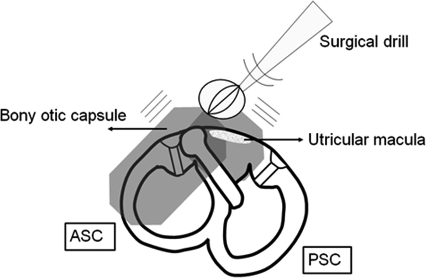
For the preparation of the experimental models, American bullfrogs weighing 110–220 g were selected. After the induction of deep anaesthesia with ether, they were decapitated and the bony labyrinth was removed, placed in a glass dish and 100 ml Ringer solution was added to the dish, according to the method described by Suzuki et al. Reference Suzuki, Harada, Hirakawa, Hirakawa and Omura1 The caudal otic capsule was chiselled so that the entire posterior semicircular canal was exposed. The remaining membranous labyrinth was left enclosed in the bony capsule. The labyrinth preparation was held by forceps with the posterior semicircular canal in the undermost position. Mechanical vibration was applied to the upper surface of the bony otic capsule using a surgical drill (BL-F5A; Osada Electric Company, Tokyo, Japan) according to the method described by Otsuka et al. (Figure 1).Reference Otsuka, Suzuki, Negishi, Shimizu, Inagaki and Konomi2 An end-cutting bur, with a tip measuring 5 mm in diameter, was used. The frequency of the vibration, as measured by a vibration analyser (VA-12; Rion, Tokyo, Japan), was 340 Hz. Care was taken not to rupture the membranous labyrinth. This allowed the otoconia to dislodge from the utricular macula and move into the posterior semicircular canal. The time required for otoconial dislodgement was measured. A dissection microscope (SZX12; Olympus, Tokyo, Japan) was used for observation.
Experimental model. Using a surgical drill, mechanical vibration was applied to the upper surface of the bony otic capsule of the labyrinth preparation. ASC = anterior semicircular canal; PSC = posterior semicircular canal

Experiment 2 (vestibular dysfunction models)
The vestibular dysfunction models were created by injecting 300 μg (7.5 μl) of gentamicin into the perilymphatic space of the bullfrog labyrinth using a palatal approach.Reference Konomi, Suzuki, Otsuka, Shimizu, Inagaki and Hasegawa3 The bullfrogs were kept alive for 7–14 days. Then, the same procedure as in experiment 1 was performed and the time required for otoconial dislodgement was measured. The dislodgement time was compared with that of the normal models. For statistical analysis, we used the Student's t-test. P values less than p = 0.05 were considered to be statistically significant. After the experiment, sections of the utricular macula were prepared for microscopic observation; they were fixed in formalin, embedded in paraffin, sectioned and stained with haematoxylin and eosin. The otoconia and the otolithic membrane were apparently lost during the staining process. We evaluated the condition of the sensory hairs and sensory cells accordingly.
Results
In all specimens, we observed that the otoconia had dislodged from the utricular macula and moved into the posterior semicircular canal when subjected to vibration (Figure 2).
Dissection microscopic image and model of otoconial dislodgement. The utricular otoconia dislodged from the macula and moved into the posterior semicircular canal (PSC) after vibration.
Experiment 1 (normal models)
In the normal models (n = 21), the dislodgement times varied from 120 to 1000 s. The average dislodgement time was 456 s (2 standard deviations (SDs) =± 248 s).
Experiment 2 (vestibular dysfunction models)
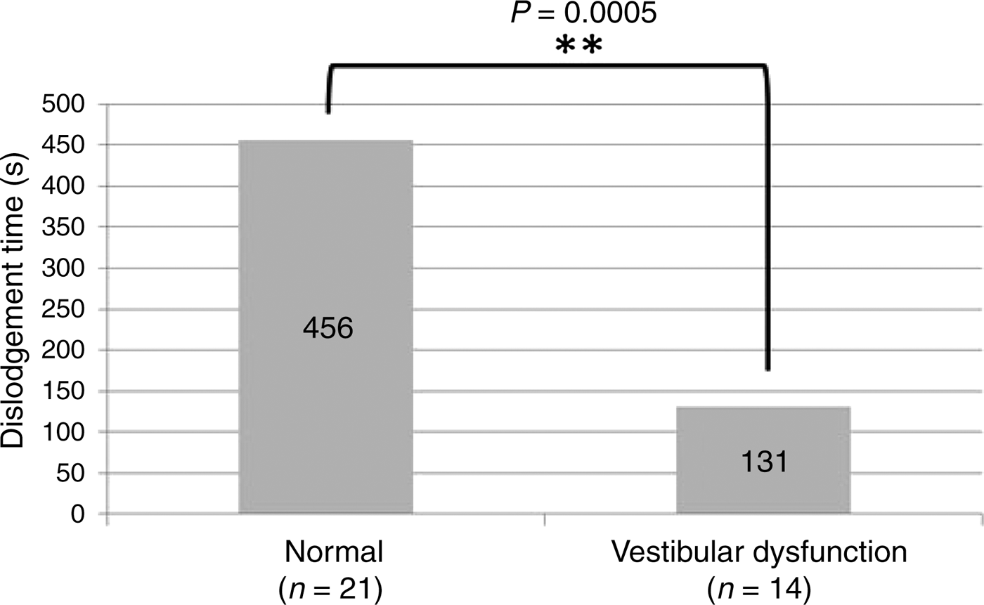
In the vestibular dysfunction models (n = 14), the dislodgement times varied from 45 to 300 s. The average dislodgement time was 131 s (2 SDs =± 85 s), which was significantly shorter than in the normal models (p = 0.0005) (Figure 3).
Average time of otoconial dislodgement. The time to otoconial dislodgement was significantly shorter in the vestibular dysfunction models than in the normal models (p = 0.0005).
The pathology of the utricular macula was investigated in nine of the vestibular dysfunction models. When compared with those of the normal model (Figure 4a), all of the sensory hairs of the vestibular dysfunction model (Figure 4b) were reduced in number. In three other vestibular dysfunction models, the otoconia were dislodged before applying vibration. In two of them, the morphological damage to the utricular macula was severe. In one specimen, the utricular sensory cells became atrophic (Figure 4c). In the other specimen, the sensory cells were missing altogether (Figure 4d).
Microscopic image of the utricular macula. When the normal model (a) was compared to the vestibular dysfunction model (b), there was a reduction in the number of sensory hairs (arrow) in the vestibular dysfunction model. In the utricular macula with dislodged otoconia before vibration, the morphological damage was severe; the sensory cells (arrow) became atrophic (c) or were missing altogether (d). (H&E; ×400)
Discussion
Ear surgery occasionally induces BPPV.Reference Viccaro, Mancini, La Gamma, De Seta, Covelli and Filipo4–Reference Dornhoffer and Colvin6 In our series of 48 tympanoplasty cases, 2 cases of direction-changing, horizontal positional nystagmus were observed after surgery.Reference Ichimura7 In all cases, the positional nystagmus resolved in 14–27 days. The mechanical vibration effect of a surgical drill on the otolithic organ was considered responsible for the emergence of positional nystagmus. There would have been no chemical effect on the endolymph since the inner ear was not disturbed during surgery.
Dental surgery also occasionally induces BPPV.Reference Andaz, Whittet and Ludman8–Reference Peñarrocha, Pérez, Garciá and Guarinos13 Chiarella et al. Reference Chiarella, Leopardi, De Fazio, Chiarella and Cassandro14 reported that BPPV arose on the operation side within 7 days after dental surgery in 8 patients, in men aged under 45 and women aged under 40. Seven out of eight patients had BPPV of the posterior semicircular canal. One patient had lateral semicircular canal BPPV. It has been suggested that vibrations are propagated throughout the bony structures reaching the labyrinth. At this level, mechanical energy would travel through the endolymphatic fluid or bone, eventually causing macular trauma. Amir et al. Reference Amir, Young and Belloso15 reported that a 44-year-old woman developed BPPV following the use of a whole-body vibration training plate, a popular form of fitness equipment widely used in sports, rehabilitation and body workout treatments.
Whole-body vibration training can potentially generate displacement or dislocation of otoconia through vibration being transmitted to the inner ear. Dan-Goor and SamraReference Dan-Goor and Samra16 reported that a 44-year-old woman developed acute and severe positional vertigo on waking up, after continuously using noise-cancelling headphones for 12 h prior to going to sleep. They suspected that repeated mechanical disruption or vibration within the vestibular system could have dislodged otoconia into the posterior semicircular canal.
We investigated whether mechanical vibration could induce otoconial dislodgement using experimental models. Fine morphological studies have shown that the otoconia of the utricle are connected to each other by means of fine fibrils and are embedded in a gelatinous substance consisting of mucopolysaccharides.Reference Kachar, Parakkal and Frex17, Reference Lins, Farina, Kurc, Riordan, Thalmann and Thalmann18 The whole otoconial mass is covered by a supra-otoconial layer.Reference Nakai, Masutani, Kato and Sugiyama19 We expected that the otoconia would not be easily dislodged from the utricular macula by weak stimulation. We directly stimulated the inner ear with the vibration of a surgical drill. The otoconia were dislodged from the utricular macula and moved into the posterior semicircular canal within 20 min in all specimens. We were able to confirm that the utricular otoconia were dislodged after vibration. The intensity of the stimulus in this experiment was strong because the vibration was applied in close proximity to the labyrinth. Although a weaker stimulation may require a longer dislodgement time, the vibratory stimulus could be one of the causes of BPPV.
Benign paroxysmal positional vertigo is usually idiopathic, but in some cases, it arises after inner-ear disease. Karlberg et al. Reference Karlberg, Hall, Quickert, Hinson and Halmagyi20 reported that 81 out of 2847 BPPV patients (2.8 per cent) had definite BPPV of the posterior semicircular canal secondary to an ipsilateral inner-ear disease. Sixteen patients had Ménière's disease, 24 had acute unilateral peripheral vestibulopathy, 12 had chronic unilateral peripheral vestibulopathy, 21 had chronic bilateral peripheral vestibulopathy and 8 had unilateral sensorineural hearing loss. Katsarkas and KirkhamReference Katsarkas and Kirkham21 found that 20 out of 255 BPPV patients (7.8 per cent) had secondary BPPV. Baloh et al. Reference Baloh, Honrubia and Jacobson22 found 80 (33.3 per cent) cases of secondary BPPV out of 240 BPPV patients. Hughes and ProctorReference Hughes and Proctor23 found 60 (39.7 per cent) cases of secondary BPPV out of 151 BPPV patients. The ratio of secondary BPPV thus varies widely. On the other hand, Inagaki et al. Reference Inagaki, Yukawa, Ichimura, Hagiwara, Ogawa and Kitajima24 analysed 123 cases of Ménière's disease, sudden deafness and vestibular neuronitis and found 14 cases (11.4 per cent) of secondary BPPV. Of 48 Ménière's disease cases, 4 (8.3 per cent) presented with BPPV. Three of 31 sudden deafness cases (9.7 per cent) and 7 of 44 vestibular neuronitis cases (15.9 per cent) presented with BPPV. Von Brevern et al. Reference von Brevern, Radtke, Lezius, Feldmann, Ziese and Lempert25 screened 4869 participants in a cross-sectional, nationally representative, neurotological survey of the general adult population in Germany. The lifetime prevalence of BPPV was found to be 2.4 per cent. Patients with inner-ear disease develop BPPV more often than the general adult population.
We investigated whether inner-ear diseases can be predisposing factors for BPPV using an experimental approach. We performed experiments using vestibular dysfunction models created by injecting gentamicin into the perilymphatic space of the bullfrog labyrinth. The dislodgement time was significantly shorter in the vestibular dysfunction models than in the normal models, and the utricular macula sustained morphological damage, such as a reduction in the number of sensory hairs and sensory cell atrophy. The relationship between the short dislodgement time and the morphological damage of sensory cells is unknown.
• Benign paroxysmal positional vertigo occasionally develops after vibration, such as during ear or dental surgery and physical exercise using a vibration device
• In this experimental study, utricular otoconia were dislodged into the posterior semicircular canal after vibration
• The time to dislodgement was significantly shorter in the vestibular dysfunction models than in the normal models
• Inner-ear diseases can be predisposing factors for benign paroxysmal positional vertigo
A reduction in the number of sensory hairs possibly indicates an association with the changes in the otoconia and otolithic membrane. If this is true, the otoconia are easily dislodged by the damage. On the other hand, the sensory hairs protrude into the otolithic membrane and possibly support the membrane. A reduction in the number of the hair cells weakens the support, thus inducing dislodgement of the otoconia. Other insults, such as ischaemia, endolymphatic hydrops or ageing, potentially change the utricular macula, thus possibly leading to easy dislodgement of the otoconia from the macula.
Conclusions
In our study, the utricular otoconia were dislodged into the posterior semicircular canal after vibration. The time to dislodgement was significantly shorter in the vestibular dysfunction models than in the normal models; the utricular macula sustained morphological damage.
Acknowledgements
The authors are indebted to Associate Professor Edward F Barroga of the Department of International Medical Communications at Tokyo Medical University for the editorial review of the English manuscript. They also thank Hiroaki Iobe of the Department of Anatomic Pathology, Tokyo Medical University and Hirotoshi Hishida of the Department of Mechanical Engineering, Faculty of Engineering, Kogakuin University. This study received financial support from a Grant-in-Aid for Scientific Research (C) from the Ministry of Education, Culture, Sports, Science and Technology, Japan, a Health and Labour Science Research Grant for Research on Specific Disease (Vestibular Disorders) from the Ministry of Health, Labour and Welfare, Japan and a follow-up grant for a Tokyo Medical University research project.