Introduction
Since ancient times, several herbal biomaterials play a vital role in the wound healing applications. Curcumin (diferuloylmethane) is a biocompatible and biodegradable yellow-orange phenolic compound, extracted from the rhizomes of Curcuma longa (turmeric).[Reference Aggarwal, Kumar and Bharti1] Typically used in Asian foods as spice ingredient, specifically in India and Eastern Asia.[Reference Goel, Kunnumakkara and Aggarwal2] Curcumin exhibits numerous therapeutic properties including anti-inflammatory, antifungal, antioxidant, antiviral, antibacterial, and anticancer.[Reference Aggarwal, Kumar and Bharti1–Reference Merrell, McLaughlin, Tie, Laurencin, Chen and Nair3] Further, it showed a potential to treat asthma, diabetes, allergies, arthritis, neurodegeneration disease, atherosclerosis, and chronic cancer.[Reference Abidi4–Reference Shanbhag10] Several clinical trials for human cancer have demonstrated the effectiveness, pharmacokinetics, and safety of curcumin.[Reference Aggarwal, Kumar and Bharti1, Reference Goel, Kunnumakkara and Aggarwal2]
However, clinical effect and bioavailability of curcumin are greatly limited due to its hydrophobic nature. Therefore, it is important to improve the solubility of curcumin by functionalization or select the appropriate method to deliver the drug in a specific tumor site.[Reference Greiner and Wendorff11] Usually, nanoparticles, cast films, hydrogels, and ultrafine nanofiber scaffolds have been used as drug transporters.[Reference Boateng, Mani and Kianfar12–Reference Thangaraju, Srinivasan, Kumar, Sehgal and Rajiv14] Among them, fiber scaffolds are potential implantable drug carriers for local delivery and controlled drug release.[Reference Kenawy, Bowlin, Mansfield, Layman, Simpson, Sanders and Wnek15] This method can minimize the side effects and provides the high concentration of antineoplastic drug by using a small quantity of the drug. Due to improved therapeutic efficiency, inferior toxicity, and sustained drug release rate, localized control drug delivery has been a subject of intensive research and drug development.[Reference Jiang, Fang, Hsiao, Chu and Chen16]
Tissue engineering and drug delivery are interconnected new research areas. Tissue regeneration efficiency can be improved by combining therapeutic drug release with implantable fiber scaffolds. Thus, fiber scaffolds have been used as promising drug carriers to deliver anticancer drugs, peptides, protein, and genes due to its higher surface area, interconnecting pores, high permeability, and versatile fabricating methods.[Reference Son, Kim and Yoo17] To improve the biologic characteristics of curcumin, bio-polymeric fiber scaffolds are utilized. Thus, curcumin-based poly(3-caprolactone)-poly(ethylene glycol)-poly(3-caprolactone) (PCL-PEG-PCL, PCEC) electrospun fibers were used to study antitumor activity in glioma 9L cells.[Reference Guo, Fu, Zhou, Liang, Fan, Luo, Qian and Wei18] Poly caprolactone/gum tragacanth/curcumin electrospun fibers were used to study prolonged drug release and its biocompatibility.[Reference Ranjbar-Mohammadi and Bahrami19] PCL/gelatin (GL) fibers were used to deliver curcumin in a sustained release.[Reference Fallah, Bahrami and Ranjbar-Mohammadi20] In addition, curcumin loaded polyvinyl alcohol and polylactic acid (PLA)/hyperbranched polyglycerol fibers were used for drug release and wound healing, respectively.[Reference Sun, Williams, Hou and Zhu21, Reference Perumal, Pappuru, Chakraborty, Maya Nandkumar, Chand and Doble22]
PLA is a biocompatible and biodegradable polymer used for drug delivery, tissue engineering, regenerative medicine, fabrication of surgical sutures, and gene transfer applications.[Reference Langer and Tirrell23, Reference Luten, van Nostrum, De Smedt and Hennink24] It was copolymerized with synthetic and natural biopolymers including polyglycolide, PCL, and GL, respectively, in electrospinning.[Reference Perumal, Pappuru, Chakraborty, Maya Nandkumar, Chand and Doble22, Reference Li, Laurencin, Caterson, Tuan and Ko25] Thus, curcumin/PLA fibers were used in wound healing and blood compatibility applications.[Reference Chen, Lin, Wan, Fei, Wang and Gao26, Reference Nguyen, Ghosh, Hwang, Tran and Park27] GL is a biopolymer extracted from collagen. It is an excellent biomaterial for cell proliferation and cell adhesion. However, water solubility, degradation, and poor tensile properties of pure GL fibers have restricted the application of GL in drug release and tissue engineering.
Therefore, recently GL was reinforced and forcespun with zein and poly(epichlorohydrin-co-ethylene oxide) to improve biologic, drug release, physicochemical properties.[Reference Mamidi, Leija Gutiérrez, Villela-Castrejón, Isenhart, Barrera and Elías-Zúñiga28, Reference Mamidi, Romo, Leija Gutiérrez, Barrera and Elías-Zúñiga29]
In the present study, aligned nanofibers are fabricated using a high-throughput Forcespinning® (FS) technology that works with the principle of centrifugal force.[Reference Mamidi, Leija Gutiérrez, Villela-Castrejón, Isenhart, Barrera and Elías-Zúñiga28, Reference Mamidi, Romo, Leija Gutiérrez, Barrera and Elías-Zúñiga29] Notably, this method allows sustained production of highly aligned ultrafine fiber mats with 80% of fabrication yield. Curcumin embedded GL and PLA fibrous scaffolds were manufactured to demonstrate the versatility of FS and studied sustained curcumin drug release. GL and PLA ultrafine fibers would be greatly advantageous for the drug delivery, wound healing, and tissue engineering applications because, they provide favorable mechanical strength and biocompatibility, respectively. Improved mechanical, sustained curcumin drug release and cell viability properties are observed.
Materials and methods
Materials
GL, PLA, curcumin and trifluoroacetic acid (TFA) were purchased from Sigma Aldrich (St. Louis, MO, USA). Cell culture consumables and supplements were obtained from Gibco Invitrogen (Carlsbad, CA, USA). CellTiter96®AQueous One-Solution Cell Proliferation Assays was bought from, Promega, (Madison, WI, USA.
Fabrication of pure GL, PLA, GL/Cur and GL/PLA/Cur nanofibers
All the nanofibers were manufactured by forcespinning® technique. To achieve this,
(a) Pure GL (30 wt .%): 6.0 g of GL (30 wt.%) was dissolved in 20 mL of TFA and stirred at 50 °C for 24 h. After obtaining a homogeneous solution, GL (30 wt.%) blend solution was forcespun at 6500 rpm for 30 s.
(b) Pure PLA (30 wt.%): 6.0 g of PLA (30 wt.%) was dissolved in 20 mL of TFA and stirred at 50° C for 24 h. After obtaining a homogeneous solution, PLA (30 wt.%) blend solution was forcespun at 6500 rpm for 30 s.
(c) GL (30 wt.%)/Cur (8.3 wt.%): 6.0 g of GL (30 wt.%) and 0. 498 g of curcumin (8.3 wt.%, relative to GL) were dissolved in 20 mL of TFA and stirred at 50 C for 24 h. After obtaining a homogeneous solution, GL (30 wt.%)/Cur (8.3 wt.%) blend solution was forcespun at 6500 rpm for 30 s.
(d) GL (7.5 wt.%)/PLA (22.5 wt.%)/Cur (8.3 wt.%): 1.5 g of GL (7.5 wt.%), 4.5 g of PLA (22.5 wt.%) and 0.498 g of curcumin (8.3 wt.%, relative to both polymers) were dissolved in 20 mL of TFA and stirred at 50 °C for 24 h. Then, a homogeneous blend solution [GL (7.5 wt.%)/PLA (22.5 wt.%)/Cur (8.3 wt.%)] was forcespun at 6500 rpm for 30 s.
(e) GL (6 wt.%)/PLA (24 wt.%)/Cur (8.3 wt.%): For this, 1.2 g of GL (6.0 wt.%), 4.8 g of PLA (24.0 wt.%) and 0.498 g of curcumin (8.3 wt.%, relative to both polymers) were dissolved in 20 mL of TFA and stirred at 50 °C for 24 h. Then, a homogeneous GL (6 wt.%)/PLA (24 wt.%)/Cur (8.3 wt.%) solution was forcespun at 6500 rpm for 30 s.
Characterization of nanofibers
The following techniques were used to characterize pure GL, curcumin, PLA, GL/Cur and GL/PLA/Cur nanofibers. Scanning electron microscopy (SEM) (ZEISS EVO® MA 25, Ostalbkreis, Baden-Württemberg, Germany) was used for the morphological investigations. Fourier transform infrared (FTIR) spectroscopy (Perkin Elmer Universal ATR Sampling Accessory Frontier) was used to analyze the functional groups and hydrogen bonding interactions of fibers. Image J analyzer software (Image J, NIH, USA) was utilized to measure the pore size of fibers. The tensile properties of fibers were measured using a uniaxial tensile machine (Instron 3365, Instron, Norwood, MA, USA). In order to do in vitro biodegradation of fiber scaffolds, the fiber was immersed in Dulbecco's Modified Eagle's medium (DMEM) and incubated for 25 days at 37 °C. At 10, 15, 20, and 25 days, the fiber scaffolds were taken out from the buffer, washed and dried in the hot oven for 24 h. Optical contact angle measurement (OCA 15EC) was used to measure the surface wettability of fibers.
Curcumin release from forcespun GL/Cur and GL/PLA/Cur nanofibers
Curcumin drug release from forcespun GL/Cur and GL/PLA/Cur fibers was measured by a UV-spectrophotometer (Agilent Technologies, 89090A). 50 mg of curcumin embedded GL/Cur and GL/PLA/Cur fiber scaffolds were immersed in 20 mL of DMEM and incubated on a shaking incubator at 37 °C. The UV absorbance of released curcumin in DMEM was determined at λ max = 426 nm and transformed into curcumin concentration, through the calibration curve. The cumulative curcumin release from GL/Cur and GL/PLA/Cur fibers against drug release time was showed. The experiments were run in triplicate.
Cell proliferation studies
Human fibroblast cells (4.8 × 10⁵ cells/cm2) were seeded on the prior sterilized ultrafine fiber scaffolds (0.1 cm2) in DMEM, supplemented with 100 µg/mL penicillin, 5% (w/v) fetal bovine serum and 100 µg/mL streptomycin. After that, the samples were incubated at 37 °C under 5% CO2 atmosphere. All experiments were carried out in triplicate. The colorimetric 3-(4,5-dimethylthiazol-2-yl)-2,5-diphenyltetrazolium bromide assay (CellTiter96®AQueous One Solution) was utilized to measure the cell proliferation and viability of ultrafine fiber scaffolds. For this, cell (4.8 × 10⁵ cells/cm2) were seeded on fiber membranes. The samples were shifted into fresh wells and CellTiter (20 µL) was added on the first day, second day, and third day, respectively. After that, the samples were incubated at 37 °C for 60 min under dark conditions. Microplate reader (Synergy HT, BioTek, Winooski, Vermont, USA) was used to measure the absorbance of each well. Viability percentage was measured from the absorbance of each well and control (without fibers). The experiments were run in triplicate.
Cell morphology studies
To measure the cell proliferation and cell attachment capability of GL (6 wt.%)/PLA (24 wt.%)/Cur (8.3 wt.%) ultrafine fibers, cell culture experiments were performed. SEM was utilized to measure the morphological characteristic of cells cultured on the surface of GL (6 wt.%)/PLA (24 wt.%)/Cur (8.3 wt.%) fibers. After day 1, day 2 and day 3 of incubation, the fiber scaffolds were rinsed with PBS and fixed with 2.5% glutaraldehyde for overnight at 4 °C. Then, the samples were dehydrated with various concentrations of ethanol, i.e., 50, 70, 90, and 100% and stored in the vacuum desiccator for 12 h. The fibers samples were gold coated and then SEM analysis was performed.
Statistical analysis
All data presented are expressed as the mean ± standard deviation. One-way analysis of variance and Tukey post hoc were used for the statistical analysis and statistical significance was considered at P ≤ 0.05.
Results and discussion
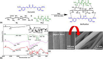
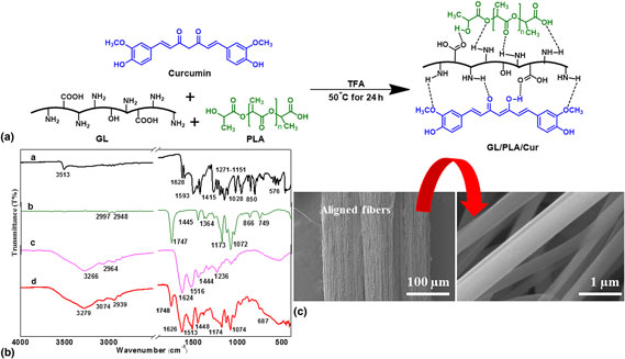
In the current study, curcumin bearing GL/PLA/Cur composites were synthesized in a single step reaction and GL/PLA/Cur ultrafine fiber scaffolds were manufactured by FS. Figure 1 shows the synthesis, FTIR, and SEM images of curcumin loaded GL/PLA/Cur composite fibers. Initially GL (7.5 wt.%)/PLA (22.5 wt.%)/Cur (8.3 wt.%) composite was prepared [Fig. 1(a)]. For this, GL (1.5 g) was treated with PLA (4.5 g) and curcumin (8.3 wt.%) in 20 mL of TFA as a solvent system and the mixture was stirred at 50 °C for 24 h. The resulted homogeneous GL (7.5 wt.%)/PLA (22.5 wt.%)/Cur (8.3 wt.%) solution was fed into the spinneret of FS and the experiment was run under several conditions. However, 6500 rpm and 30 s were found to be the best-operating conditions. Under similar conditions, GL (6 wt.%)/PLA (24 wt.%)/Cur (8.3 wt.%) composite was prepared [Fig. 1(a)]. The homogeneous solution of GL (6 wt.%)/PLA (24 wt.%)/Cur (8.3 wt.%) was loaded in spinneret and obtained align fibers under the same operating parameters (6500 rpm and 30 s). The orientation of the fibers are established by SEM analysis [Fig. 1(c)].
Development of GL/PLA/Cur composites: (a) synthesis of curcumin embedded GL/PLA/Cur composites, (b) FTIR spectra of (a) pure curcumin, (b) pure PLA, (c) pure GL, and (d) GL (6 wt.%)/PLA (24 wt.%)/Cur (8.3 wt.%) composite fibers and (c) SEM image of GL (6 wt.%)/PLA (24 wt.%)/Cur (8.3 wt.%) aligned fiber scaffolds fabricated at 6500 rpm in 30 s, respectively (arrow indicates zoom in SEM image). Scale bar = 100 and 1 µm, respectively.
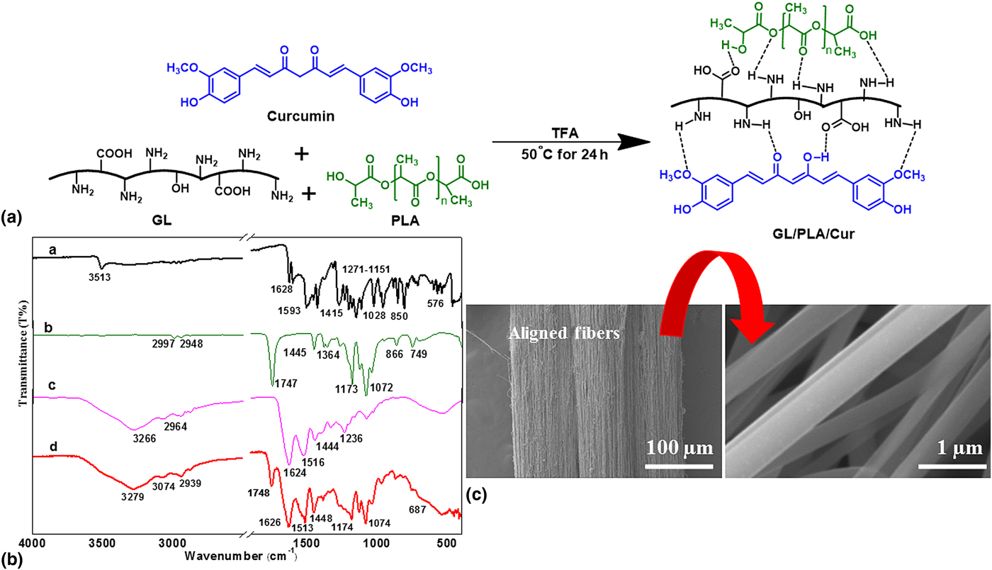
To scrutinize the chemical structure and interfacial interactions of GL/PLA/Cur composites, FTIR analysis was carried out and compared with FTIR spectra of pristine PLA, GL, and curcumin [Fig. 1(b)]. The major peaks of interest for PLA and curcumin are 1747 and 1628 cm−1 for carbonyl (C = O stretching) and 3513–3000 cm−1 for hydroxyl functional groups (O–H stretching).[Reference Perumal, Pappuru, Chakraborty, Maya Nandkumar, Chand and Doble22, Reference Ranjbar-Mohammadi and Bahrami19, Reference Fallah, Bahrami and Ranjbar-Mohammadi20] GL bands appeared at around 1624 cm−1 for C = O stretching, 1516 cm−1 for N–H bending and 3266 cm−1 for N–H stretching.[Reference Fallah, Bahrami and Ranjbar-Mohammadi20] The FTIR spectrum of curcumin showed a peak at 3513 cm−1 denoted as a phenolic O–H stretching. Sharp peaks at 1628 cm−1 (indicated C = O group), 1593 cm−1 for benzene stretching vibrations and C = C vibrations, 1415 cm−1 for bending vibration of olefinic C–H, 1271–1151 cm−1 for C–O stretching of benzene ring and 1028/576 cm−1 for C–O–C stretching of curcumin were observed.[Reference Ranjbar-Mohammadi and Bahrami19, Reference Fallah, Bahrami and Ranjbar-Mohammadi20]
However, the peaks at 1628 cm−1 for the carbonyl (C = O stretching) and at 3513 cm−1 for the OH functional groups of curcumin were no longer observed in GL (6 wt.%)/PLA (24 wt.%)/Cur (8.3 wt.%) fibers. Rather, the fiber formation generated peaks at 3279 and 1626 cm−1, respectively. These peaks indicate the generation of hydrogen bonds between curcumin and GL. In addition, C = O stretching (1747 cm−1) and C–O–C stretching (1364 cm−1) peaks of pristine PLA are clearly perceptible in the FTIR spectrum of PLA [Fig. 1(b)].[Reference Perumal, Pappuru, Chakraborty, Maya Nandkumar, Chand and Doble22] These peaks are shifted to 1748 and 1448 cm−1 in GL (6 wt.%)/PLA (24 wt.%)/Cur (8.3 wt.%), respectively. It indicates the formation of hydrogen bonds between PLA and GL [dotted black lines in Fig. 1(a)]. Further, The O–H peaks of pristine PLA were vanished in GL (6 wt.%)/PLA (24 wt.%)/Cur (8.3 wt.%) composite fibers and generated a broad peak at 3279 cm−1. This could be due to the formation of hydrogen bonds between PLA and GL [dotted black lines in Fig. 1(a)]. As anticipated, FTIR analysis of PLA, GL, curcumin, and GL (6 wt.%)/PLA (24 wt.%)/Cur (8.3 wt.%) fibers showed an indication of hydrogen bonding.
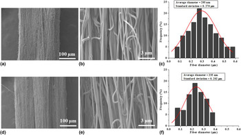
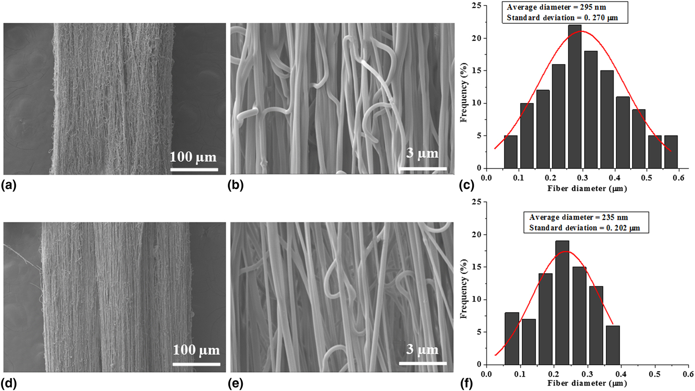
Morphology, diameter, and structure of forcespun GL/PLA/Cur fibers were investigated using SEM analysis. Figure 2 shows the SEM images of GL (7.5 wt.%)/PLA (22.5 wt.%)/Cur (8.3 wt.%) and GL (6 wt.%)/PLA (24 wt.%)/Cur (8.3 wt.%) ultrafine aligned fibers. SEM micrographs revealed that the fiber scaffolds were homogeneous and bead free interconnected aligned fibers. The amalgamation of curcumin into GL/PLA/Cur fibers slightly decreased their average fiber diameter and diameter distribution of forcespun ultrafine fibers. The average diameter of GL (7.5 wt.%)/PLA (22.5 wt.%)/Cur (8.3 wt.%) and GL (6 wt.%)/PLA (24 wt.%)/Cur (8.3 wt.%) was found 295.0 nm and 235.0 nm, respectively. The incorporation of curcumin did not influence the diameter of GL/PLA/Cur fibers significantly. However, diameter was slightly altered from 195.0 nm for GL (7.5 wt.%)/PLA (22.5 wt.%)/Cur (8.3 wt.%) fibers to 235.0 nm for GL (6 wt.%)/PLA (24 wt.%)/Cur (8.3 wt.%) fibers, respectively. Further, the morphology of GL/PLA/Cur fibers was not effected and produced continuous ultrafine fiber scaffolds without any crashes. Surface drug loading, roughness and random orientation of fibers were not observed on the surface of both GL (7.5 wt.%)/PLA (22.5 wt.%)/Cur (8.3 wt.%) and GL (6 wt.%)/PLA (24 wt.%)/Cur (8.3 wt.%) ultrafine fibers. It indicates that curcumin was involved in the formation of hydrogen bonds with GL. The fiber scaffolds could be tolerably porous to release the drug molecules captured within nanofibers.
(a, b) SEM images and (c) diameter distribution of GL (7.5 wt.%)/PLA (22.5 wt.%)/Cur (8.3 wt.%) fibers. (d, e) SEM images and (f) diameter distribution of GL (6 wt.%)/PLA (24 wt.%)/Cur (8.3 wt.%). Scale bar = 100 and 3 µm, respectively.
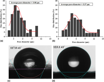
Burst release would occur if the porosity is too high. Thus, an optimization of porosity is an important factor for the sustained drug release. Therefore, poresize distribution of GL (7.5 wt.%)/PLA (22.5 wt.%)/Cur (8.3 wt.%) and GL (6 wt.%)/PLA (24 wt.%)/Cur (8.3 wt.%) fiber scaffolds was studied and results were illustrated in Figs. 3(a) and 3(b). Average pore diameter of 3.98 µm was observed in GL (7.5 wt.%)/PLA (22.5 wt.%)/Cur (8.3 wt.%) fiber scaffolds, whereas 5.37 µm was found in GL (6 wt.%)/PLA (24 wt.%)/Cur (8.3 wt.%) fiber scaffolds [Figs. 3(a) and 3(b)]. These results revealed that the poresize distribution of GL (6 wt.%)/PLA (24 wt.%)/Cur (8.3 wt.%) fiber scaffolds were increased at higher concentration of PLA.
(a, b) pore size distribution of GL (7.5 wt.%)/PLA (22.5 wt.%)/Cur (8.3 wt.%) and GL (6 wt.%)/PLA (24 wt.%)/Cur (8.3 wt.%) fibers, respectively. (c, d) water contact angles of GL (7.5 wt.%)/PLA (22.5 wt.%)/Cur (8.3 wt.%) and GL (6 wt.%)/PLA (24 wt.%)/Cur (8.3 wt.%) fibers, respectively.
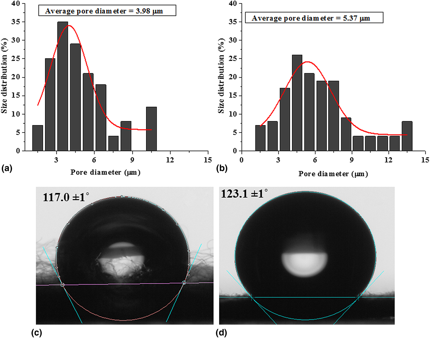
The wettability of ultrafine fibers is a unique and vital property; it regulates the cell proliferation and adhesion. In addition, it shows a significant impact on the drug release profile. For example, lower wettability leads to greater trapping of the drug, whereas higher wettability leads to vast initial burst release of the drug. Therefore, the wettability of GL/PLA/Cur fibers was studied and presented in Figs. 3(c) and 3(d). GL (7.5 wt.%)/PLA (22.5 wt.%)/Cur (8.3 wt.%) fibers showed 117.0 ± 1 of water contact angle and GL (6 wt.%)/PLA (24 wt.%)/Cur (8.3 wt.%) fibers exhibited 123.1 ± 1 of contact angle [Figs. 3(c) and (d)]. The contact angle values were proportional to the concentration of PLA. Thus, these results revealed that the surface wettability of fiber scaffolds was slightly increased; this could be due to a hydrophobic nature of PLA and curcumin. The forcespun fiber scaffolds manufactured from GL (6 wt.%)/PLA (24 wt.%)/Cur (8.3 wt.%) composites exhibited morphological variations including improvement in porosity, which enhances the wettability of the fibers.
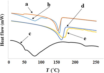
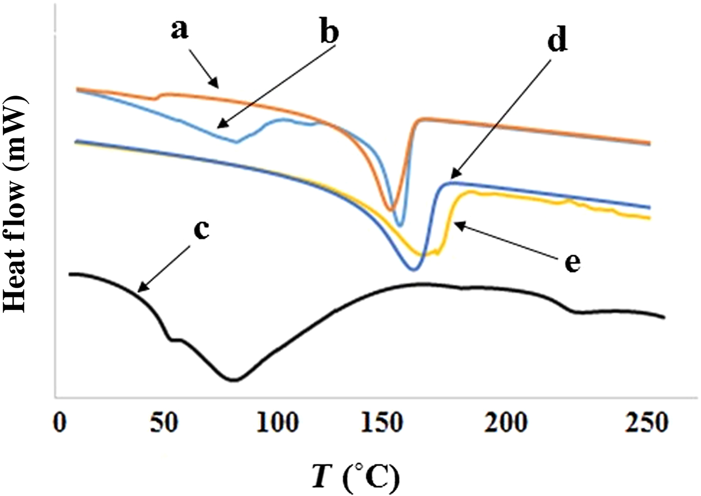
Thermal properties of pure curcumin, GL, PLA, and newly developed GL/PLA/Cur composite fibers were measured and presented in Fig. 4. Pure curcumin showed a peak at 166 °C that shows a melting temperature of curcumin.[Reference Ranjbar-Mohammadi and Bahrami19, Reference Fallah, Bahrami and Ranjbar-Mohammadi20] GL protein showed an endothermic peak at around 94 °C, this peak might be due to loss of water or denaturation of protein during helix transition to coil transition. The second peak of GL was at around 220 °C, which was melt temperature of GL. Pristine PLA exhibited a sharp endothermic peak at 169 °C.[Reference Chen, He, Yu, Jia and Meng30] Further, GL (7.5 wt.%)/PLA (22.5 wt.%)/Cur (8.3 wt.%) composite showed a melting temperature at 173 °C. Whereas, an endothermic peak was observed at 175 °C for GL (6 wt.%)/PLA (24 wt.%)/Cur (8.3 wt.%) fibers. Overall, DSC results indicate that the melting temperatures of GL/PLA/Cur composite fibers were increased in the presence of curcumin. This could be due to the formation of hydrogen bonds and intermolecular interactions between curcumin, GL, and PLA.
DSC curves of (a) pure curcumin, (b) pure PLA, (c) pure GL, (d) GL (7.5 wt.%)/PLA (22.5 wt.%)/Cur (8.3 wt.%), and (e) GL (6 wt.%)/PLA (24 wt.%)/Cur (8.3 wt.%) fibers.
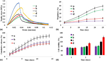
To measure the tensile properties of GL/PLA/Cur fiber scaffolds, a non-linear stress/strain curve was obtained and the graph was presented in Fig. 5a. It is important to tolerate sustain deformation and high loads to use the fiber scaffolds in tissue engineering applications. As PLA concentration increased, the tensile strength of GL/PLA/Cur fiber scaffolds was increased. Particularly, GL (6 wt.%)/PLA (24 wt.%)/Cur (8.3 wt.%) fiber scaffolds showed 1.49 MPa of tensile strength. Whereas, GL (7.5 wt.%)/PLA (22.5 wt.%)/Cur (8.3 wt.%) fibers exhibited 1.17 MPa of tensile strength. The tensile results revealed that GL (6 wt.%)/PLA (24 wt.%)/Cur (8.3 wt.%) fiber scaffolds showed higher tensile strength and lower strain than GL (7.5 wt.%)/PLA (22.5 wt.%)/Cur (8.3 wt.%) fiber scaffolds. It was anticipated that the stiffness of PLA and hydrogen bonding interactions between Cur, GL, and PLA played a vital role in improving the tensile properties of GL (6 wt.%)/PLA (24 wt.%)/Cur (8.3 wt.%) fibers. The lower concentration of PLA leads to decrease the tensile strength and increase the tensile strain. The tensile properties of GL/PLA/Cur nanofibers were compared with pristine GL (0.54 MPa), PLA (0.67 MPa) and GL (30 wt.%)/Cur (8.3 wt.%) fibers (0.72 MPa) and enhanced tensile properties were observed. This might be due to the formation of hydrogen bonds between curcumin, GL, and PLA. Overall, these results reveal that PLA concentration could be ideal (24 wt.%) to acquire enhanced tensile properties. Therefore, the tensile results of GL/PLA/Cur fibers suggested the formation of hydrogen bonding interactions between curcumin, GL, and PLA.
(a) Tensile graph of (a) pure GL, (b) pure PLA, (c) GL (30 wt.%)/Cur (8.3 wt.%), (d) GL (7.5 wt.%)/PLA (22.5 wt.%)/Cur (8.3 wt.%), and (e) GL (6 wt.%)/PLA (24 wt.%)/Cur (8.3 wt.%) fibers; (b) in vitro biodegradation of (a) pure GL, (b) GL (30 wt.%)/Cur (8.3 wt.%), (c) GL (7.5 wt.%)/PLA (22.5 wt.%)/Cur (8.3 wt.%) and (d) GL (6 wt.%)/PLA (24 wt.%)/Cur (8.3 wt.%) fibers; (c) cumulative drug release of (a) GL (30 wt.%)/Cur (8.3 wt.%), (b) GL (7.5 wt.%)/PLA (22.5 wt.%)/Cur (8.3 wt.%), and (c) GL (6 wt.%)/PLA (24 wt.%)/Cur (8.3 wt.%) fibers; and (d) cell viability of (a) pure GL, (b) GL (30 wt.%)/Cur (8.3 wt.%), (c) GL (7.5 wt.%)/PLA (22.5 wt.%)/Cur (8.3 wt.%) and (d) GL (6 wt.%)/PLA (24 wt.%)/Cur (8.3 wt.%) fibers. Data represent mean ± SD (n = 3, Tukey's post hoc test). Statistically significant difference (P < 0.05) was observed.
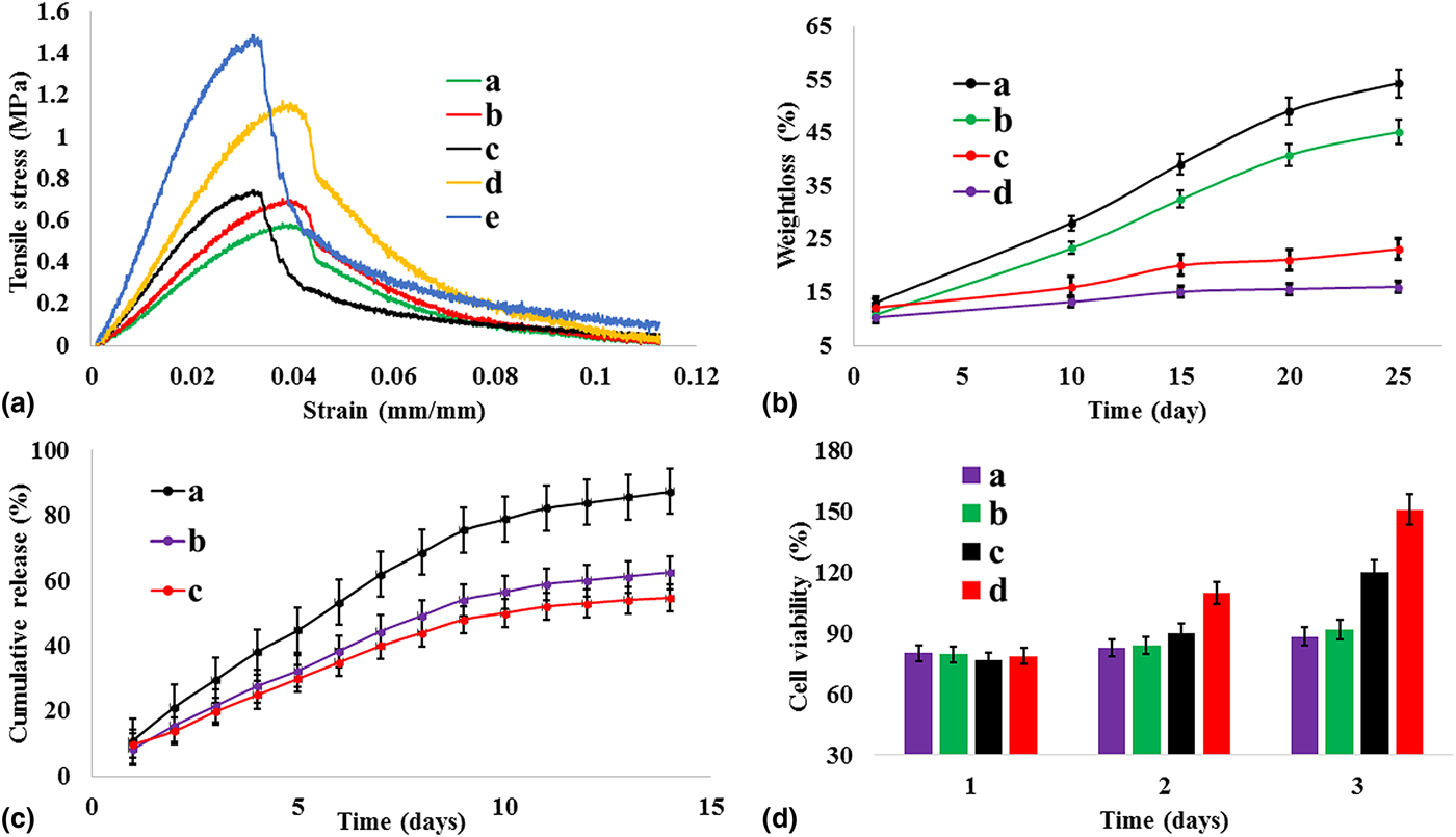
Figure 5(b) shows the in vitro degradation results of pure GL, GL (30 wt.%)/Cur (8.3 wt.%), GL (7.5 wt.%)/PLA (22.5 wt.%)/Cur (8.3 wt.%) and GL (6 wt.%)/PLA (24 wt.%)/Cur (8.3 wt.%) fibers. The degradation rate was varied with respect to PLA concentration. Pristine GL fibers showed 54.2% of degradation after 25 days. GL (30 wt.%)/Cur (8.3 wt.%) fibers exhibited 45.1% of degradation after 25 days. GL (7.5 wt.%)/PLA (22.5 wt.%)/Cur (8.3 wt.%) fibers revealed approximately 23.1% degradation after 25 days. Whereas, GL (6 wt.%)/PLA (24 wt.%)/Cur (8.3 wt.%) presented as a least degradation 16.0% after 25 days. This could be due to the greater hydrophobicity and higher tensile strength of GL (6 wt.%)/PLA (24 wt.%)/Cur (8.3 wt.%) fiber scaffolds. These degradation characteristics are important in the cell attachment and cell proliferation process.
Disparate tissue engineering and drug delivery need a specific and selective prolonged drug release, which is accomplished by nanofiber scaffolds.[Reference Ranjbar-Mohammadi and Bahrami19] It is also important to control the initial burst drug release, which reduces the damage of the cells during the tissue/cell culturing. Because effectual chemotherapy depends on the drug concentration between the minimum effective therapeutic level and tolerable long periods. Due to the poor water solubility and less bioavailability, curcumin has limited in drug delivery applications. Therefore, curcumin loaded nanofibers have been reported as drug carriers. Thus, GL (7.5 wt.%)/PLA (22.5 wt.%)/Cur (8.3 wt.%) and GL (6 wt.%)/PLA (24 wt.%)/Cur (8.3 wt.%) fiber scaffolds were fabricated as appropriate drug carriers for the prolonged release of curcumin. The cumulative drug release profile of GL (30 wt.%)/Cur (8.3 wt.%), GL (7.5 wt.%)/PLA (22.5 wt.%)/Cur (8.3 wt.%) and GL (6 wt.%)/PLA (24 wt.%)/Cur (8.3 wt.%) was presented in Fig. 5(c). GL (30 wt.%)/Cur (8.3 wt.%) fibers showed ~89.0% of curcumin release in 15 days, whereas GL (7.5 wt.%)/PLA (22.5 wt.%)/Cur (8.3 wt.%) fiber scaffolds displayed ~63.2% of curcumin release rate in 15 days. In addition, GL (6 wt.%)/PLA (24 wt.%)/Cur (8.3 wt.%) fibers exhibited ~54.6% of curcumin release in 15 days. Initial burst curcumin release was observed in the GL (30 wt.%)/Cur (8.3 wt.%) fibers but not in GL/PLA/Cur fibers. It could be due to more hydrophilicity and higher degradation rate of GL (30 wt.%)/Cur (8.3 wt.%) fibers. Generally, initial burst release occurs due to the surface drug loading of fibers. However, in the present study, curcumin drug was directly attached into the backbone of biodegradable polymers through the hydrogen bonds, it controlled initial burst drug release from GL/PLA/Cur fibers.
The forcespun GL (7.5 wt.%)/PLA (22.5 wt.%)/Cur (8.3 wt.%) fibers showed higher curcumin release than GL (6 wt.%)/PLA (24 wt.%)/Cur (8.3 wt.%) fibers. Forcespun of GL (6 wt.%)/PLA (24 wt.%)/Cur (8.3 wt.%) fibers showed a smooth slope and sustained curcumin release over 15 days and finally reached a plateau. Similarly, GL (7.5 wt.%)/PLA (22.5 wt.%)/Cur (8.3 wt.%) fibers also exhibited prolonged curcumin release in 15 days. These in vitro drug release experiments confirmed that the curcumin release profile was controlled and prolonged. Therefore, drug release results demonstrated that the GL/PLA/Cur fibers would potential drug carriers for the drug delivery and biomedical applications.
Good progress of cell proliferation, adhesion, spreading and migration on the surface of nanofibers is an important property to use fibers as tissue regeneration scaffolds. Therefore, the proficiency of cell viability of curcumin embedded pure GL, GL/Cur, and GL/PLA/Cur fiber scaffolds was investigated using CellTiter96®AQueous One Solution. Human fibroblast cell lines were cultivated on fiber scaffolds for 1–3 days and results were presented in Fig. 5(d). On day 1, pristine GL, GL (30 wt.%)/Cur (8.3 wt.%), GL (7.5 wt.%)/PLA (22.5 wt.%)/Cur (8.3 wt.%) and GL (6 wt.%)/PLA (24 wt.%)/Cur (8.3 wt.%) fibers did not show any significant difference in cell viability. But on the second day, cell viability was slightly increased in both GL (7.5 wt.%)/PLA (22.5 wt.%)/Cur (8.3 wt.%) and GL (6 wt.%)/PLA (24 wt.%)/Cur (8.3 wt.%) fibers, whereas cell viability, was constant in pure GL and GL (30 wt.%)/Cur (8.3 wt.%) fibers. On day 3, the cell viability was significantly increased in GL (6 wt.%)/PLA (24 wt.%)/Cur (8.3 wt.%) fibers than pristine GL, GL (30 wt.%)/Cur (8.3 wt.%) and GL (7.5 wt.%)/PLA (22.5 wt.%)/Cur (8.3 wt.%) fibers. This could be due to the higher hydrophilicity and degradation rate of pure GL, GL (30 wt.%)/Cur (8.3 wt.%), and GL (7.5 wt.%)/PLA (22.5 wt.%)/Cur (8.3 wt.%) fibers. Overall, these results reveal that GL (6 wt.%)/PLA (24 wt.%)/Cur (8.3 wt.%) fibers demonstrated higher cell viability compared with pristine GL and GL (30 wt.%)/Cur (8.3 wt.%) fibers. This could be due to good biocompatibility, hydrophobicity, honed tensile strength, and higher porosity of GL (6 wt.%)/PLA (24 wt.%)/Cur (8.3 wt.%) fibers.
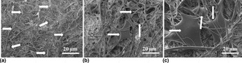
The cellular morphology and interactions of human fibroblast cells on the surface of GL (6 wt.%)/PLA (24 wt.%)/Cur (8.3 wt.%) fibers were studied for day 1 to day 3 (Fig. 6). The cellular attachments on the surface of GL (6 wt.%)/PLA (24 wt.%)/Cur (8.3 wt.%) fiber scaffolds were improved along with the culturing time [day 1 to day 3, Figs. 6(a)–6(c)]. Particularly, cells were adhered with stretched morphology on the surface of the fiber scaffolds. On day 1, the size of cells was very tiny throughout the fiber scaffolds [Fig. 6(a)], however, the cells were grownup on day 2 and day 3 [Figs. 6(b) and 6(c)]. On day 3, improved cellular extensions were observed on the surface of GL (6 wt.%)/PLA (24 wt.%)/Cur (8.3 wt.%) fiber scaffolds [white arrows in Fig. 6(c)]. The size, colonization and cell population of extensions were enhanced from day 1 to day 3. Due to the high porosity nature of GL (6 wt.%)/PLA (24 wt.%)/Cur (8.3 wt.%) fibers, some of the cells were grownup on top and the bottom surface of fiber scaffolds [Fig. 6(c)]. These results reveal the good cell attachment, proliferation, and biocompatibility of GL (6 wt.%)/PLA (24 wt.%)/Cur (8.3 wt.%) fibers.
Cellular interactions and morphology of fibroblast cells on GL (6 wt.%)/PLA (24 wt.%)/Cur (8.3 wt.%) fibers on (a) day 1; (b) day 2 and (c) day 3. Scale bar = 20 µm.

Conclusion
For the first time, anti-cancer drug curcumin incorporated GL/PLA/Cur composites were successfully developed and aligned nanofiber scaffolds were fabricated by Forcespinning®. FTIR, DSC, and tensile results confirmed the formation of hydrogen bonds between curcumin, GL, and PLA in GL/PLA/Cur composites. In vitro drug release, porosity, water absorption, biodegradation, thermal, tensile properties, cell morphology, and cell viability studies were evaluated. GL (7.5 wt.%)/PLA (22.5 wt.%)/Cur (8.3 wt.%) and GL (6 wt.%)/PLA (24 wt.%)/Cur (8.3 wt.%) fibers were demonstrated sustained release of curcumin over 15 days. Biodegradation results showed their original structure of GL/PLA/Cur fibers even after 25 days. Hydrophobicity results indicated that the hydrophobicity of GL/PLA/Cur fibers was improved by the addition of PLA and curcumin. Further, honed mechanical properties were observed in GL/PLA/Cur fibers. Cytotoxicity studies demonstrated excellent cell viability over 3 days. Nevertheless, prolonged drug release and good biologic features of GL/PLA/Cur fiber scaffolds suggested that curcumin embedded GL/PLA/Cur fibers may show potential application for drug delivery, wound healing, healthcare, and biomedicine.
Conflict of interest
The authors declare no competing financial interest.
Acknowledgment
The authors acknowledge the financial support from the Consejo Nacional de Ciencia y Tecnología de México (CONACyT), Project Number 242269.