Introduction
Schizophrenia can be considered a developmental disorder (Insel, Reference Insel2010). While the clinical identification of most patients with schizophrenia starts with the manifestation of the first psychotic episode in late adolescence, deviations from the normal developmental trajectory are demonstrated to occur much earlier (Insel, Reference Insel2010; Kahn & Keefe, Reference Kahn and Keefe2013; Sommer et al. Reference Sommer, Bearden, van Dellen, Breetvelt, Duijff, Maijer, van Amelsvoort, de Haan, Gur, Arango, Díaz-Caneja, Vinkers and Vorstman2016). In recent years, the importance of cognitive impairment as a core feature of schizophrenia has gained considerable attention (Kahn & Keefe, Reference Kahn and Keefe2013). Not only do schizophrenia patients show decreased intellectual and cognitive performance compared with healthy controls (O'Carroll, Reference O'Carroll2000), a decline in cognitive abilities precedes the onset of the first psychotic episode by several years (Reichenberg et al. Reference Reichenberg, Caspi, Harrington, Houts, Keefe, Murray, Poulton and Moffitt2010).
Advanced stage schizophrenia is marked by alterations in white matter microstructural properties (for an extensive review see Fitzsimmons et al. Reference Fitzsimmons, Kubicki and Shenton2013), in particular by decreases in fractional anisotropy (FA). In comparison, relatively few studies have investigated white matter microstructural properties in the early preclinical stage (ultra-high risk, UHR) (Hoptman et al. Reference Hoptman, Nierenberg, Bertisch, Catalano, Ardekani, Branch and DeLisi2008; Peters et al. Reference Peters, de Haan, Dekker, Blaas, Becker, Dingemans, Akkerman, Majoie, van Amelsvoort, den Heeten and Linszen2008; Reference Peters, Schmitz, Dingemans, van Amelsvoort, Linszen, de Haan, Majoie and den Heeten2009; Reference Peters, Dingemans, Dekker, Blaas, Akkerman, van Amelsvoort, Majoie, den Heeten, Linszen and de Haan2010; Karlsgodt et al. Reference Karlsgodt, Niendam, Bearden and Cannon2009; Bloemen et al. Reference Bloemen, de Koning, Schmitz, Nieman, Becker, de Haan, Dingemans, Linszen and van Amelsvoort2010; Carletti et al. Reference Carletti, Woolley, Bhattacharyya, Perez-Iglesias, Fusar Poli, Valmaggia, Broome, Bramon, Johns, Giampietro, Williams, Barker and McGuire2012; Clemm Von Hohenberg et al. Reference Clemm Von Hohenberg, Pasternak, Kubicki, Ballinger, Vu, Swisher, Green, Giwerc, Dahlben, Goldstein, Woo, Petryshen, Mesholam-Gately, Woodberry, Thermenos, Mulert, McCarley, Seidman and Shenton2014; O'Hanlon et al. Reference O'Hanlon, Leemans, Kelleher, Clarke, Roddy, Coughlan, Harley, Amico, Hoscheit, Tiedt, Tabish, McGettigan, Frodl and Cannon2015; Bakker et al. Reference Bakker, Caan, Schluter, Bloemen, da Silva-Alves, de Koning, Boot, Vingerhoets, Nieman, de Haan, Booij and van Amelsvoort2016). Assessments prior to the UHR stage are scarce and current literature shows inconsistent results of both increased and decreased FA (Gilmore et al. Reference Gilmore, Kang, Evans, Wolfe, Smith, Lieberman, Lin, Hamer, Styner and Gerig2010; Francis et al. Reference Francis, Bhojraj, Prasad, Montrose, Eack, Rajarethinam, van Elst and Keshavan2013; Samartzis et al. Reference Samartzis, Dima, Fusar-Poli and Kyriakopoulos2014; Satterthwaite et al. Reference Satterthwaite, Wolf, Calkins, Vandekar, Erus, Ruparel, Roalf, Linn, Elliott, Moore, Hakonarson, Shinohara, Davatzikos, Gur and Gur2016).
Indeed, the cognitive deficit in schizophrenia has been associated with alterations in white matter microstructure (Nazeri et al. Reference Nazeri, Chakravarty, Felsky, Lobaugh, Rajji, Mulsant and Voineskos2013; Roalf et al. Reference Roalf, Gur, Verma, Parker, Quarmley, Ruparel and Gur2015). Also, it has been shown that decreases in cognitive performance and in white matter microstructure constitute a genetic risk factor for schizophrenia (Bohlken et al. Reference Bohlken, Brouwer, Mandl, Van den Heuvel, Hedman, De Hert, Cahn, Kahn and Hulshoff Pol2015). Currently, little is known about the interplay between cognitive ability and white matter integrity preceding psychosis. In order to better understand these neurodevelopmental risk factors of schizophrenia, it is important to examine the stages preceding psychosis. However, studying the early stages of schizophrenia, i.e. before the onset of the first psychotic symptoms, is challenging because of the low occurrence of the disorder with prevalence and incidence rates of respectively 1 in 100 and 1 in 5000 persons in the general population (Regier et al. Reference Regier, Narrow, Rae, Manderscheid, Locke and Goodwin1993; van der Werf et al. Reference van der Werf, Hanssen, Köhler, Verkaaik, Verhey, van Winkel, van Os and Allardyce2014).
Approximately one in four patients with the 22q11.2 deletion syndrome (22q11DS) develop schizophrenia or other psychotic disorders (Schneider et al. Reference Schneider, Debbané, Bassett, Chow, Fung, van den Bree, Owen, Murphy, Niarchou, Kates, Antshel, Fremont, McDonald-McGinn, Gur, Zackai, Vorstman, Duijff, Klaassen, Swillen, Gothelf, Green, Weizman, Van Amelsvoort, Evers, Boot, Shashi, Hooper, Bearden, Jalbrzikowski, Armando, Vicari, Murphy, Ousley, Campbell, Simon and Eliez2014). Consistent with findings in schizophrenia, it was recently shown that cognitive decline, most pronounced in verbal IQ, precedes the onset of psychosis by several years in patients with 22q11DS (Vorstman et al. Reference Vorstman, Breetvelt, Duijff, Eliez, Schneider, Jalbrzikowski, Armando, Vicari, Shashi, Hooper, Chow, Fung, Butcher, Young, McDonald-McGinn, Vogels, van Amelsvoort, Gothelf, Weinberger, Weizman, Klaassen, Koops, Kates, Antshel, Simon, Ousley, Swillen, Gur, Bearden, Kahn and Bassett2015). Specifically, patients who showed a decline in verbal IQ had a threefold increased risk of developing a psychotic disorder (Vorstman et al. Reference Vorstman, Breetvelt, Duijff, Eliez, Schneider, Jalbrzikowski, Armando, Vicari, Shashi, Hooper, Chow, Fung, Butcher, Young, McDonald-McGinn, Vogels, van Amelsvoort, Gothelf, Weinberger, Weizman, Klaassen, Koops, Kates, Antshel, Simon, Ousley, Swillen, Gur, Bearden, Kahn and Bassett2015). 22q11DS is an appealing model to investigate the early trajectory of schizophrenia (Bassett & Chow, Reference Bassett and Chow1999; Insel, Reference Insel2010), as patients with 22q11DS are often identified very early in life due to the presence of somatic symptoms, including cardiac, pharyngeal or facial abnormalities (see McDonald-McGinn et al. Reference McDonald-McGinn, Sullivan, Marino, Philip, Swillen, Vorstman, Zackai, Emanuel, Vermeesch, Morrow, Scambler and Bassett2015 for a detailed overview). Herein lies an important research opportunity; by virtue of their identification early in life, the trajectory of schizophrenia can be followed prospectively in this group of individuals, starting from birth (or even in utero) (Insel, Reference Insel2010). Another important advantage over other UHR studies is that these individuals are not selected based on subjectively assessed symptoms, required to define an at-risk mental state, but based on the 1.5- to 3-Mb hemizygous deletion at the long (q) arm of chromosome 22 (Edelmann, Reference Edelmann1999) that causes 22q11.2 deletion syndrome. At present, 22q11DS is the strongest known single genetic risk factor for schizophrenia (Karayiorgou et al. Reference Karayiorgou, Simon and Gogos2010; Marshall et al. Reference Marshall, Howrigan, Merico, Thiruvahindrapuram, Wu, Greer, Antaki, Shetty, Holmans, Pinto, Gujral, Brandler, Malhotra, Wang, Fajarado, Maile, Ripke, Agartz, Albus, Alexander, Amin, Atkins, Bacanu, Belliveau, Bergen, Bertalan, Bevilacqua, Bigdeli, Black, Bruggeman, Buccola, Buckner, Bulik-Sullivan, Byerley, Cahn, Cai, Cairns, Campion, Cantor, Carr, Carrera, Catts SV Chambert, Cheng, Cloninger, Cohen, Cormican, Craddock, Crespo-Facorro, Crowley, Curtis, Davidson, Davis, Degenhardt, Del Favero, DeLisi, Dikeos, Dinan, Djurovic, Donohoe, Drapeau, Duan, Dudbridge, Eichhammer, Eriksson, Escott-Price, Essioux, Fanous, Farh, Farrell, Frank, Franke, Freedman, Freimer, Friedman, Forstner, Fromer, Genovese, Georgieva, Gershon, Giegling, Giusti-Rodríguez, Godard, Goldstein, Gratten, de Haan, Hamshere, Hansen, Hansen, Haroutunian, Hartmann, Henskens, Herms, Hirschhorn, Hoffmann, Hofman, Huang, Ikeda, Joa, Kähler, Kahn, Kalaydjieva, Karjalainen, Kavanagh, Keller, Kelly, Kennedy, Kim, Knowles, Konte, Laurent, Lee, Lee, Legge, Lerer, Levy, Liang, Lieberman, Lönnqvist, Loughland, Magnusson, Maher, Maier, Mallet, Mattheisen, Mattingsdal, McCarley, McDonald, McIntosh, Meier, Meijer, Melle, Mesholam-Gately, Metspalu, Michie, Milani, Milanova, Mokrab, Morris, Müller-Myhsok, Murphy, Murray, Myin-Germeys, Nenadic, Nertney, Nestadt, Nicodemus, Nisenbaum, Nordin, O'Callaghan, O'Dushlaine, Oh, Olincy, Olsen, O'Neill, Van Os, Pantelis, Papadimitriou, Parkhomenko, Pato, Paunio, Perkins, Pers, Pietiläinen, Pimm, Pocklington, Powell, Price, Pulver, Purcell, Quested, Rasmussen, Reichenberg, Reimers, Richards, Roffman, Roussos, Ruderfer, Salomaa, Sanders, Savitz, Schall, Schulze, Schwab, Scolnick, Scott, Seidman, Shi, Silverman, Smoller, Söderman, Spencer, Stahl, Strengman, Strohmaier, Stroup, Suvisaari, Svrakic, Szatkiewicz, Thirumalai, Tooney, Veijola, Visscher, Waddington, Walsh, Webb, Weiser, Wildenauer, Williams, Williams, Witt, Wolen, Wormley, Wray, Wu, Zai, Adolfsson, Andreassen, Blackwood, Bramon, Buxbaum, Cichon, Collier, Corvin, Daly, Darvasi, Domenici, Esko, Gejman PV Gill, Gurling, Hultman, Iwata, Jablensky AV Jönsson, Kendler, Kirov, Knight, Levinson, Li, McCarroll, McQuillin, Moran, Mowry, Nöthen, Ophoff, Owen, Palotie, Pato, Petryshen, Posthuma, Rietschel, Riley, Rujescu, Sklar, St Clair, Walters, Werge, Sullivan, O'Donovan, Scherer, Neale and Sebat2016). Vice versa, the 22q11DS can be identified in approximately 1–2% of the general population of schizophrenia patients (Bassett & Chow, Reference Bassett and Chow2008), as compared to 0.05% in the general population (Regier et al. Reference Regier, Narrow, Rae, Manderscheid, Locke and Goodwin1993; van der Werf et al. Reference van der Werf, Hanssen, Köhler, Verkaaik, Verhey, van Winkel, van Os and Allardyce2014).
Several studies have investigated white matter structure in 22q11DS. Alterations in FA have been reported in 22q11DS patients, compared to healthy controls. Reduced FA is found in the superior longitudinal fasciculus (Sundram et al. Reference Sundram, Campbell, Azuma, Daly, Bloemen, Barker, Chitnis, Jones, van Amelsvoort, Murphy and Murphy2010; Kikinis et al. Reference Kikinis, Asami, Bouix, Finn, Ballinger, Tworog-Dube, Kucherlapati, Kikinis, Shenton and Kubicki2012), cingulum bundle (Jalbrzikowski et al. Reference Jalbrzikowski, Villalon-Reina, Karlsgodt, Senturk, Chow, Thompson and Bearden2014; Kates et al. Reference Kates, Olszewski, Gnirke, Kikinis, Nelson, Antshel, Fremont, Radoeva, Middleton, Shenton and Coman2015; Roalf et al. Reference Roalf, Eric Schmitt, Vandekar, Satterthwaite, Shinohara, Ruparel, Elliott, Prabhakaran, McDonald-McGinn, Zackai, Gur, Emanuel and Gur2017), and uncinate fasciculus (Kikinis et al. Reference Kikinis, Asami, Bouix, Finn, Ballinger, Tworog-Dube, Kucherlapati, Kikinis, Shenton and Kubicki2012; Radoeva et al. Reference Radoeva, Coman, Antshel, Fremont, McCarthy, Kotkar, Wang, Shprintzen and Kates2012). However, increased FA is found in regions of the corpus callosum (Barnea-Goraly et al. Reference Barnea-Goraly, Menon, Krasnow, Ko, Reiss and Eliez2003; Bakker et al. Reference Bakker, Caan, Schluter, Bloemen, da Silva-Alves, de Koning, Boot, Vingerhoets, Nieman, de Haan, Booij and van Amelsvoort2016), the anterior limb of the internal capsule (Perlstein et al. Reference Perlstein, Chohan, Coman, Antshel, Fremont, Gnirke, Kikinis, Middleton, Radoeva, Shenton and Kates2014), the corona radiata (Sundram et al. Reference Sundram, Campbell, Azuma, Daly, Bloemen, Barker, Chitnis, Jones, van Amelsvoort, Murphy and Murphy2010), the anterior thalamic radiation and the inferior fronto-occitpital fasciculus (Bakker et al. Reference Bakker, Caan, Schluter, Bloemen, da Silva-Alves, de Koning, Boot, Vingerhoets, Nieman, de Haan, Booij and van Amelsvoort2016), and the superior longitudinal fasciculus (Simon et al. Reference Simon, Ding, Bish, McDonald-McGinn, Zackai and Gee2005). These findings in 22q11DS show some overlap with findings in UHR within the general population and suggest that altered white matter microstructural properties may constitute a risk factor for schizophrenia. However, it has not yet been investigated whether such alterations are related to other known risk factors such as cognitive decline.
As 22q11DS is the strongest known genetic risk factor for developing schizophrenia, and it was observed that cognitive decline in 22q11DS additionally increases this risk we hypothesize that cognitive decline in 22q11DS is accompanied by alterations in white matter microstructural properties. Such evidence would inform the hypothesis that neurodevelopmental risk for schizophrenia may be expressed in prodromal white matter microstructural alterations accompanied by loss of cognitive abilities. Specifically, we examined whether early cognitive decline observed in individuals with 22q11DS is associated with alterations of white matter microstructural directionality. To test this, IQ-trajectories of 22q11DS patients with and without cognitive decline prior to MRI acquisition were compared on whole-brain and atlas-based measures of white matter FA.
Methods
Participants
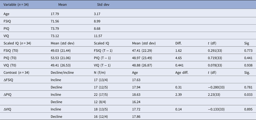
Analyses were performed on a subsample of the 22q11DS cohort studied at the University Medical Center Utrecht, the Netherlands. Recruitment and assessment of this cohort have been reported previously (Vorstman et al. Reference Vorstman, Breetvelt, Duijff, Eliez, Schneider, Jalbrzikowski, Armando, Vicari, Shashi, Hooper, Chow, Fung, Butcher, Young, McDonald-McGinn, Vogels, van Amelsvoort, Gothelf, Weinberger, Weizman, Klaassen, Koops, Kates, Antshel, Simon, Ousley, Swillen, Gur, Bearden, Kahn and Bassett2015; Fiksinski et al. Reference Fiksinski, Breetvelt, Duijff, Bassett, Kahn and Vorstman2017). We acquired MRI data in a total of 35 patients for this study. One patient was excluded because of significant scan artefacts. Descriptive statistics of the remaining 34 participants are displayed in Table 1. Thirteen patients were experiencing psychotic symptoms at the time of the scanning (T0). Of this group, six were already diagnosed with a psychotic disorder (four with cognitive decline preceding the scan, two without cognitive decline preceding the scan). Assessment of diagnosis of a psychotic disorder (according to the Diagnostic and Statistical Manual of Mental Disorders, DSM-IV) and/or psychotic symptomatology was performed by trained clinicians in a multidisciplinary setup, using the Schedule for Affective Disorders and Schizophrenia for School Age Children [K-SADS (Kaufman et al. Reference Kaufman, Birmaher, Brent, Rao, Flynn, Moreci, Williamson and Ryan1997)]. Prior to the scan, two patients received antipsychotic medication.
General descriptives of the total sample, IQ scores, scaled IQ scores & IQ contrasts

Std dev, standard deviation; FSIQ, full-scale intelligence quotient; PIQ, Performance IQ; VIQ, Verbal IQ; T0, time point zero, time of the scan; T − 1, T minus one, time point preceding the scan; Diff, difference in IQ scores between T0 and T − 1; t(df), t-statistic (degrees of freedom); Sig., p value; Δ, contrast incline/decline; Age diff, difference in age between decline/no decline; f/m, number of females/number of males.
The first part of the table displays information about the total sample's age, IQ scores. The second part shows the gender distribution within the sample. The third part displays the descriptives of the scaled IQ scores at T0 and T − 1, including information about the difference between T0 and T1. The fourth part shows the different contrasts used in the sample and information about these groups.
The chromosomal deletion at the 22q11.2 region was confirmed in every patient by fluorescent in situ hybridization (FISH) or Multiplex Ligation-dependent Probe Amplification [MLPA (Vorstman et al. Reference Vorstman, Jalali, Rappaport, Hacker, Scott and Emanuel2006)]. Participants were scanned between May 2010 and October 2015. This study has been approved by the local research ethics board (Dutch Central Committee on Research Involving Human Subjects; C.C.M.O) and all participants (and/or their legal guardians) provided written informed consent.
Assessment of intellectual features and percentile conversion
Certified assessors obtained all IQ scores using age-appropriate versions of the Wechsler intelligence scale (e.g. Wechsler Intelligence Scale for Children or the Wechsler Adult Intelligence Scale). Table 1 shows IQ scores at the time of the scan. We analysed IQ trajectories by comparing IQ at the time of the scan (T0, see Table 1) to IQ score as assessed by the preceding IQ measurement (T − 1). To establish cognitive decline, individual IQ trajectories were plotted against the previously constructed IQ normative chart specific for the 22q11DS population (Vorstman et al. Reference Vorstman, Breetvelt, Duijff, Eliez, Schneider, Jalbrzikowski, Armando, Vicari, Shashi, Hooper, Chow, Fung, Butcher, Young, McDonald-McGinn, Vogels, van Amelsvoort, Gothelf, Weinberger, Weizman, Klaassen, Koops, Kates, Antshel, Simon, Ousley, Swillen, Gur, Bearden, Kahn and Bassett2015). In short, the IQ scores of 34 22q11DS patients were converted into percentiles, which were calculated using a 4-year sliding bin of mean IQ scores of 22q11DS patients at different ages. In essence, this strategy allows identifying subjects who show a decline in IQ beyond what would be expected in this specific population. Subjects with IQ decline in our cohort were those displaying at least one percentile point negative deviation from T − 1 to T0. We examined three comparison contrasts: decline v. no-decline in verbal IQ trajectory (ΔVIQ), Full-Scale IQ trajectory (ΔFSIQ), and performance IQ trajectory (ΔPIQ).
Neuroimaging and image processing
All brain scans were acquired on a 3T Philips Achieva magnetic resonance imaging (MRI) scanner using an eight-channel SENSE head-coil. For each participant two diffusion-weighted imaging (DWI) scans were acquired using the following parameters: single-shot EPI-DTI with 30 diffusion-weighted volumes (b = 1000 s/mm2) with non-collinear gradient directions and five diffusion unweighted volumes (b = 0 s/mm2), TR/TE = 7035/68 ms, field of view = 240 mm, EPI factor 35, SENSE factor 3, no cardiac gating, no gap, 128 × 128 matrix, and 75 slices of 2 mm thickness. To correct for susceptibility effects of the scanner one diffusion weighted scan was acquired in the anterior–posterior direction, and the other one in the opposite plane (posterior-anterior).
All (pre)processing of the MRI scans was done with different FSL tools (FSL, version 5.0.6, Oxford (Smith et al. Reference Smith, Jenkinson, Woolrich, Beckmann, Behrens, Johansen-Berg, Bannister, De Luca, Drobnjak, Flitney, Niazy, Saunders, Vickers, Zhang, De Stefano, Brady and Matthews2004)). See online Supplementary Materials (S1) for an overview of the analysis pipeline.
Region of interest
Using 40 binary WM masks based on the Johns Hopkins University International Consortium of Brain Mapping 81 [JHU-ICBM 81 (Mori et al. Reference Mori, Oishi, Jiang, Jiang, Li, Akhter, Hua, Faria, Mahmood, Woods, Toga, Pike, Neto, Evans, Zhang, Huang, Miller, van Zijl and Mazziotta2008)] atlas (see online Supplementary S2 for an overview of regions included) we extracted mean FA, MD, AD and RD per tract per individual.
Statistical analyses
Statistical analyses are performed in R (version 3.1.1) and SPSS (IBM, version 22). FA, MD, AD and RD were imported in SPSS. Using a t test, differences between patients who declined in verbal IQ (ΔVIQ), performance IQ (ΔPIQ) and full-scale IQ (ΔFSIQ) and patients who showed no decline, within WM regions of interest (ROIs) after correcting for age and gender were tested. Post hoc analyses were performed in order to study the contributions of different diffusivity measures to the effect found in FA, these included axial diffusivity (AD), mean diffusivity (MD) and radial diffusivity (RD) (see online Supplementary S3). Whole brain white matter diffusivity was investigated by averaging the FA values extracted from the ROIs of the JHU-ICBM 81 atlas.
To correct for multiple comparisons, false discovery rates (FDR) were computed using the Benjamini and Hochberg (BH) procedure (Benjamini & Hochberg, Reference Benjamini and Hochberg1995) (as implemented in R, version 3.1.1) where a p value is considered significant as FDR value (p.adjust i) ⩽0.05, where p.adjust i is determined by the rank (R i) of the p value (p i) and the number of tests (n): p.adjusti = p i*(n/R i).
Results
Sample
At time of the scan (T0) mean age of participants (n = 34, f/m = 25/9) was 17.79 (years; s.d. = 3.17 years; 12.3–24.9), at the time point preceding the scan (T − 1, read as T minus 1) the mean age was 11.79 (years; s.d. = 2.93 years; 7.0–17.0). The mean interval between the time points was 5.99 (years; s.d. = 2.30 years), there was a trend towards a significant difference for those with and those without cognitive decline (p = 0.052). The distribution of age within the three different contrasts is presented in Table 1. We observed no statistically significant differences in distribution of age (Table 1) or gender for the IQ trajectories [ΔFSIQ [χ2(1) = 0.151, p = 0.697], ΔPIQ [χ2(1) = 0.449, p = 0.503] and ΔVIQ [χ2(1) = 0.034, p = 0.855]. Within the three different contrasts, the distribution of psychotic symptoms, and diagnosis is not significantly different (see online Supplementary S4).
IQ trajectories
To ensure that possible differences found in FA were not due to overall group differences in IQ, we assessed potential group differences at baseline (T − 1), at the time of the scan (T0) and between these time points. Table 1 represents the scaled IQ scores of the sample at T0 and T − 1 and the difference between T0 and T − 1. The difference in scaled IQ scores between T0 and T − 1 is not statistically significant (Table 1; see online Supplementary S5 for comparisons between time points T0 and T − 1 between decline and non-decline). We found no statistically significant differences in FSIQ, VIQ and PIQ at T − 1 and T0 between the decline and the non-decline group (see online Supplementary S6).
Region of interest
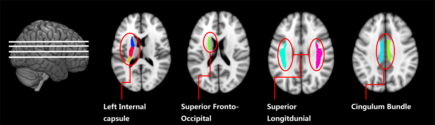
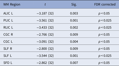
Subjects that declined in VIQ showed higher FA values in the bilateral superior longitudinal fasciculus, the anterior limb of the left internal capsule, the rentrolenticular part of the left internal capsule, the posterior limb of the left internal capsule, the bilateral cingulum bundle and the left superior fronto-occipital fasciculus. All of these regions withstood correction for multiple comparisons (see Table 2 for corrected and uncorrected p values, see Fig. 1 for illustration of WM ROIs). Cognitive decline was not associated with decreased FA. For VIQ, 16 patients showed a decline of -18.34 (s.d. = 15.83) percentile points over 6.80 (s.d. = 2.39) years, and 18 patients showed an incline of 17.71 (s.d. = 20.17) VIQ percentile points over 5.27 (s.d. = 2.03) years. Furthermore, patients with VIQ decline showed higher whole brain FA values compared with patients without decline (t(32) = −2.511, p = 0.017). We did not find statistically significant differences that survived multiple comparisons correction in any of the WM ROIs nor in whole brain FA on the contrasts ΔTIQ and ΔPIQ. In addition, we did not find any significant correlations that survived corrections for multiple comparisons between FA values in each ROI and scaled IQ scores at T0 (see online Supplementary S7).
Transverse sections of the brain showing WM regions where FA is higher in decline v. no decline.

Corrected and uncorrected p values on the contrast ΔVIQ

Sig, significant value, 2-tailed; ALIC L, anterior limb of the internal capsule, left; PLIC L, posterior limb of the internal capsule, left; RLIC L, rentrolenticular part of the internal capsule, left; CGC R; cingulum bundle, around cingulate gyrus, right; CGC L, cingulum bundle, around cingulate gyrus, left; SLF R, superior longitudinal fasciculus, right; SLF L, superior longitudinal fasciculus, left; SFO L, superior fronto-occipital gyrus, left; df, degrees of freedom; FDR, false discovery rate.
This table displays the corrected and uncorrected p values of the comparison of FA in the corresponding regions between 22q11DS patients showing a verbal cognitive decline to 22q11DS patients without verbal cognitive decline.
Post hoc analysis
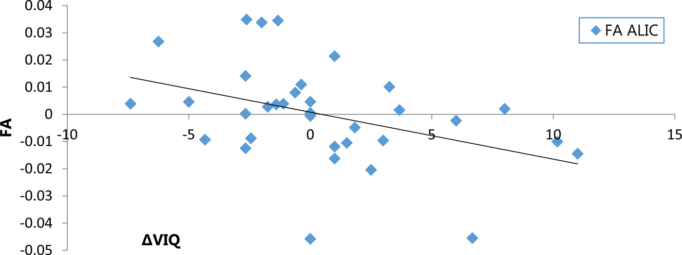
We explored whether the magnitude of IQ change per year was associated with FA values in the significant WM ROIs. We found a significant correlation between change in VIQ per year and FA in the anterior limb of the left internal capsule (r = −0.395 p = 0.021, uncorrected, see Fig. 2). The other WM tracts follow the same trend, albeit not in a statistically significant manner (see online Supplementary S8 for correlation coefficients and p values).
Association between FA in ALIC and ΔVIQ. ALIC, anterior limb of the internal capsule, left; FA, fractional anisotropy; Horizontal axis, ΔVIQ mean change per individual per year, negative values indicate decline; Vertical axis, unstandardized residuals of FA values where effects of age and gender are regressed out.

Furthermore, we performed several post-hoc analyses to check for possible confounding effects of: (1) Whole-brain FA, (2) diagnosis of a psychotic disorder (3) switch in IQ test (4) head movement in the scanner. All effects remained significant after correcting for any of these variables (see online Supplementary S9 and S10). Furthermore, we checked which of the other diffusivity measures (MD/RD/AD) differed between the groups, the results of which are shown in the online Supplementary S3. Finally, to ensure that the results found were not influenced by baseline IQ and the amount of time between the first measurement and the second, baseline IQ and measurement interval (in years) were included as covariates in the model. Including these variables did not alter the results.
Discussion
We investigated microstructural properties of white matter in relation to cognitive decline in 22q11DS patients. Our main finding is that FA is increased in several major white matter regions in those individuals with 22q11DS who displayed a cognitive decline in the years preceding MRI acquisition, as compared to those without cognitive decline. This finding confirms our hypothesis that cognitive decline in 22q11DS is accompanied by alterations in white matter microstructural properties.
Interestingly, our findings show overlap with previous studies investigating white matter alterations related to 22q11DS. However, the literature is not always consistent with the direction of the effect, as both increased and decreased FA was reported in 22q11DS patients compared to healthy controls (for review see Squarcione et al. Reference Squarcione, Torti, Di and Biondi2013; Scariati et al. Reference Scariati, Padula, Schaer and Eliez2016). These studies show decreased FA in 22q11DS in the superior longitudinal fasciculus (Sundram et al. Reference Sundram, Campbell, Azuma, Daly, Bloemen, Barker, Chitnis, Jones, van Amelsvoort, Murphy and Murphy2010), uncinate fasciculus (Kikinis et al. Reference Kikinis, Asami, Bouix, Finn, Ballinger, Tworog-Dube, Kucherlapati, Kikinis, Shenton and Kubicki2012; Radoeva et al. Reference Radoeva, Coman, Antshel, Fremont, McCarthy, Kotkar, Wang, Shprintzen and Kates2012), and the cingulum bundle (Jalbrzikowski et al. Reference Jalbrzikowski, Villalon-Reina, Karlsgodt, Senturk, Chow, Thompson and Bearden2014; Kates et al. Reference Kates, Olszewski, Gnirke, Kikinis, Nelson, Antshel, Fremont, Radoeva, Middleton, Shenton and Coman2015). Increased FA is found in the corpus callosum (Bakker et al. Reference Bakker, Caan, Schluter, Bloemen, da Silva-Alves, de Koning, Boot, Vingerhoets, Nieman, de Haan, Booij and van Amelsvoort2016), the anterior limb of the internal capsule (ALIC)/anterior thalamic radiation (Perlstein et al. Reference Perlstein, Chohan, Coman, Antshel, Fremont, Gnirke, Kikinis, Middleton, Radoeva, Shenton and Kates2014; Bakker et al. Reference Bakker, Caan, Schluter, Bloemen, da Silva-Alves, de Koning, Boot, Vingerhoets, Nieman, de Haan, Booij and van Amelsvoort2016), the superior longitudinal fasciculus (Simon et al. Reference Simon, Ding, Bish, McDonald-McGinn, Zackai and Gee2005), the bilateral inferior fronto-occipital fasciculus and the right cingulum bundle (Bakker et al. Reference Bakker, Caan, Schluter, Bloemen, da Silva-Alves, de Koning, Boot, Vingerhoets, Nieman, de Haan, Booij and van Amelsvoort2016; Olszewski et al. Reference Olszewski, Kikinis, Gonzalez, Coman, Makris, Gong, Rathi, Zhu, Antshel, Fremont, Kubicki, Bouix, Shenton and Kates2017) and the inferior longitudinal fasciculus (Tylee et al. Reference Tylee, Kikinis, Quinn, Antshel, Fremont, Tahir, Zhu, Gong, Glatt, Coman, Shenton, Kates and Makris2017). Interestingly, we show that the ALIC, cingulum bundle and the superior longitudinal fasciculus have increased FA in 22q11DS with cognitive decline. Our observation suggests that cognitive decline may exacerbate white matter abnormalities in 22q11DS.
Recently, a study comparing 22q11DS patients to subjects at UHR for developing psychosis reported higher FA in the corpus callosum and the anterior thalamic radiation in 22q11DS patients (Bakker et al. Reference Bakker, Caan, Schluter, Bloemen, da Silva-Alves, de Koning, Boot, Vingerhoets, Nieman, de Haan, Booij and van Amelsvoort2016). However, decreased FA in the cingulum bundle and left inferior longitudinal fasciculus was reported in 22q11DS patients with psychotic symptoms compared to 22q11DS patients without psychotic symptoms (Padula et al. Reference Padula, Scariati, Schaer, Sandini, Ottet, Schneider, Van De Ville and Eliez2017; Roalf et al. Reference Roalf, Eric Schmitt, Vandekar, Satterthwaite, Shinohara, Ruparel, Elliott, Prabhakaran, McDonald-McGinn, Zackai, Gur, Emanuel and Gur2017). This suggests that psychosis onset in 22q11DS may be associated with a complex pattern of alterations in the white matter microstructure. Our findings may add to the existing literature that cognitive decline in 22q11DS is an important preclinical risk factor implicated in white matter alterations, which are at least partly also observed after psychosis onset.
Furthermore, our findings are consistent with several studies in UHR samples reporting increased FA compared to healthy controls (Hoptman et al. Reference Hoptman, Nierenberg, Bertisch, Catalano, Ardekani, Branch and DeLisi2008; Bloemen et al. Reference Bloemen, de Koning, Schmitz, Nieman, Becker, de Haan, Dingemans, Linszen and van Amelsvoort2010; O'Hanlon et al. Reference O'Hanlon, Leemans, Kelleher, Clarke, Roddy, Coughlan, Harley, Amico, Hoscheit, Tiedt, Tabish, McGettigan, Frodl and Cannon2015; de Leeuw et al. Reference de Leeuw, Bohlken, Mandl, Hillegers, Kahn and Vink2017). Specifically, we found that FA was both globally and locally increased and that local WM regions overlap with WM tracts found to be affected in schizophrenia and individuals with 22q11DS and psychosis. These regions include the cingulum bundle the superior longitudinal fasciculus, the left internal capsule and fronto-occipital fasciculus (Fitzsimmons et al. Reference Fitzsimmons, Kubicki and Shenton2013). However, decreased FA, or absence of case-control differences are also reported (Hoptman et al. Reference Hoptman, Nierenberg, Bertisch, Catalano, Ardekani, Branch and DeLisi2008; Peters et al. Reference Peters, de Haan, Dekker, Blaas, Becker, Dingemans, Akkerman, Majoie, van Amelsvoort, den Heeten and Linszen2008; Reference Peters, Dingemans, Dekker, Blaas, Akkerman, van Amelsvoort, Majoie, den Heeten, Linszen and de Haan2010; Karlsgodt et al. Reference Karlsgodt, Niendam, Bearden and Cannon2009; Clemm Von Hohenberg et al. Reference Clemm Von Hohenberg, Pasternak, Kubicki, Ballinger, Vu, Swisher, Green, Giwerc, Dahlben, Goldstein, Woo, Petryshen, Mesholam-Gately, Woodberry, Thermenos, Mulert, McCarley, Seidman and Shenton2014; Samartzis et al. Reference Samartzis, Dima, Fusar-Poli and Kyriakopoulos2014). Interestingly, the mean age of the high-risk samples reporting decreased FA in UHR (or no difference) is 20.79 years of age (weighted for sample size) (Hoptman et al. Reference Hoptman, Nierenberg, Bertisch, Catalano, Ardekani, Branch and DeLisi2008; Peters et al. Reference Peters, de Haan, Dekker, Blaas, Becker, Dingemans, Akkerman, Majoie, van Amelsvoort, den Heeten and Linszen2008, Reference Peters, Schmitz, Dingemans, van Amelsvoort, Linszen, de Haan, Majoie and den Heeten2009, Reference Peters, Dingemans, Dekker, Blaas, Akkerman, van Amelsvoort, Majoie, den Heeten, Linszen and de Haan2010; Bloemen et al. Reference Bloemen, de Koning, Schmitz, Nieman, Becker, de Haan, Dingemans, Linszen and van Amelsvoort2010; Carletti et al. Reference Carletti, Woolley, Bhattacharyya, Perez-Iglesias, Fusar Poli, Valmaggia, Broome, Bramon, Johns, Giampietro, Williams, Barker and McGuire2012; Clemm Von Hohenberg et al. Reference Clemm Von Hohenberg, Pasternak, Kubicki, Ballinger, Vu, Swisher, Green, Giwerc, Dahlben, Goldstein, Woo, Petryshen, Mesholam-Gately, Woodberry, Thermenos, Mulert, McCarley, Seidman and Shenton2014), whereas the mean age of the samples reporting increased FA in UHR is 17.34 (17.4 including the current study) years of age (Hoptman et al. Reference Hoptman, Nierenberg, Bertisch, Catalano, Ardekani, Branch and DeLisi2008; Bloemen et al. Reference Bloemen, de Koning, Schmitz, Nieman, Becker, de Haan, Dingemans, Linszen and van Amelsvoort2010; O'Hanlon et al. Reference O'Hanlon, Leemans, Kelleher, Clarke, Roddy, Coughlan, Harley, Amico, Hoscheit, Tiedt, Tabish, McGettigan, Frodl and Cannon2015). In addition, one study reporting decreased FA in UHR (mean age = 17.41) described a group-by-age interaction, demonstrating increased FA values in patients below the age of twenty (Karlsgodt et al. Reference Karlsgodt, Niendam, Bearden and Cannon2009). Of note, the current sample falls in the same age-range (M = 17.8 s.d. = 3.2).
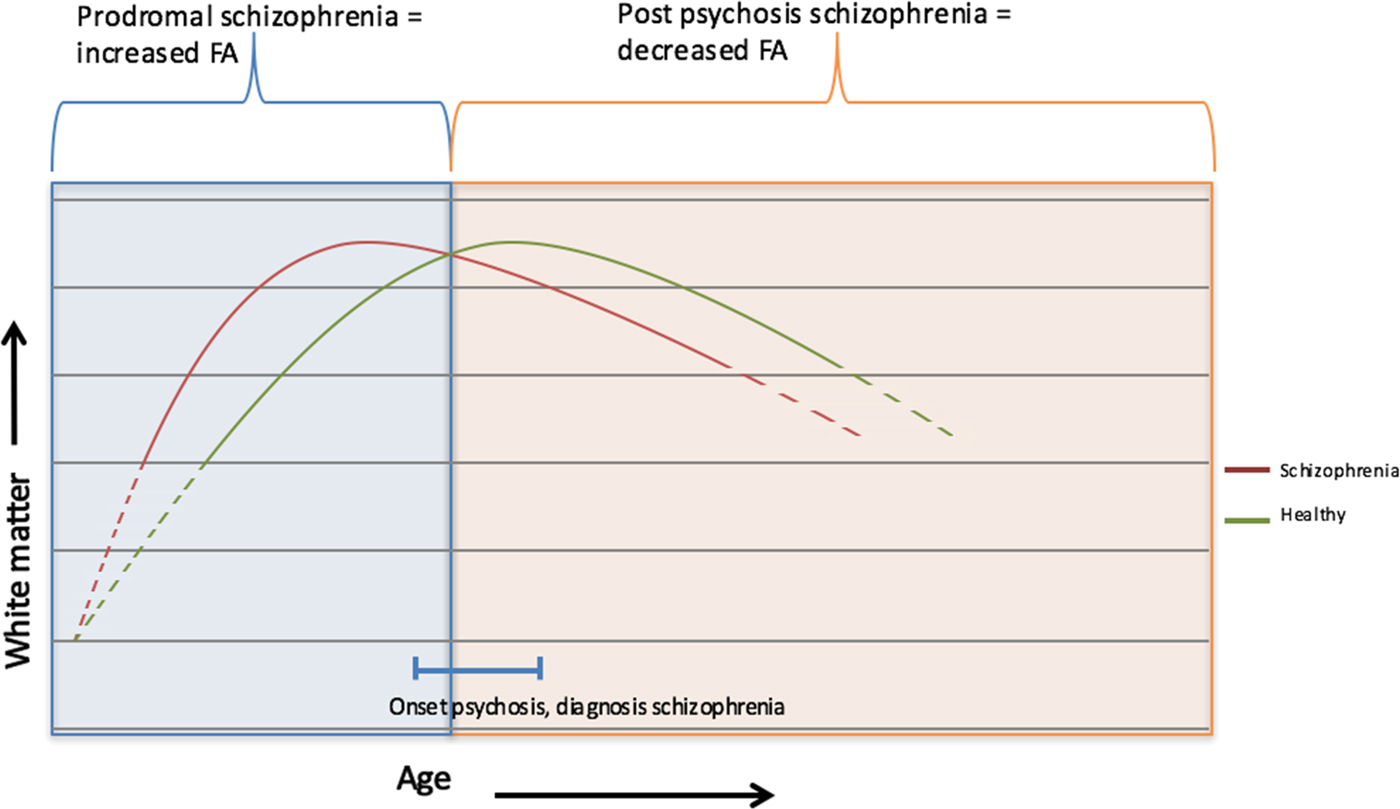
Studies of typical white matter development indicate that FA increases from childhood to puberty, plateaus at early adulthood and decreases in later adulthood (Schmithorst & Yuan, Reference Schmithorst and Yuan2010; Lebel et al. Reference Lebel, Gee, Camicioli, Wieler, Martin and Beaulieu2012; Krogsrud et al. Reference Krogsrud, Fjell, Tamnes, Grydeland, Mork, Due-Tønnessen, Bjørnerud, Sampaio-Baptista, Andersson, Johansen-Berg and Walhovd2016). Evidence for this pattern was recently presented in a review looking into typical and atypical brain development (Dennis & Thompson, Reference Dennis and Thompson2013). Interestingly, in three studies this downwards slope in adulthood was more pronounced in schizophrenia patients (Mori et al. Reference Mori, Ohnishi, Hashimoto, Nemoto, Moriguchi, Noguchi, Nakabayashi, Hori, Harada, Saitoh, Matsuda and Kunugi2007; Kochunov et al. Reference Kochunov, Glahn, Rowland, Olvera, Winkler, Yang, Sampath, Carpenter, Duggirala, Curran, Blangero and Hong2013; Cropley et al. Reference Cropley, Klauser, Lenroot, Bruggemann, Sundram, Bousman, Pereira, Di Biase, Weickert, Weickert, Pantelis and Zalesky2017). The regression lines of the association between FA and age, corresponding to schizophrenia patients and healthy controls, intersected between the ages of 20–30 (Mori et al. Reference Mori, Ohnishi, Hashimoto, Nemoto, Moriguchi, Noguchi, Nakabayashi, Hori, Harada, Saitoh, Matsuda and Kunugi2007; Kochunov et al. Reference Kochunov, Glahn, Rowland, Olvera, Winkler, Yang, Sampath, Carpenter, Duggirala, Curran, Blangero and Hong2013; Cropley et al. Reference Cropley, Klauser, Lenroot, Bruggemann, Sundram, Bousman, Pereira, Di Biase, Weickert, Weickert, Pantelis and Zalesky2017), suggesting that before the age of 20, not lower, but higher FA may be expected in patients developing schizophrenia. This finding suggests that during adolescence, increased FA may reflect a vulnerability to develop psychosis (as was suggested by O'Hanlon et al. Reference O'Hanlon, Leemans, Kelleher, Clarke, Roddy, Coughlan, Harley, Amico, Hoscheit, Tiedt, Tabish, McGettigan, Frodl and Cannon2015) and subsequently, decreased FA at a higher age may be the result of accelerated white matter ageing in schizophrenia patients. Tentative evidence suggests that this model may also apply for the trajectory of schizophrenia in individuals with 22q11DS (Jalbrzikowski et al. Reference Jalbrzikowski, Villalon-Reina, Karlsgodt, Senturk, Chow, Thompson and Bearden2014). In this study, we showed an interaction between FA and cognitive decline, but not a three-way interaction with psychosis. This may be due to our limited sample size, as the current study is a subset of a larger sample in which cognitive decline was found to predict psychosis onset (Vorstman et al. Reference Vorstman, Breetvelt, Duijff, Eliez, Schneider, Jalbrzikowski, Armando, Vicari, Shashi, Hooper, Chow, Fung, Butcher, Young, McDonald-McGinn, Vogels, van Amelsvoort, Gothelf, Weinberger, Weizman, Klaassen, Koops, Kates, Antshel, Simon, Ousley, Swillen, Gur, Bearden, Kahn and Bassett2015). Furthermore, the interaction between FA and cognitive decline was observed in white matter tracts often found to be implicated in schizophrenia. Therefore, although our finding of increased FA in 22q11DS patients with cognitive decline does not provide direct evidence, it is consistent with the accelerated ageing/early maturation theory. Based on our observations we speculate that cognitive decline in combination with increased FA reflects a vulnerability marker of schizophrenia (see Fig. 3; Kirkpatrick et al. Reference Kirkpatrick, Messias, Harvey, Fernandez-Egea and Bowie2008). Studies with a larger sample size would be needed to find further supporting evidence for this interpretation of our findings.
Hypothetical, simplified, model of accelerated ageing. The curve with its peak on the right side of the vertical line in the figure indicates white matter development in the healthy population. The curve with its peak on the left side indicates white matter development in schizophrenia patients. This curve is shifted to the left assuming that white matter development in schizophrenia peaks earlier and decreases earlier. Diagnosis of schizophrenia is indicated by the horizontal line in the bottom of the figure. For obvious reasons, the majority of studies on schizophrenia report on data collected after diagnosis (i.e. right area) finding decreased FA. The current study investigated white matter alterations before diagnosis (i.e. the left area) which may explain the observed increased FA in those with cognitive decline compared to those without.

Our study has several limitations. Some of the subjects in our study were already diagnosed with a psychotic disorder (four with cognitive decline, and two without cognitive decline). Removing these subjects from the sample did not alter the results. In fact, some ROIs showed a stronger effect, suggesting that our findings may pertain in particular to the prodromal phase of psychosis (online Supplementary S9).
Several neurobiological processes may explain changes in FA. For example, crossing fibre architecture may increase FA values due to degeneration of WM bundles in one of the crossing fibres (De Santis et al. Reference De Santis, Drakesmith, Bells, Assaf and Jones2014). To further investigate the neurobiological process underlying alterations in FA, complementary white matter measures such as magnetization transfer imaging are needed (Mandl et al. Reference Mandl, Pasternak, Cahn, Kubicki, Kahn, Shenton and Hulshoff Pol2015).
The cross-sectional nature of our MRI data precluded the investigation of individual FA trajectories in relation to individual IQ trajectories, nor could we investigate whether the group showing cognitive decline shows a higher transition rate to psychosis later in life. Longitudinal assessment of the interactions between white matter microstructure, IQ and psychosis onset (in 22q11DS as well as in the general population) may allow to more comprehensively investigate how cognitive decline and white matter alterations are implicated in psychosis onset.
Furthermore, movement in the scanner could influence the diffusion tensor model resulting in attenuated or exaggerated FA values depending on the tissue measured (Ling et al. Reference Ling, Merideth, Caprihan, Pena, Teshiba and Mayer2012). However, including translation and rotation movement parameters in the regression model did not alter the findings (see online Supplementary 10).
Due to the longitudinal IQ assessment, within-subject differences existed in the IQ tests used pertaining to the children's or adult version of the Wechsler Intelligence Scales. It has been reported that the transition between the two scales may be accompanied by a slight increase/decrease in several scores (Usner & Fitzgerald, Reference Usner and Fitzgerald1999). However, our groups did not significantly differ in the distribution of those who were tested twice with the same test and those who switched between different tests. Moreover, including a dichotomous variable (dividing the sample in a group with a switch in IQ test and in a group without a switch) in the regression model did not alter the results.
Lastly, one could argue that cognitive decline is not clinically relevant when there is only a small negative difference between T − 1 en T0. However, when dividing the sample with a more clinically relevant cut-off score of 5 percentile points decline, all regions of the left internal capsule and the left superior longitudinal fasciculus showed significantly higher FA values in the decline group compared to the non-decline group at uncorrected p < 0.05 (see online Supplementary S11). In addition, we observed a significant correlation between the degree of decline per year and FA. This suggests that the effect is not solely a group-effect, which could arguably be confounded by the choice of assuming one percentile point as true decline.
In this study, we show that alternate IQ trajectories are associated with differences in white matter microstructure in patients with 22q11DS. This finding withstood correction for multiple comparisons and could not be attributed to variation in gender, age, baseline intellectual ability, the interval between IQ measurements, psychiatric status, head motion or type of IQ test. The overlap between the current results of white matter regions implicated in cognitive decline in 22q11DS and previously reported white matter regions involved in psychosis onset in 22q11DS, suggests that cognitive decline may be crucially implicated in mediating psychosis risk in 22q11DS.
Supplementary material
The supplementary material for this article can be found at https://doi.org/10.1017/S0033291717003142
Declaration of Interest
None.
Ethical standards
This study has been approved by the local research ethics board (Dutch Central Committee on Research Involving Human Subjects; C.C.M.O) and all participants (and/or their legal guardians) provided written informed consent.