Introduction
Tympanoplasty is a common procedure for addressing perforation and retraction of the tympanic membrane. Several types of tissue for the tympanic membrane graft have been used historically.Reference Dornhoffer1 Surgeons have found cartilage grafts attractive because of the advantages of a low rate of post-operative perforations and the improved ability to address atrophic tympanic membranes, all while obtaining satisfactory hearing results.Reference Dornhoffer1
Various techniques of cartilage tympanoplasty have been proposed.Reference Dornhoffer1–Reference Ozdemir, Ozgur, Akgul, Celebi, Mehel and Yemis15 We use an over-under technique, where the cartilage–perichondrial island graft is placed lateral to the malleus and medial to the annulus. We also use a cartilage shim between the native tympanic membrane and the cartilage graft when the graft does not make contact with the tympanic membrane, and we believe this additional step is unique. We have found over-under cartilage tympanoplasty to be a flexible and versatile approach in addressing the tympanic membrane, but the available articles on this only briefly describe the approach.Reference Kirazli, Bilgen, Midilli and Ogut2–Reference Bedri, Korra, Redleaf and Worku6,Reference Balci, Islek and Ciger10–Reference Ozdemir, Ozgur, Akgul, Celebi, Mehel and Yemis15
The objectives of this article are to describe the technique we use for the microscopic over-under cartilage tympanoplasty and to report the hearing outcomes and complications. We hypothesised that hearing would be shown to improve across the hearing spectrum and that reperforation would be rare.
Materials and methods
The authors assert that all procedures contributing to this work comply with the ethical standards of the relevant national and local institutional guidelines on human experimentation and with the Helsinki Declaration of 1975, as revised in 2008. This study was approved by the institutional review board of Our Lady of the Lake Regional Medical Center, Baton Rouge, USA (institutional review board number: 19-082-OLOL).
Patient selection
A retrospective review of all microscopic over-under cartilage tympanoplasty procedures performed by the senior author between January 2015 and January 2019 at three tertiary care facilities was undertaken.
Cases were excluded if an ossicular prosthesis was used, if a mastoidectomy or encephalocele repair was performed during the procedure, or if cholesteatoma was encountered at the time of or prior to the tympanoplasty.
Patients were not excluded if a pressure equalisation tube was placed in the same setting, if scar bands were removed in the middle ear or if the indication for the tympanoplasty was for an atrophic tympanic membrane. These criteria were evaluated based on the patient's operative reports.
Data collection
Patients were included if both a pre-operative audiogram and an audiogram two months or later were available. Air conduction at 250, 500, 1000, 2000, 3000, 4000, 6000 and 8000 Hz and bone conduction at 500, 1000, 2000, 3000 and 4000 Hz were collected. Word recognition scores were also collected. Decibel levels at 3000 and 6000 Hz were interpolated if necessary.Reference Gurgel, Popelka, Oghalai, Blevins, Chang and Jackler16 Air and bone pure-tone averages were evaluated at 500, 1000, 2000 and 3000 Hz. To meet American Academy of Otolaryngology Head and Neck Surgery (AAO-HNS) 1995 minimal reporting guidelines, ‘high pure-tone bone-conduction averages’ of 1000, 2000 and 4000 Hz were also evaluated.Reference Monsell, Balkany, Gates, Goldenberg, Meyerhoff and House17 The first available post-operative audiogram was used, typically obtained at the three-month point. If an audiogram was available at the one-year point or later, this was also collected. Primary versus revision tympanoplasty was determined from notes available. Post-operative complications were evaluated and considered clinically significant if surgery was recommended.
Data analysis
Data were analysed in R (version 3.5.3; R Foundation for Statistical Computing; Vienna, Austria) and RStudio (version 1.1.463; RStudio, Boston, USA) along with the additional R packages tidyverse, psych and Toster. Post-operative changes in word score and pure tones were evaluated with paired, two-sided t-tests. Equivalence testing for pre- and post-operative bone pure-tone averages was assessed using ‘two one-sided t-tests’ (‘TOST’) using the equivalence bounds of 3 dB. These bounds were based on the just-noticeable difference in hearing in signal-to-noise ratio.Reference McShefferty, Whitmer and Akeroyd18 The scattergram plots were created with assistance from the Stanford web-based tool.Reference Oghalai and Jackler19
Over-under cartilage tympanoplasty
The procedure was performed with an operating microscope. Canal incisions were made in the 6 and 12 o'clock positions, and a posterior horizontal incision was made between them about 3 mm from the annulus. A post-auricular incision was made, and the ear was reflected anteriorly.
An anterior canalplasty was frequently performed. Bony anterior canal bulges were drilled until the entire fibrous annulus was seen in one microscopic view.
The tympanic membrane and middle ear were then prepared for tympanoplasty (Figure 1). The edges of the perforation were incised and removed. The tympanomeatal flap was raised and the middle ear entered. The tympanic membrane was elevated from the bony tympanic ring from the 6 o'clock position inferiorly and to the 2 o'clock position antero-superiorly. The tympanic membrane was elevated from the malleus with a sickle knife. If there was any evidence or concern of squamous tissue remaining on the malleus, an argon laser at 1500 mW for 200 ms per pulse was used. The distance between the malleus and the bony tympanic ring anteriorly, inferiorly and posteriorly was measured using a right-angle hook.
Preparing the tympanic membrane and middle ear. (a) An anterior inferior perforation of the right ear. (b) The tympanic membrane is elevated from the malleus with a sickle knife, resulting in (c) an excellent view of the entire mesotympanum. Note the laser marks present on the handle of malleus.

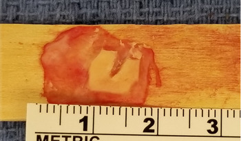
Cartilage was harvested, most often from the tragus. A linear 1–1.5 cm incision was made on the posterior surface of the tragus through skin and cartilage, leaving a dome of tragal cartilage laterally for cosmesis. An approximately 1.5 × 1 cm section of cartilage and overlying perichondrium was harvested. The cartilage-perichondrial island graft was then prepared (Figure 2). For control of the graft and the depth of incisions into the graft, we placed the graft on a sterile, wooden tongue blade. The thicker perichondrium from the concave side of the graft was removed. Using a House round knife, incisions were made into the cartilage but not through the perichondrium on the other side of the graft. Cartilage was removed from the perichondrium, usually in the shape of a horseshoe, using the previous measurements with the right-angle hook.
The cartilage-perichondrial island graft. The graft is created from harvested tragal cartilage. Perichondrium has been elevated from the concave side, and a round knife is used to remove cartilage to the desired shape while leaving the contralateral perichondrium intact. A right-angle pick can be used to measure the resultant cartilage island to ensure that it fits the dimensions of the bony annulus in Figure 1c. Note that the graft shown was fashioned for a left ear.

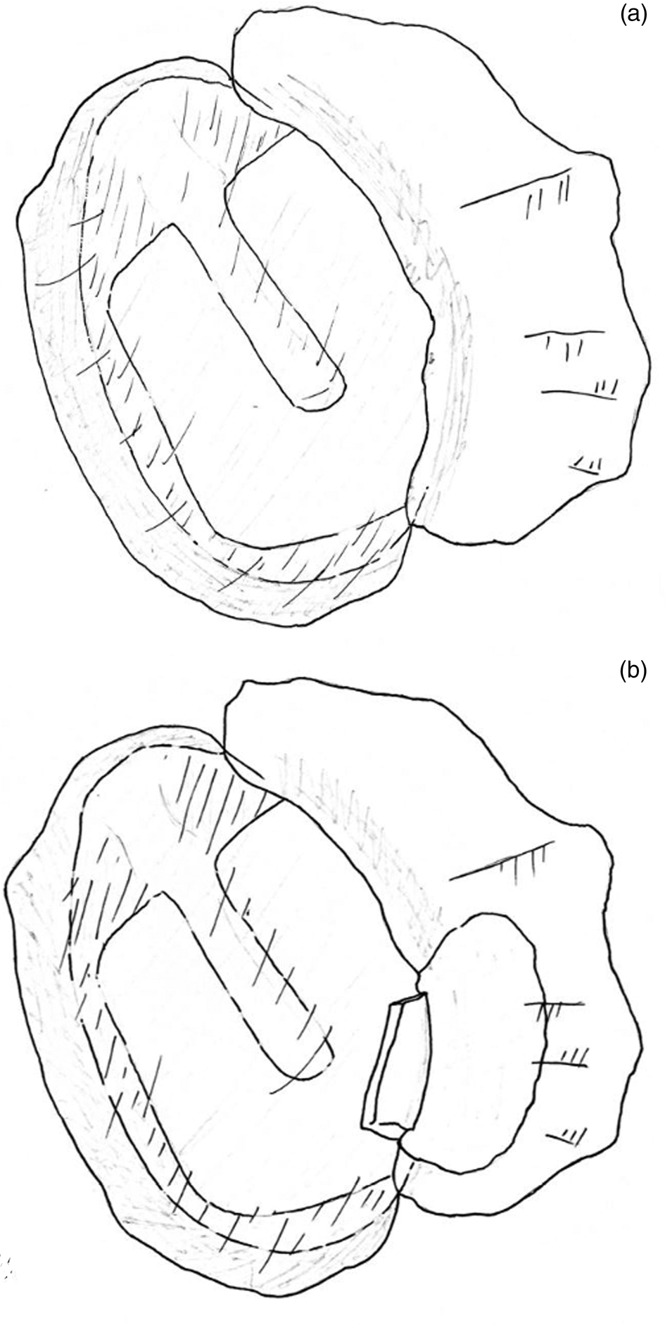
Once fashioned, the graft was placed into the ear (see the short video, available on The Journal of Laryngology & Otology website; Appendix 1), with the cartilage side facing medially and the perichondrial surface facing laterally. It was placed on the malleus, with the malleus fitting in the groove made in the cartilage. The perichondrium was draped on the posterior external auditory canal. The tympanomeatal flap was re-draped, and the area of the perforation evaluated to determine if the graft reached the same lateral height as the tympanic membrane, especially in the case of a medially rotated malleus. If it did not, a shim was placed medially to the perforation and laterally to the graft. The shim was fashioned with (1) the previously harvested perichondrium placed under the native tympanic membrane and (2) an additional small piece of cartilage, obtained when fashioning the island graft, placed between the island graft and the perichondrium (Figure 3).
Cartilage shim, if required. After the graft is placed (a), perichondrium is placed on the undersurface of the tympanomeatal flap, and (b) a bit of cartilage is placed between the graft and the perichondrium.

The external auditory canal, tragal and post-auricular incisions were closed per surgeon preference.
Results
In this study, 68 cases of microscopic over-under cartilage tympanoplasty were identified as meeting inclusion criteria. An additional six cases of over-under cartilage tympanoplasty were identified in the study period but were lost to follow up. Median follow-up length was 168 days, and the average was 321 days.
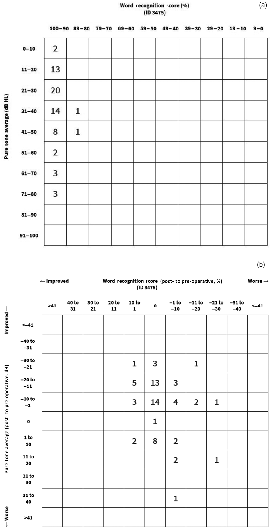
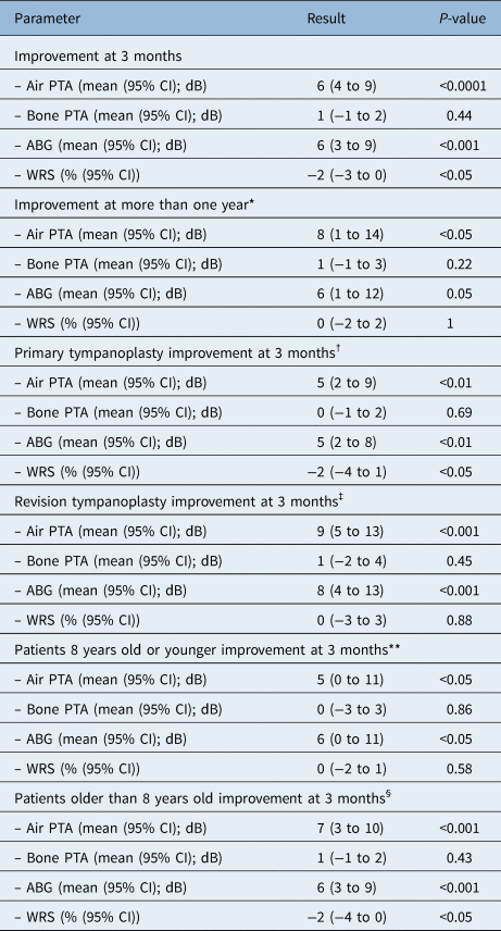
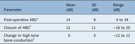
Hearing results are summarised in Table 1. Results meeting the minimum AAO-HNS 1995 and 2012 reporting guidelines are shown in Table 2 and Figure 4. Overall improvement in air pure-tone average was 6 dB (p < 0.0001) at the time of the initial post-operative audiogram. Word score declined modestly by 2 per cent (p < 0.05). Post-operative bone pure-tone averages demonstrated equivalence compared to pre-operative levels (p < 0.001); this was also true for ‘high pure-tone bone-conduction average’ (p < 0.0001).
American Academy of Otolaryngology scattergram plots. (a) Pre-operative and (b) post-operative changes.

Hearing results

Total patients = 68. Air–bone gap measured as an average of 500, 1000, 2000 and 3000 Hz. For CIs, positive values indicate post-operative improvement and negative values indicate post-operative decline. *n = 19; †n = 49; ‡n = 19; **n = 17; §n = 51. PTA = pure tone average; CI = confidence interval; ABG = air–bone gap; WRS = word recognition score
AAO-HNS 1995 Minimum Reporting Guidelines

Air–bone gap is measured as the difference of air and bone pure tones averaged at 500, 1000, 2000 and 3000 Hz. High-tone bone-conduction is defined as the bone conduction averages of 1000, 2000 and 4000 Hz. *n = 19; †n = 19; ‡n = 68. AAO-HNS = American Academy of Otolaryngology Head and Neck Surgery; SD = standard deviation; ABG = air–bone gap
Improvement was noted regardless of age or whether the procedure was a primary tympanoplasty or revision. Neither statistical equivalence (p = 0.24) or difference (p = 0.79) was found in the improvement in air pure tone averages when comparing patients eight years or younger versus older patients. When comparing revision tympanoplasty to primary tympanoplasty, revision tympanoplasty had equivalent or better improvement in air pure tone average (p < 0.01).
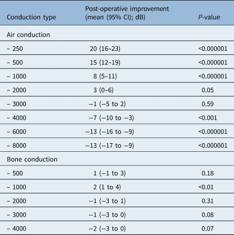
When evaluating individual frequencies with air conduction, there was a statistically significant improvement at the low frequencies (250, 500 and 1000 Hz) and statistically significant loss at the high frequencies (4000, 6000 and 8000 Hz; Table 3). Post-operative non-inferiority was found at 2000 Hz (p < 0.0001), and pre-operative non-inferiority was found at 3000 Hz (p < 0.05).
Post-operative improvement of hearing by frequency

Positive values indicate post-operative improvement, negative values indicate post-operative decline. CI = confidence interval
Among 19 patients with audiograms at the one-year point or later, there was statistically significant post-operative improvement in air pure tone averages at both the three-month point (8 dB, 95 per cent confidence interval: 2–14 dB; p < 0.05) and their later audiogram (8 dB, 95 per cent confidence interval: 1–14 dB; p < 0.05). There was also equivalence in air pure tone average between the three-month point and their later audiogram (p < 0.05).
Clinically significant complications were noted in six patients. Two had post-operative perforations, both of which occurred next to the umbo. One had a cholesteatoma of the middle ear. Three had external auditory canal scar bands, of which two developed a cholesteatoma of the external auditory canal. Among patients not requiring additional surgical intervention, a perforation was encountered, which spontaneously resolved, and there were two patients with a keratin pearl found on their tympanic membrane. Neither lateralisation nor anterior blunting were encountered. There was no statistically significant difference in complications for patients with only shorter follow up available (n = 2) and those who were followed for longer (n = 4; p = 0.07).
Discussion
The microscopic over-under cartilage tympanoplasty technique presented in this article demonstrates hearing improvement at frequencies considered clinically significant. Hearing improvement is noted in both primary tympanoplasties and revisions, and in patients both older and younger than eight years old. The post-operative perforation rate is low, and the most frequent clinically important complication encountered was external auditory canal scarring.
The ability to confidently close a perforation is a strong reason to consider this technique, particularly in patients undergoing revision. The 97 per cent closure rate presented compares well to the 84–95 per cent rate in over-under temporalis fascia grafts,Reference Kartush, Michaelides, Becvarovski and LaRouere20–Reference Yigit, Alkan, Topuz, Uslu, Unsal and Dadas22 and the rate of 88–98 per cent in other over-under cartilage tympanoplasty procedures.Reference Mohanty, Manimaran, Umamaheswaran, Jeyabalakrishnan and Chelladurai3,Reference Solmaz, Akduman, Haksever, Gundogdu, Yanilmaz and Mescioglu4,Reference Bedri, Korra, Redleaf and Worku6,Reference Ciger, Balci, Islek and Onal11–Reference Ulku14 This was reliable in younger patients as well, though we avoided performing tympanoplasty on young patients with active Eustachian tube dysfunction. The closure rate presented in this article is likely due to the use of cartilage for grafting material, the placing of the graft snugly onto the malleus and the addition of the cartilage shim, which, when required, brings the graft in contact with the native tympanic membrane.
This technique has several other strengths. Similar to arguments in other studies,Reference Kartush, Michaelides, Becvarovski and LaRouere20,Reference Babu, Luryi and Schutt21,Reference Yawn, Carlson, Haynes and Rivas23 raising the tympanic membrane from the malleus gives excellent visualisation of the entire mesotympanum, allowing for both the improved identification of pathology and the ability to address perforations of the anterior superior quadrant. Additionally, in patients with narrow middle-ear spaces from retraction or medially rotated mallei, placing a cartilage graft lateral to the malleus can be easier than placing it as an underlay, while still preserving some middle-ear space. Finally, the technique is versatile enough that it can be used with slight modification for lateral grafts, tympanomastoidectomy, ossiculoplasty, cartilage embedded pressure equalisation tubes and endoscopic ear surgery.
This study showed hearing loss in the higher frequencies. These frequencies are at higher tones than what are typically considered clinically significant for reasons discussed below. The possible causes for this loss are the material used for the graft, the location of the graft, trauma from elevating the drum from the ossicles or acoustic trauma from drilling the anterior canal.Reference Kazikdas, Onal and Yildirim24,Reference Domenech, Carulla and Traserra25 Although we do not have bone conductive values in the high frequencies, we believe that this is a conductive loss. Cadaveric studies support the possibility of high frequency loss from both materialReference Eldaebes, Landry and Bance26 and location of the graft.Reference Eldaebes, Landry and Bance27 Sensorineural loss from ossicular or acoustic trauma is less likely because this study found equivalence between pre- and post-operative bone conduction in the frequencies obtained. Similarly, Morrison et al. did not find changes in bone conduction in their series of drilled anterior canalplasty procedures, although they did not report any high frequency results.Reference Morrison, O'Connell and Lambert28
Comparing our findings of high frequency loss to the literature is challenging. Concurrent articles on cartilage tympanoplasty frequently report hearing thresholds or averages using the highest frequency of 2000 Hz,Reference Mohanty, Manimaran, Umamaheswaran, Jeyabalakrishnan and Chelladurai3,Reference Genc5,Reference Parelkar, Thorawade, Marfatia and Shere13,Reference Kim, Park, Suh and Song29 3000 Hz1,Reference Salviz, Bayram, Bayram, Balikci, Chatzi and Paltura8,Reference Morrison, O'Connell and Lambert28,Reference Demirpehlivan, Onal, Arslanoglu, Songu, Ciger and Can30–Reference Kolethekkat, Al Abri, Al Zaabi, Al Marhoobi, Jose and Pillai32 or 4000 Hz.Reference Kirazli, Bilgen, Midilli and Ogut2,Reference Solmaz, Akduman, Haksever, Gundogdu, Yanilmaz and Mescioglu4,Reference Bedri, Korra, Redleaf and Worku6,Reference Karaman, Duman, Isildak and Enver12,Reference Ulku14,Reference Ozdemir, Ozgur, Akgul, Celebi, Mehel and Yemis15,Reference Khan and Parab33–Reference Vashishth, Mathur, Choudhary and Bhardwaj36 Were we to narrow our reporting to these same levels or simply present a pure tone average, the high frequency loss presented here would have been missed, since statistical significance was only noted at 4000, 6000 and 8000 Hz.
Rare reports of high frequency data in cartilage tympanoplasty suggest that our findings are not unique. Shishegar et al. report large high frequency air–bone gaps with both cartilage and fascia tympanoplasty procedures.Reference Shishegar, Faramarzi and Taraghi37 Vadiya et al. showed a smaller air–bone gap improvement at 8000 Hz with cartilage than fascia by a statistically significant margin, and this statistically significant difference was not present at lower frequencies reported.Reference Vadiya, Parikh, Shah, Pandya and Kansara38
The reason for not reporting frequencies higher than 3000 Hz is well supported in the literature. Current guidelines for air conduction values recommend reporting 500, 1000, 2000 and 3000 Hz.Reference Gurgel, Jackler, Dobie and Popelka39–Reference Kim41 This captures clinically significant loss, and audiometric loss at these frequencies has been validated to self-perceived hearing handicap.Reference Dobie42 Current AAO-HNS guidelines for conductive loss also include reporting the post-operative air–bone gap at one year, the change in air–bone gap at one year and bone conduction average of 1000, 2000 and 4000 Hz at six weeks.Reference Monsell, Balkany, Gates, Goldenberg, Meyerhoff and House17 Reporting beyond these values is ‘discretionary’.Reference Monsell, Balkany, Gates, Goldenberg, Meyerhoff and House17,Reference Gurgel, Jackler, Dobie and Popelka39
However, there may be some value in obtaining and reporting these additional frequencies. Although lower frequencies are important for intelligibility,Reference Dobie42 there is evidence that high frequencies may be important for sound localisation and understanding speech in noisy environments.Reference Monson, Hunter, Lotto and Story43,Reference Levy, Freed, Nilsson, Moore and Puria44 Air conduction values above 4000 Hz are also routinely obtained in clinical practice, despite their absence in the literature. If our findings of high frequency loss are reproduced by other authors, it would be reasonable to consider reporting these routinely obtained values. This would allow for a more thoughtful evaluation of the risks and benefits of differing tympanoplasty techniques.
The other major risks encountered were middle-ear cholesteatoma and the external auditory canal scar banding. In the one case of middle-ear cholesteatoma, the use of the laser to ablate squamous tissue was not reported in the operative report. This is now a step that is routinely employed. The scar banding may be a result of the steps around the anterior canalplasty. Every effort is made to stagger canal incisions to prevent a circular incision, but this may be a factor. We also believe that some scarring may be due to the use of gelatin foam to pack the ear canal. Since obtaining this data, we have replaced most of the canal packing with antibiotic ointment instead.
This article has several strengths. It describes the over-under cartilage tympanoplasty technique in detail, and it is one of the larger series available focusing solely on the over-under cartilage tympanoplasty. This series includes a wide range of patient ages, representing what a general otolaryngologist might encounter. The article also reports and evaluates several details that many articles omit, including bone conduction results, air conduction frequencies outside the pure tone averages, changes in word recognition scores and post-operative complications.
However, there are several limitations to this study. This is a retrospective case series. Follow up was somewhat limited, both by study design and loss to follow up: our catchment area is large enough that patients may return to their local otolaryngologist instead of travelling several hours to return to us for their later follow-up appointments. Finally, there was some degree of selection bias, given the intra-operative decision for cartilage lateral grafts in subtotal perforations and for cartilage butterfly inlay grafts in select small perforations.
• Microscopic over-under cartilage tympanoplasty is a reliable technique to close perforations and evaluate the entire middle ear
• Perforations were closed in 97 per cent of cases
• The addition of a tissue shim to allow the graft to contact the native tympanic membrane may have improved perforation closure rates
• Surgeons employing this technique should be aware of possible external auditory canal scar bands, which were found in 4 per cent of cases
• Hearing improvement in air pure tone averages was 6 dB, and hearing improvement was found regardless of young age or revision surgery
• Hearing loss was noted at high frequencies, but there is a paucity of comparative literature presenting high frequency data
Conclusion
The microscopic over-under cartilage tympanoplasty procedure has acceptable hearing improvement and 97 per cent rate of tympanic membrane closure. The literature would be improved with additional reporting of high frequencies.
Supplementary material
The supplementary material for this article can be found at https://doi.org/10.1017/S0022215120001978.
Appendix 1. Supplementary video material
A short video demonstrating the cartilage-perichondrial island graft after placement on the malleus. This video is available online at The Journal of Laryngology & Otology website, at https://protect-eu.mimecast.com/s/WFQpCGRrRH08pQGU7RUJ-?domain=click.email.vimeo.com"https://vimeo.com/396355694