




No CrossRef data available.
Article contents
Neural substrates of syntactic mapping treatment: An fMRI study of two cases
Published online by Cambridge University Press: 23 January 2006
Abstract
Two patients (G01, J02) with chronic nonfluent aphasia and sentence production deficits received syntactic mapping treatment to improve sentence production. The patients had dramatically different outcomes in that improved syntax production generalized to nontreatment tasks for G01, but not for JO2. To learn how treatment influenced the neural substrates for syntax production, both patients underwent pre- and posttreatment functional magnetic resonance imaging (fMRI) of sentence generation. G01 showed more robust activity posttreatment than pretreatment in Broca's area; ventral temporal activity decreased slightly from pre- to posttreatment. Comparison of J02's pretreatment and posttreatment images revealed little change, although activity was more diffuse pre- than posttreatment. Findings suggest that for G01, rehabilitation led to engagement of an area (Broca's area) used minimally during the pretreatment scan, whereas for J02, rehabilitation may have led to more efficient use of areas already involved in sentence generation during the pretreatment scan. fMRI findings are discussed in the context of sentence-production outcome and generalization. (JINS, 2006, 12, 132–146.)
Keywords
Information
- Type
- NEUROBEHAVIORAL GRAND ROUNDS
- Information
- Journal of the International Neuropsychological Society , Volume 12 , Issue 1 , January 2006 , pp. 132 - 146
- Copyright
- © 2006 The International Neuropsychological Society
INTRODUCTION
Emerging functional neuroimaging studies regarding plasticity in aphasia secondary to stroke indicate that good language recovery most frequently is supported by left perilesional areas, but that patients with significant, persistent aphasias demonstrate more right-hemisphere activity than patients with good recovery (Basso et al., 1989; Heiss et al., 1997, 1999; Karbe et al., 1998; Cao et al., 1999; Rosen et al., 2000; Perani et al., 2003). In at least some cases of rehabilitative intervention, left perilesional regions are mobilized during recovery of nonfluent aphasia (Belin et al., 1996; Cornelissen et al., 2003), while other reports suggest that right-hemisphere structures also contribute to language recovery (Musso et al., 1999; Crosson et al., 2005; Peck et al., 2004a). The majority of neuroimaging studies investigating language recovery poststroke have focused on word production or receptive language; unfortunately, little information is available regarding cortical regions mobilized by rehabilitation of syntax deficits in sentence production.
Although there is considerable symptom variability among nonfluent aphasias, a deficit in thematic role assignment manifested via word-order deficits (i.e., accurate placement of the arguments around the verb) is a frequent form of syntax impairment (Caramazza & Miceli, 1991). This deficit is commonly referred to as a mapping deficit (Schwartz et al., 1980, 1994a). Difficulty mapping thematic roles reflects underlying problems communicating knowledge of who is doing what to whom. Performance is often worsened with reversible sentences, that is, ones in which it is plausible to reverse the actor and the recipient of the action. Mapping problems may be explained by a deficit at the early stage of sentence production, during which the thematic relationship of sentence elements is established (Garrett, 1980, 1984; Berndt, 1998; Chatterjee & Maher, 2000). Mapping therapies have been designed to address assignment of thematic roles in sentences and to explicitly strengthen the association between thematic roles and word order (Jones, 1986; Byng, 1988; Byng et al., 1994; Schwartz et al., 1994b; Chatterjee et al., 1995; Maher et al., 1995). The effectiveness of this treatment approach has been variable. In some instances, treatment gains are specific to treated sentences, with little generalization (Byng et al., 1994; Schwartz et al., 1994b). It remains unclear which factors contribute to the relative success or failure in a given patient, and the mechanisms by which treatment influences the underlying neural substrates of recovery are not well understood.
The heterogeneity of syntax deficits (i.e., problems with word order, thematic role assignment, and morphology) resulting from various lesion sites suggests that a discrete grammar center in the brain is unlikely. Recent functional neuroimaging studies have implicated Broca's area (Brodmann's Areas 44, 45) in a variety of receptive syntactic functions, including comprehending lexical-semantic information and syntactic structure (Dapretto & Bookheimer, 1999), processing syntactic ambiguities (Stowe et al., 1998), detecting syntactic anomalies and errors (Embick et al., 2000; Ni et al., 2000), and processing syntactically more complex sentences (Just et al., 1996; Caplan & Waters, 1999; Caplan et al., 2000). Posterior perisylvian regions, including the superior temporal gyrus, angular gyrus, supramarginal gyrus, and superior parietal lobe, also have been implicated in syntactic processing (Just et al., 1996; Carpenter et al., 1999; Caplan et al., 2001; Newman et al., 2001). Studies that have investigated sentence production have found activity in and around Broca's area, including the left anterior Rolandic operculum (Indefrey et al., 2001, 2004; Peck et al., 2004b) as well as in left posterior perisylvian regions (Peck et al., 2004b).
In the current study, two patients with residual nonfluent aphasia and sentence production deficits (G01 and J02) received an experimental syntactic mapping treatment to target thematic role assignment in sentence production (i.e., word order). The purpose of the study was to investigate the neural substrates that support recovery of function following treatment of thematic role assignment. Based on the research reviewed earlier, we developed the following hypotheses: (1) patients will show an improvement in producing syntactically correct sentences following treatment, and (2) because treatment emphasizes word order, we expect to see increased activation in cortical regions associated with syntax. Specifically, fMRI activity during sentence production is expected to increase from pre- to posttreatment in the intact perilesional regions of the left inferior frontal gyrus (including Broca's area).
METHOD
Research Participants
Two patients (G01, J02) with chronic nonfluent aphasia received syntactic mapping treatment under two different conditions as part of a larger study, and underwent fMRI pre- and posttreatment. Pretreatment assessment suggested that both G01 and J02 demonstrated aphasia profiles most consistent with transcortical motor aphasia, with repetition, naming, and auditory comprehension scores relatively higher than fluency scores (see Table 1).
Pretreatment language assessment

Patient G01
G01 was a 72-year-old right-handed woman treated 53 months postonset of a left hemisphere cerebral vascular accident (CVA). She obtained 12 years of education and had been a homemaker prior to her stroke. She reportedly had received 6 months of speech and language therapy following her stroke. An MRI scan revealed a lesion consistent with a proximal occlusion of the left middle cerebral artery, with damage to the left temporal and parietal lobes, basal ganglia, thalamus, sensorimotor area, premotor cortex, and centrum semiovale (Figure 1).

T1-weighted axial magnetic resonance images in radiologic space (left is on the right) showing the extent of left cerebral vascular infarction for G01 and J02.
In addition to deficits in fluency and word retrieval on the Western Aphasia Battery (WAB; Kertesz, 1982), results of the pretreatment Action Naming Test (ANT; Obler & Albert, unpublished) and Boston Naming Test (BNT; Kaplan et al., 2000) indicated that G01 had greater difficulty with verb than noun retrieval (see Table 1). Verb retrieval errors tended to be the use of nonspecific or “light” verbs (such as “using a saw” for “sawing”). Performance on the Circles and Squares Test (Schwartz et al., 1980) revealed auditory comprehension of reversible active sentences was quite good (23/24 correct), however, comprehension of passive sentences, particularly when the action was depicted from left to right, was impaired (16/24 correct).
Sentence production in response to reversible action pictures highlighted the difficulty in generating accurate active and passive sentences. A 90-sentence subset of the pretreatment responses was coded for type of error, and half (45) of these sentences were coded by a second examiner. Interjudge agreement for presence of error was 100%, and for type of errors was 83%. Disagreements in error coding were resolved by consensus. Errors were predominantly word order errors (e.g., beginning a passive sentence with the agent or “actor” noun), use of a light verb in place of the target verb (e.g., “went after” for the target “follow”), and incorrect verb selection (e.g., “teasing” for “biting”). Pretreatment performance on the baseline and probe measures revealed a stable deficit in sentence generation (z = 1.04; p > .05). Analysis of narrative discourse using the Quantitative Production Analysis (Berndt et al., 2000) and additional linguistic measures revealed persistent deficits with word order and morphology, decreased representation of verbs, and reduced syntactic accuracy and complexity.
Patient J02
J02 was a 58-year-old right-handed man seen 8 months postonset of a left hemisphere CVA. He completed 18 years of education and prior to his stroke had been employed in law enforcement. He had received approximately one month of speech and language therapy following his stroke. An MRI scan revealed a lesion consistent with a proximal occlusion of the left middle cerebral artery, with damage to the anterior temporal lobe, insular region, basal ganglia, and premotor and sensorimotor cortex (Figure 1). Results of J02's pretreatment ANT and BNT indicated he had greater difficulty with noun retrieval than verb retrieval (see Table 1). Predominant word retrieval errors included no responses, perseverations, and semantic paraphasias. Performance on the Circles and Squares test revealed relatively preserved comprehension of active (19/24) and passive (23/24) reversible sentences.
Much like G01, J02's performance on active and passive sentence generation revealed marked difficulty in generating active and passive sentences. Again, a 90-sentence subset of the pretreatment responses was coded for type of error, and half (45) were coded by a second examiner. Interjudge agreement for presence of error was 100%, and for type of errors it was 76%. Disagreements in error coding were resolved by consensus. The types of errors he produced were predominantly in verb retrieval (e.g., incorrect or absent verbs), word order (e.g., active syntax for a passive sentence), and morphologic (e.g., omission of functors). Pretreatment performance on the baseline and probe measures revealed a stable deficit in sentence generation (z = 1.24, p > .05). J02's narrative discourse also revealed persistent deficits with word order and morphology, marked decrease in verb use, and reduced syntactic accuracy and complexity.
Syntactic Mapping Treatment
The syntactic mapping treatment differed from what has been most often reported in the literature in that the intervention focused on sentence production rather than comprehension of thematic roles. Sentence production was paired with a color and spatially coded mapping template focusing on thematic role positions (see Figure 2). The patients were encouraged to use the illustration in the template to guide the placement of the lexical items in the sentence to accurately reflect the thematic roles. Errors in morphological endings and article omissions were ignored for scoring purposes, with the exception of the passive morphology for passive sentence construction (e.g., “was + verb + by”).

Illustration of color and spatially coded mapping template used to train active and passive sentences in both phases (Errorless Mapping Treatment, Errorful Mapping Treatment) of the intervention.
The treatment was administered in two phases. In the first phase, Errorless Learning Mapping (ELM) utilized constant time delay as an errorless training procedure for sixteen 90-minute sessions, occurring 3–4 times per week (Wolery et al., 1992; Maher et al., 2002). This was followed by a second phase, Errorful Mapping (EFM), which utilized the more traditional approach for the same number and frequency of sessions. Active sentences were trained first (8 sessions), followed by passive sentence training (8 sessions) in each of the phases. In ELM, sentence production errors were prevented by providing an immediate model for repetition of the target sentence. The patient was instructed to only produce the sentence if he/she was certain it would be correct. Otherwise, the therapist would provide the model and the patient would repeat the target sentence. After 80% of the sentences were produced correctly at this stage, then a time-delay between the presentation of the picture and the model was introduced, allowing the patient increasing independence in producing the sentence. However, if an error was either produced or anticipated, the model for the correct response was provided yielding a near errorless learning environment. In contrast, in EFM, the patients first generated their independent responses to the target, and errors were corrected through a query approach of the lexical items to fill the respective thematic roles of agent (actor) and patient (recipient of action) in the target sentence (Schwartz et al., 1994b). In both the ELM and EFM phases, the color-coded template of the sentence constituents (and when appropriate the passive morphology) was present. In the ELM phase, written words corresponding to the agent, recipient, and verb were placed in the appropriate locations to “fill in” the map prior to the response (or after a specified time delay). In EFM, the map was filled in as the patient produced the lexical items, either independently or with cues for the therapist. The comparative impact of ELM and EFM has been reported elsewhere (Maher et al., 2002).
For each participant, a corpus of reversible sentences was probed to yield three different sets of transitive verbs that were balanced for word frequency (Kucera & Francis, 1967) for each patient. All of the verbs required only an agent (actor) and a patient or goal (recipient) to be syntactically correct. Within each set, no two verbs began with the same initial phoneme. These verbs were paired with high frequency animate nouns to yield reversible sentences that were pictured in simple line drawings (see Figure 2). Multiple baselines of active and passive sentence production in response to the three sets of reversible action pictures were obtained prior to the initiation of treatment. These same sentences were probed throughout the two phases of treatment to determine treatment and generalization effects. Again, errors in morphologic endings and article omissions were ignored for scoring purposes, with the exception of the passive morphology for passive sentence construction (e.g., “was + verb + by”). Synonyms for the lexical items (e.g., “lady” for woman; “bashing” for hitting) were scored as correct. Each response was transcribed and scored offline by the therapist and by a second examiner who was unfamiliar with the patients for reliability. Interjudge agreement exceeded 90%; disagreements were resolved by a third examiner.
Following the determination of a stable baseline as indicated by the C-statistic (Tryon, 1982), ELM treatment was initiated using the first set of pictures, while the other two sets were not treated but were probed with the same frequency as the treated set. The second set of pictures was used for training during EFM, and the third set of pictures was never treated, to allow for a generalization comparison for both phases of treatment.
Functional Magnetic Imaging Task
G01 and J02 underwent fMRI scanning both pre- and postmapping treatment. A silent sentence generation task alternated with a passive viewing task in a block paradigm. For the baseline task, patients passively viewed nonsense objects thought to have minimal semantic representation (Kroll & Potter, 1984). A similar paradigm has been used for detecting the neural substrates of simple sentence production in healthy adults (Peck et al., 2004b). The use of perceptual tasks during baseline states is recommended to interrupt ongoing neural activity that may involve the same brain regions activated during linguistic retrieval (Binder et al., 1999). For the sentence production task, patients were instructed to silently produce sentences describing events depicted in black-and-white line drawings of an action occurring between two nouns (one actor and one recipient). Because stimuli presented in the scanner were similar to the items presented during treatment probes, performance of patients in the scanner could be estimated based on their performance during the treatment-related sentence generation tasks. Although often erroneous in lexical selection and/or thematic role assignment, even prior to treatment, G01 produced active sentences containing an argument structure (characterized by the presence of noun/verb combinations) 99% of the time, and for J02, 94% of the active sentences he produced contained an argument structure, indicating generation of a syntactic frame during each attempt. Therefore, we are confident that both patients engaged in syntactic processing during the active blocks of the paradigm.
Silent sentence production was chosen, despite its drawbacks, to minimize imaging motion artifacts brought about by extended speech. Although artifacts from overt language production can be averaged away across participants in group studies of healthy adults, Barch et al. (1999) advised against individual participant analyses using overt production. Given the variability in lesion size, shape, and location, studies of stroke patients are constrained to within-subject designs to avoid grossly underestimating perilesional activity. Such underestimation can result with group averages when perilesional activity for some patients falls within the lesion territory of other patients. Methods to reduce motion-related signal change by discarding motion-corrupted images obtained while speaking (Birn et al., 1999) cannot easily be applied to overt sentence production in aphasic patients because of the variable onsets and variable durations of their overt responses. Furthermore, those early images are crucial for capturing processes related to ordering sentence elements because those must occur prior to spoken sentence production (Garrett, 1980, 1984; Berndt, 1998). Lastly, comparisons of overt and covert language production reveal similar patterns of activity regardless of response modality, with the addition of regions associated with motor aspects of speech production in overt responding (Palmer et al., 2001; Ackermann & Riecker, 2004; Kan & Thompson-Schill, 2004).
Each 26.4-second active (silent sentence production) block contained 3 line drawings, each presented for 8.8 seconds to allow time for patients to respond. A run comprised 6 active blocks interleaved with 7 passive viewing intervals that varied in length. Each experimental run began and ended with a passive viewing block. Passive viewing intervals were pseudorandomly varied between 17.6 sec (4 images), 22 sec (5 images), and 26.4 sec (6 images) to mitigate low frequency artifacts (Zarahn et al., 1997). The minimum interval was long enough to allow hemodynamic responses to return to baseline before the subsequent active block began. Each run lasted 316.8 sec during which 72 whole-brain functional images were acquired. Each patient completed 3 runs for 216 images total. Stimuli were projected onto a translucent screen at the patients' feet via a Solo 2500 LS Laptop Computer (Gateway) and an XG-NV7XU LCD Projector (Sharp Electronics) using E-Prime Version 1 software.
Image Acquisition
Images were acquired on a 1.5T GE Signa MR scanner using a dome-shaped quadrature radio frequency head coil (MRI Devices). Head motion was minimized using foam padding. For functional images, 24 contiguous sagittal slices covering the whole brain were acquired using a gradient echo 2-shot spiral scan sequence (TE = 40 ms; TR = 2200 ms; FA = 90°; FOV = 180 mm) (Macovski, 1985; King et al., 1995; Noll et al., 1995). Slice thickness differed slightly (less than 4%) between sessions to accommodate slight differences in head angle within the field of view between sessions (6.0 mm pre- and 6.2 mm posttreatment for G01; 6.6 mm pre- and 6.4 mm posttreatment for J02). In a 2-shot spiral sequence, each spiral is acquired during a separate TR; thus, the total time for acquisition of a single whole-brain volume of functional images was 4.4 seconds. For purposes of analysis, data were interpolated to a 128 × 128 matrix. Subsequent to functional images, structural T1-weighted images were acquired for 124 × 1.3 mm thick sagittal slices, using a 3D spoiled GRASS volume acquisition (TE = 7 ms; TR = 27 ms; NEX = 1; FOV = 240 mm; FA = 45°; matrix size = 256 × 192). Dual-echo T2-weighted images were also acquired (TEeff = 34 ms and 85 ms; TR = 3000 ms; NEX = 1; FOV = 240 mm; matrix size = 256 × 128) using a fast spin echo sequence.
Image Analysis
Functional images were analyzed and overlaid onto structural images with the Analysis of Functional Neuroimaging (AFNI) program from the Medical College of Wisconsin (Cox, 1996). To minimize the effects of head motion, time series images were spatially registered in 3-dimensional space. Images were visually inspected for gross artifacts and viewed in a cine loop to detect residual motion. Neither patient's data contained enough images with gross artifacts or uncorrected head motion in pre- or posttreatment scans to warrant exclusion from the study. To mitigate large vessel effects and other sources of error, voxels where the standard deviation of the signal change exceeded 5% of the mean signal intensity were set to a value of zero. This threshold was developed in our laboratory for use with the current instrument and spiral pulse sequences as a convenient way to eliminate nonbrain voxels and voxels in large veins identified on MR angiograms, while at the same time preserving brain activity outside of major veins. Linear trends were removed from individual imaging runs by orthogonalization (i.e., detrending) (Birn et al., 2001).
Imaging runs were concatenated into a time series of 216 serial images. Hemodynamic responses for the active task were deconvolved from the time series on a voxel-by-voxel basis. Deconvolution was based on a 44-sec or 10-image lag comprising the active block length (6 images) plus 4 images (the shortest baseline interval). Deconvolution estimates the hemodynamic response by empirically determining the optimum shape of waveform matching the data and finding its goodness-of-fit or R2 (Ward, 1998). Because the shape of the hemodynamic response in a given area for a particular task can vary considerably from one individual to another (Aguirre et al., 1998), deconvolution analysis should offer greater sensitivity between patients than methods making a priori assumptions about the shape of the hemodynamic response.
Signal-to-noise ratios (SNRs) were calculated based on the method of Weisskoff (1996), using the formula SNR = (0.655 × Sbrain)/Sbackground, with Sbrain being the mean of the signal in a 5 × 5 region of the periventricular white matter in a medial slice and Sbackground being the standard deviation in a 5 × 5 region of the signal in the background of the image (i.e., outside the head) in a region free from motion-related signal change. Although for G01 the SNRs were similar between sessions (pre: 45.2; post: 46.7), the SNRs for J02 were 59.7 pretreatment and 48.1 posttreatment. Since the noise structure varied between these scans, prior to analysis of pre- and posttreatment functional MR images, images were equated for sensitivity to BOLD response across sessions on a voxel-by-voxel basis using a procedure developed in our laboratory. In brief, this procedure uses residual variances from the deconvolution analyses to estimate the noise structures of pre- and posttreatment images. Different known amounts of signals were added to those noise structures. Probability density curves were generated for each noise structure at each signal level, showing the fraction of voxels as a function of R2. Detection probability was then used to equate the pretreatment and posttreatment R2s for sensitivity. This procedure was applied to both patients' images.
After equating for sensitivity, each patient's pre- and posttreatment images were spatially coregistered in 3-dimensional space and converted to 1-mm cubic voxels having the same spatial coordinates for both scans. Activation was defined by statistical thresholds of R2 ≥ .30 for G01 and R2 ≥ .40 for J02. To compare amplitude of response in areas activated in pre- or posttreatment images, a composite image was created such that a voxel would be declared activated if statistical threshold was met either pre- or posttreatment (or both). A contiguity threshold of volume ≥ 200 microliters was used to select areas of activity for this union of pre- and posttreatment scans, so that the possibility of interpreting false positive activity was minimized. Hemodynamic response waveforms for each voxel in an area so identified were extracted from pre- and posttreatment deconvolution analyses. Conversion to Talairach coordinate space (Talairach & Tournoux, 1988) was performed to assist in finding the general regions in which these clusters of activity were located. Because locations of structures vary between individuals and mislocalization could result from using atlas coordinates alone, clusters were localized by reference to anatomic landmarks, using the atlas as a guide when necessary. A smaller contiguity threshold (volume > 100 microliters) was adopted for interpretation of the results of individual scans.
Finally, differences in volume of the active clusters could be observed by comparing pre- to posttreatment scans. The likelihood that these differences occurred by chance was evaluated by testing with binomial statistics whether there were different proportions of active voxels within a larger volume containing the pre- and posttreatment active clusters. That is, an active voxel was counted as a success, and the probability of success was estimated from the average of pre- and posttreatment counts. There were two caveats for these calculations. First, voxel counts were based on acquisition voxels (not 1 mm cubic voxels), because the former are more nearly independent, an important assumption about binomial trials. Second, there was no ideal way to determine the size of the larger volume within which the active clusters were embedded, that is, by analogy, the total number of binomial trials within which proportions of successes could be evaluated. Therefore, a wide range of volumes were used in the calculations, from the smallest possible for embedding the clusters (min) to larger than the whole brain, in steps of 2 × min, 4 × min, 10 × min, 100 × min, 1,000 × min, 10,000 × min, and 1,000,000 × min. We accepted as reliably different only those clusters for which, over the whole range of embedding volumes, p < .01 that the observed number of active voxels either pre- or posttreatment resulted from the probability of being active, estimated from their average.
RESULTS
Patient G01
Mapping treatment
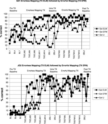
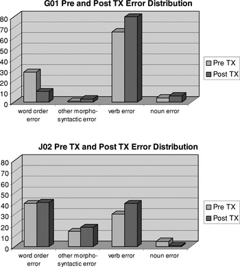
Outcomes of the intervention are depicted in Figures 3 and 4. Significant change from baseline performance for the treated set was determined using the C-statistic (Tryon, 1982). Significant change on the untreated set (set U) was interpreted as a positive generalization effect. Functional generalization to narrative discourse was evaluated comparing performance on repeated story-retelling samples pre- and posttreatment. An examination of the treatment effects for G01 (Figure 3) demonstrates that a significant improvement occurred for treated sentences (Phase 1: z = 3.45; Phase 2: z =4.03, p < .01). Separate review of active versus passive sentence production (see Table 2) revealed an average gain of 56% for treated active sentences and over 66% for treated passive sentences collapsed over the two phases of treatment. However, G01 did not show significant change on the untreated sentences (Phase 1: z = 1.26; Phase 2: z = .66, p > .05), with performance on untreated actives and passives yielding no appreciable change. An analysis of the types of errors made at the end of treatment suggests that the failure to generalize accurate production to the untreated set of sentences is accounted for by a persistent difficulty with verb retrieval. At the conclusion of treatment, G01 made relatively few errors in word order (see Figure 4), which was the target of treatment. Despite this persistent deficit in verb retrieval, G01 did demonstrate generalization of improved sentence production in narrative discourse, as indicated by increased proportion of well-formed sentences to within normal range of .98, (SD = .05) (Berndt et al., 2000), suggesting that a functional impact of the intervention was achieved (see Table 3).

Time-series graphs of treatment and generalization response for G01 and J02 during the course of the study. BL = Baseline, TXELM = Errorless Mapping Treatment, TXEFM = Errorful Mapping Treatment. Numbers indicate the session.

Change in error distribution for G01 and J02 sentence probes pre- and posttreatment (TX). A ratio of the frequency of occurrence of each error type over the total number of errors made was used to calculate the proportion of each error relative to the total sample.
Mean percent correct of baselines for active and passive sentence production pre- and post-Errorless Mapping Therapy and pre- and post-Errorful Mapping Therapy

Linguistic analyses of narrative discourse (story retelling) prior to and upon completion of mapping for G01 and J02

Functional magnetic resonance imaging
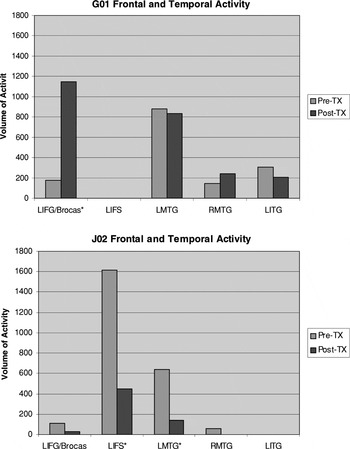
Volumes of significant activity in various regions are graphed in Figure 5a. For G01, pretreatment sentence production resulted in a volume of activity (176 μl) in the left inferior frontal gyrus including Broca's area along the diagonal sulcus and within pars opercularis (Brodmann's Area 44; Figure 6, upper left). Additionally, a large volume of activity was found in the left posterior middle temporal gyrus, and a moderately large volume was found in the left inferior temporal gyrus and the right middle temporal gyrus (Figure 5a). Following treatment, G01 exhibited more robust activity (1146 μl; p < .01, binomial test) in Broca's area and adjacent left inferior frontal cortex, including pars opercularis (Brodmann's Area 44) along the anterior ascending ramus and extending medially within pars triangularis (Brodmann's Area 45) and pars orbitalis (Brodmann's Area 47) along the anterior horizontal ramus of the Sylvian fissure (Figure 6, upper right). Posttreatment there was a small (nonsignificant) decrease in activity in the left inferior temporal gyrus (pre: 307 μl; post: 205 μl). Activity in the posterior left middle temporal gyrus was also relatively stable in extent from pre- to posttreatment imaging (pre: 879 μl; post: 832 μl). Thus, treatment effects for syntax generation by G01 may be attributed to greater recruitment of Broca's area (see Figure 5a).

Volumes (in microliters) of Activity Pre- and Post-Syntactic Mapping Treatment. Note: * indicates areas of significant change between pre- and posttreatment based on the binomial equation (p < .01). Volumes of significant activity in microliters (R2 ≥ .30 for G01 pre- and posttreatment, R2 ≥ .40 for J02 pre- and posttreatment, volume ≥ 100 microliters) are shown for presyntactic mapping treatment fMRI and postsyntactic mapping treatment fMRI. From pre- to posttreatment scans for G01, activity increased in the left inferior frontal gyrus including Broca's area and remained relatively consistent in inferior temporal regions (a). For J02, activity was predominantly frontally mediated both pre- and posttreatment, with a decrease in activity along the inferior frontal sulcus and left middle temporal gyrus from pre- to posttreatment (b). L = left, R = right, IFG = inferior frontal gyrus, IFS = inferior frontal sulcus, MTG = middle temporal gyrus, ITG = inferior temporal gyrus.

Deconvolved waveforms averaged over voxels within regions of interest in the frontal and temporal lobes. Regions of interest were identified by means of composite images uniting activations observed pre- and posttreatment. For G01, Broca's area and left middle temporal gyrus are depicted. For J02, left inferior frontal sulcus and left middle temporal gyrus are depicted.
A similar picture emerges when the time courses of activations are examined. Figure 7 shows waveforms recovered by deconvolution averaged over voxels in G01's frontal (upper left panel) and temporal (upper right panel) regions of activation. As expected, the MR signal rises during active sentence production (first 6 images) and falls during passive viewing (last 4 images). The rise is smaller pre- than posttreatment frontally but larger pre- than posttreatment temporally, consistent with changes in the extent of activity exceeding threshold, as noted earlier.

Pre- and Post-Mapping Therapy fMRI Images for G01 and J02 during sentence generation. Frontal activity during naming for group comparison. Pretreatment images are shown for G01 and J02 on the left side of the figure and posttreatment images for both patients are shown on the right side of the figure. Images for G01 show changes in activity in Broca's area, red = R2 ≥ .30; yellow = R2 ≥ .35 for pre- and posttreatment. Prior to the mapping treatment, G01 showed some activity in this region (BA 44, 45). Posttreatment, activity in Broca's area increased substantially to include pars opercularis (BA 44) along the anterior ascending ramus and extended medially within pars triangularis (BA 45) and pars orbitalis (BA 47) along the anterior horizontal ramus of the Sylvian fissure. Images for J01 show the left lateral frontal and temporal cortices, red = R2 ≥ .40; yellow = R2 ≥ .45 for pre- and posttreatment. His pattern of activation remained relatively consistent pre- and posttreatment, with a decrease in activity in the left inferior frontal sulcus and left middle temporal gyrus.
Patient J02
Mapping treatment
J02 (Figure 4) demonstrated significant improvement at the .01 level for both treated (ELM, z = 3.39; p < .01) and untreated (z = 3.21; p < .01) sentences. Assessment of Phase 2 intervention could not be made, as there was generalization from the first phase to the sentences for Phase 2, as well as the untreated sentence set. However, visual inspection of the graph suggests this performance was somewhat variable, especially during the second phase of treatment. While J02 demonstrated improvement in untreated sentences, these gains did not appear to impact narrative discourse, as indicated by the lack of improvement in the proportion of words in sentences or well-formed sentences during story retelling (Table 3). The variability in sentence production during treatment, and the lack of generalization to more functional narrative discourse, suggests improvement in the process of sentence generation specific to the treatment task (i.e., active or passive sentence production in response to picture description) rather than to the process of sentence generation per se. This notion is supported by the continued heavy distribution of word-order errors, as well as verb retrieval errors, at the end of treatment (see Figure 4).
Functional magnetic resonance imaging
FMRI findings for J02 were substantially different than for G01. In the two largest areas of activity from J02's pretreatment images, activity from posttreatment images decreased significantly. This decrease in posttreatment activity was especially evident in the inferior frontal sulcus (pre: 1616 μl, post: 449 μl; p < .01, binomial test) (Figure 5b), but also happened in the left middle temporal gyrus (pre: 641 μl, post: 140 μl; p < .01, binomial test). Other areas of activity from pre- or posttreatment images in the left inferior frontal gyrus, right pre-supplemental motor area (pre-SMA), left SMA, or right middle temporal gyrus did not meet the contiguity threshold for interpretation.
The time courses of activations for J02 are shown in the lower panels of Figure 7. The rise is larger pre- than posttreatment both frontally and temporally. Although these reductions in amplitude are superficially what could be expected from the lower SNR in the posttreatment scan, there were other brain regions with the inverse relationship (e.g., right pre-SMA, left SMA), which would be inconsistent with this interpretation. This pattern of decreased activity in major ROIs therefore suggests greater efficiency in utilizing areas posttreatment that were active during pretreatment imaging.
DISCUSSION
In summary, these two patients with chronic nonfluent aphasia and syntactic language deficits prior to rehabilitation underwent the same intervention with the same intensity and methodology. Treatment yielded rather different outcomes, as indicated by both their response to probes during treatment and generalization to nontreatment tasks. G01 did not demonstrate generalization from treated to untreated sentences when both word-finding and syntactic (word order) errors were considered, but error analysis revealed a decrease in thematic role assignment errors on treated active and passive sentences posttreatment. Further, her narrative discourse showed an increase in well-formed sentences, although she continued to make lexical retrieval errors for verbs, which accounts for her lack of generalization to the untreated sentences. On the other hand, J02 showed a rapid improvement on treated and untreated items, but demonstrated variability in accuracy at the end of treatment. Error analysis reveals that he continued to make word order errors and did not show changes in the proportion of well-formed sentences in narrative discourse from pre- to posttreatment. In other words, he did not learn procedures for producing proper thematic role assignment during treatment.
The differences in response to treatment for G01 and J02 were paralleled by the differences in their functional neuroimaging results. Pre- versus postmapping treatment changes in neural activity during sentence generation showed different patterns in these two patients. For G01, rehabilitation led to a significant expansion in the extent of activity in Broca's area both medially and anteriorly along the inferior frontal gyrus. In posterior regions, G01 demonstrated a slight reduction of activation in inferior and lateral temporal cortices from pre- to posttreatment images, but continued to recruit regions responsible for the semantic processing of items (Ungerleider & Haxby, 1994; Foundas et al., 1998; Chao et al., 1999; Ishai et al., 1999) within a sentence, even with improvement in thematic mapping ability. On the other hand, J02 demonstrated little change in the location of activity from pre- to posttreatment except for a significant reduction in the volume of activity for the left inferior frontal sulcus from pre- to posttreatment scans. Recent findings from our laboratory indicate that healthy adults activate a network of structures including, but not limited to left Broca's area, inferior frontal sulcus, angular gyrus, superior temporal gyrus, and fusiform gyrus during passive-voice sentence generation. Both of our patients activated only partial syntactic networks compared to healthy adults.
Differences in functional activity reflecting neuroplasticity demonstrated by G01 and J02 shed light on the mechanisms underlying language rehabilitation. The patients' behavioral performance at the end of treatment informs interpretation of functional activity. For G01, expansion of activity within Broca's area may reflect increased syntactic abilities, given that an increase in the intensity and extent of brain activation in nondemented older adults during learning tasks has been interpreted as reflecting a compensatory mechanism wherein individuals utilize additional cognitive resources to bring performance to a normal level (Cabeza et al., 2002; Reuter-Lorenz, 2002). If this is the case, then G01's ability in thematic role assignment may have been supported by the relative preservation and reengagement of Broca's area, since Broca's area plays a role in connecting noun phrases to distinct grammatical positions that determine their thematic roles (Zurif et al., 1993; Swinney et al., 1996; Grodzinsky, 2000). Thus, remobilization of this region is consistent with G01's improvement in syntactic production of treated sentences, and may have contributed to her functional improvement in less constrained sentence production. Generalization of syntax processing posttreatment is consistent with previous studies of syntax treatment (Thompson et al., 1997). It also should be noted that treatment did not address word retrieval; therefore, G01's persistent verb retrieval deficit could be expected.
On the other hand, the decreased activity in lateral frontal regions for J02 corresponding to treatment improvement may reflect an alternative form of neuroplasticity. Given that a reduction in activity was found to correspond to practice effects in healthy adults (Petersen et al., 1998), the reduction in activity posttreatment for J02 may reflect increased efficiency in the use of cortical areas that were already involved in sentence generation prior to treatment. Although J02 demonstrated improvement for both treated and untreated sentences following mapping treatment, the variability in his performance at the end of treatment suggests that ultimately he did not learn the process of thematic role assignment necessary to produce well-formed sentences. Instead, his performance suggests that he may have adopted a strategy in which he learned a syntactic template that he applied to treatment items, but this strategy was inadequate for less constrained discourse, accounting for the lack of generalization to narrative production. Overall, for J02, findings suggest that a failure to recruit new brain regions may yield restricted results, with limited generalization outside the template of the treatment paradigm.
That being said, it is unclear what mechanisms are responsible for different responses to treatment and apparent differences in brain reorganization. Factors such as education level, length of time since CVA onset, age of onset, length of immediate poststroke speech and language therapy, and gender cannot be ruled out. Even so, a plausible reason for different neural substrates supporting recovery in these two patients is that their lesion sites and extents differentially impacted neural response to treatment. Although J02's lesion was a smaller, relatively circumscribed lesion affecting primarily the left sensorimotor and premotor cortex, insular region, the superior temporal gyrus extending medially, and the basal ganglia, it is possible that damage to white matter pathways prevented the communication between anterior and posterior regions responsible for syntax processing (Naeser et al., 1989, 1998). In contrast, G01 sustained a larger lesion involving a greater portion of the left perisylvian cortex, and extending more laterally and posteriorly than J02's lesion. Preservation of anterior insular cortex medially and posterior inferior temporal gyrus may have facilitated recovery through a neural network similar to that used in healthy sentence production, with recruitment of Broca's area to participate again in syntactic production (Naeser et al., 1998). Further research is needed to elucidate mechanisms that contribute to or hinder rehabilitation success.
In conclusion, the two patients in this study appeared to benefit from syntactic mapping treatment in different ways. Likewise, their posttreatment fMRI data reveal different patterns of activation and suggest that the neural mechanisms of rehabilitative change can differ between participants. The hypotheses generated from these data can act as the basis for further exploration of the different neural mechanisms of rehabilitative change and their cognitive correlates based on lesion characteristics and treatment response. Knowledge of these mechanisms will enable development of conceptually driven rehabilitation strategies based on physiological principles of recovery to maximize treatment efficacy.
ACKNOWLEDGMENTS
This work was supported by NIDCD Grant #P50-DC03888 and the Department of Veteran Affairs Rehabilitation R&D Center of Excellence Grant #F2182C. We thank Floris Singletary, Diane Kendall, Jennifer Swearengin, and Cathy Clayton for their valuable contributions.
References
REFERENCES
Ackermann, H. &
Riecker, A.
(2004).
The contribution of the insula to motor aspects of speech
production: A review and a hypothesis.
Brain and Language,
89,
320–328.CrossRefGoogle Scholar
Aguirre, G.K.,
Zarahn, D., &
D'Esposito, M.
(1998).
The variability of human BOLD hemodynamic responses.
NeuroImage,
8,
360–369.CrossRefGoogle Scholar
Barch, D.M.,
Sabb, F.W.,
Carter, C.S.,
Braver, T.S.,
Noll, D.C., &
Cohen, J.D.
(1999).
Overt verbal responding during fMRI scanning: Empirical
investigations of problems and potential solutions.
NeuroImage,
10,
642–657.CrossRefGoogle Scholar
Basso, A.,
Gardelli, M.,
Grassi, M.P., &
Mariotti, M.
(1989).
The role of the right hemisphere in recovery from aphasia: Two case
studies.
Cortex,
25,
555–566.CrossRefGoogle Scholar
Belin, P.,
Van Eeckhout, P.,
Zilbovicius, M.,
Remy, P.,
Francois, C.,
Guillaume, S.,
Chain, F.,
Rancurel, G., &
Samson, Y.
(1996).
Recovery from nonfluent aphasia after melodic intonation therapy: A
PET study.
Neurology,
47,
1504–1511.Google Scholar
Berndt, R.S.
(1998).
Sentence processing in aphasia. In
M.T. Sarno (Ed.),
Acquired aphasia
(3rd ed.) (pp.
229–268).
New York:
Academic Press.
Berndt, R.S.,
Wayland, S.,
Rochon, E.,
Saffran, E., &
Schwartz, M.
(2000).
Quantitative production analysis.
Philadelphia, PA:
Psychology Press.
Binder, J.R.,
Frost, J.A.,
Hammeke, T.A.,
Bellgowan, P.S.F.,
Rao, S.M., &
Cox, R.W.
(1999).
Conceptual processing during the conscious resting state: A
functional MRI study.
Journal of Cognitive Neuroscience,
1,
80–93.CrossRefGoogle Scholar
Birn, R.M.,
Bandettini, P.A.,
Cox, R.W., &
Shaker, R.
(1999).
Event-related fMRI of tasks involving brief motion.
Human Brain Mapping,
7,
106–114.3.0.CO;2-O>CrossRefGoogle Scholar
Birn, R.M.,
Saad, Z.S., &
Bandettini, P.A.
(2001).
Spatial heterogeneity of the nonlinear dynamics in the fMRI BOLD
response.
NeuroImage,
14,
817–826.CrossRefGoogle Scholar
Byng, S.
(1988).
Sentence processing deficits: Theory and therapy.
Cognitive Neuropsychology,
5,
629–676.Google Scholar
Byng, S.,
Nickels, L., &
Black, M.
(1994).
Replicating therapy for mapping deficits in agrammatism: Remapping
the deficit?
Aphasiology,
8,
315–341.Google Scholar
Cabeza, R.,
Anderson, N.D.,
Locantore, J.K., &
McIntosh, A.R.
(2002).
Aging gracefully: Compensatory brain activity in high-performing
older adults.
NeuroImage,
17,
1394–1402.CrossRefGoogle Scholar
Cao, Y.,
Vikingstad, E.M.,
George, K.P.,
Johnson, A.F., &
Welch, K.M.A.
(1999).
Cortical language activation in stroke patients recovering from
aphasia with functional MRI.
Stroke,
30,
2331–2340.CrossRefGoogle Scholar
Caplan, D.,
Albert, N.,
Waters, G., &
Olivieri, A.
(2000).
Activation of Broca's area by syntactic processing under
conditions of concurrent articulation.
Human Brain Mapping,
9,
65–71.3.0.CO;2-4>CrossRefGoogle Scholar
Caplan, D.,
Vijayan, S.,
Kuperberg, G.,
West, C.,
Waters, G.,
Greve, D., &
Dale, A.M.
(2001).
Vascular responses to syntactic processing: Event-related fMRI study
of relative clauses.
Human Brain Mapping,
15,
26–38.Google Scholar
Caplan, D. &
Waters, G.S.
(1999).
Verbal working memory and sentence comprehension.
Behavioral and Brain Sciences,
22,
77–94.Google Scholar
Caramazza, A. &
Miceli, G.
(1991).
Selective impairment of thematic role assignment in sentence
processing.
Brain and Language,
41,
402–436.Google Scholar
Carpenter, P.A.,
Just, M.A.,
Keller, T.A.,
Eddy, W.E., &
Thulborn, K.R.
(1999).
Time course of fMRI-activation in language and spatial networks
during sentence comprehension.
NeuroImage,
10,
216–224.CrossRefGoogle Scholar
Chatterjee, A. &
Maher, L.
(2000).
Grammar and agrammatism. In
S. Nadeau,
L.J.G. Rothi, &
B. Crosson (Eds.),
Aphasia and language: Theory to practice (pp.
133–156).
New York:
Guilford Press.
Chatterjee, A.,
Maher, L.M.,
Rothi, L.J.G., &
Heilman, K.M.
(1995).
Asyntactic thematic role assignment: The use of temporal-spatial
strategy.
Brain and Language,
49,
125–139.Google Scholar
Chao, L.,
Haxby, J., &
Martin, A.
(1999).
Attribute-based neural substrates in temporal cortex for perceiving
and knowing about objects.
Nature Neuroscience,
2,
913–919.Google Scholar
Cornelissen, K.,
Laine, M.,
Tarkiainen, A.,
Järvensivu, T.,
Martin, N., &
Salmelin, R.
(2003).
Adult brain plasticity elicited by anomia treatment.
Journal of Cognitive Neuroscience,
15,
444–461.CrossRefGoogle Scholar
Cox, R.W.
(1996).
AFNI: Software for analysis and visualization of functional magnetic
resonance neuroimages.
Computers in Biomedical Research,
29,
162–173.Google Scholar
Crosson, B.,
Moore, A.B.,
Gopinath, K.,
White, K.D.,
Wierenga, C.E.,
Gaiefsky, M.E.,
Fabrizio, K.R.,
Peck, K.K.,
Soltysik, D.,
Milstead, C.,
Briggs, R.W.,
Conway, T.W., &
Rothi, L.J.G.
(2005).
Role of the right and left hemispheres in recovery of function
during treatment of intention in aphasia.
Journal of Cognitive Neuroscience,
17,
392–406.Google Scholar
Dapretto, M. &
Bookheimer, S.Y.
(1999).
Form and content: Dissociating syntax and semantics in sentence
comprehension.
Neuron,
24,
427–432.CrossRefGoogle Scholar
Embick, E.,
Marantz, A.,
Miyashita, Y.,
O'Neil, W., &
Sakai, K.L.
(2000).
A syntactic specialization for Broca's area.
Proceedings of the National Academy of Science,
97,
6150–6154.CrossRefGoogle Scholar
Foundas, A.L.,
Daniels, S.K., &
Vasterling, J.J.
(1998).
Anomia: Case studies with lesion localization.
NeuroCase,
4,
35–43.CrossRefGoogle Scholar
Garrett, M.F.
(1980).
Levels of processing in sentence production. In
B. Butterworth (Ed.),
Language production (pp.
177–221).
New York:
Academic Press.
Garrett, M.F.
(1984).
The organization of processing structure for language production:
Application to aphasic speech. In
D. Caplan,
A.R. Lecours, &
A. Smith (Eds.),
Biological perspectives on language (pp.
172–193).
Cambridge, MA:
MIT Press.
Grodzinsky, Y.
(2000).
The neurology of syntax: Language use without Broca's
area.
Behavioral and Brain Sciences,
23,
1–71.Google Scholar
Heiss, W.,
Karbe, H.,
Weber-Luxenburger, G.,
Herholz, K.,
Kessler, J.,
Pietrzyk, U., &
Pawlik, G.
(1997).
Speech-induced cerebral metabolic activation reflects recovery from
aphasia.
Journal of the Neurological Sciences,
145,
213–217.CrossRefGoogle Scholar
Heiss, W.D.,
Kessler, J.,
Thiel, A.,
Ghaemi, M., &
Karbe, H.
(1999).
Differential capacity of left and right hemispheric areas for
compensation of poststroke aphasia.
Annals of Neurology,
45,
430–438.3.0.CO;2-P>CrossRefGoogle Scholar
Indefrey, P.,
Brown, C.M.,
Hellwig, F.,
Amunts, K.,
Herzog, H.,
Seitz, R.J., &
Hagoort, P.
(2001).
A neural correlate of syntactic encoding during speech
production.
Proceedings of the National Academy of Science,
98,
5933–5936.CrossRefGoogle Scholar
Indefrey, P.,
Hellwig, F.,
Herzog, H.,
Seitz, R.J., &
Hagoort, P.
(2004).
Neural responses to the production and comprehension of syntax in
identical utterances.
Brain and Language,
89,
312–319.Google Scholar
Ishai, A.,
Ungerleider, L.,
Martin, A.,
Schouten, J., &
Haxby, J.
(1999).
Distributed representation of objects in the human ventral visual
pathway.
Proceedings of the National Academy of Science,
96,
9379–9384.CrossRefGoogle Scholar
Jones, E.V.
(1986).
Building the foundations for sentence production in a non-fluent
aphasic.
British Journal of Disorders of Communication,
21,
63–82.CrossRefGoogle Scholar
Just, M.A.,
Carpenter, P.A.,
Keller, T.A.,
Eddy, W.F., &
Thulborn, K.R.
(1996).
Brain activation modulated by sentence comprehension.
Science,
274,
114–116.CrossRefGoogle Scholar
Kan, I.P. &
Thompson-Schill, S.L.
(2004).
Effect of name agreement on prefrontal activity during overt and
covert picture naming.
Cognitive, Affective, & Behavioral Neuroscience,
4,
43–57.Google Scholar
Kaplan, E.,
Goodglass, H., &
Weintraub, S.
(2000).
The Boston Naming Test.
Philadelphia:
Lee & Febiger.
Karbe, H.,
Thiel, A.,
Weber-Luxenburger, G.,
Herholz, K.,
Kessler, J., &
Heiss, W.D.
(1998).
Brain plasticity in postroke aphasia: What is the contribution of
the right hemisphere?
Brain and Language,
64,
215–230.Google Scholar
Kertesz, A.
(1982).
Western Aphasia Battery.
San Antonio, TX:
Psychological Corporation.
King, K.F.,
Foo, T.K.F., &
Crawford, C.R.
(1995).
Optimized gradient waveforms for spiral scanning.
Magnetic Resonance in Medicine,
34,
156–160.CrossRefGoogle Scholar
Kroll, J.F. &
Potter, M.C.
(1984).
Recognizing words, pictures, and concepts: A comparison of lexical,
object, and reality decisions.
Journal of Verbal Learning and Verbal Behavior,
23,
39–66.CrossRefGoogle Scholar
Kucera, H. &
Francis, W.
(1967).
Computational Analysis of Present-Day American English.
Providence:
Brown University Press.
Macovski, A.
(1985).
Volumetric NMR imaging with time-varying gradients.
Magnetic Resonance Imaging in Medicine,
2,
29–40.CrossRefGoogle Scholar
Maher, L.,
Chatterjee, A.,
Rothi, L.J.G., &
Heilman, K.
(1995).
Agrammatic sentence production: The use of a temporal-spatial
strategy.
Brain and Language,
49,
105–124.CrossRefGoogle Scholar
Maher, L.M.,
Singletary, F.,
Swearengin, J.A.,
Moore, A.B.,
Wierenga, C.E.,
Crosson, B.,
Clayton, M.C.,
Butler, L.V.,
Kendall, D., &
Rothi, L.J.G.
(2002).
An errorless learning approach to sentence generation in
aphasia. Program & Abstracts of the Annual
International Neuropsychological Society Meeting,
177.
Musso, M.,
Weiller, C.,
Kiebel, S.,
Muller, S.P.,
Bulau, P., &
Rijntjes, M.
(1999).
Training-induced brain plasticity in aphasia.
Brain,
122,
1781–1790.CrossRefGoogle Scholar
Naeser, M.A.,
Baker, E.H.,
Palumbo, C.L.,
Nicholas, M.,
Alexander, M.P.,
Samaraweera, R.,
Prete, M.N.,
Hodge, S.N., &
Weissman, T.
(1998).
Lesion site patterns in severe, nonverbal aphasia to predict outcome
with a computer-assisted treatment program.
Archives of Neurology,
55,
1438–1448.CrossRefGoogle Scholar
Naeser, M.A.,
Palumbo, A.L.,
Helm-Estabrooks, N.,
Stiassny-Eder, D., &
Albert, M.L.
(1989).
Severe nonfluency in aphasia: Role of the medial subcallosal
fasciculus and other white matter pathways in recovery of spontaneous
speech.
Brain,
112,
1–38.Google Scholar
Newman, A.J.,
Pancheva, R.,
Ozawa, K.,
Neville, H.J., &
Ullman, M.T.
(2001).
An event-related fMRI study of syntactic and semantic
violations.
Journal of Psycholinguistic Research,
30,
339–364.CrossRefGoogle Scholar
Ni, W.,
Constable, R.T.,
Mencl, W.E.,
Pugh, K.R.,
Fulbright, R.K.,
Shaywitz, S.E.,
Shaywitz, B.A.,
Gore, J.C., &
Shankweiler, D.
(2000).
An event-related neuroimaging study distinguishing form and content
in sentence processing.
Journal of Cognitive Neuroscience,
12,
120–133.CrossRefGoogle Scholar
Noll, D.C.,
Cohen, J.D.,
Meyer, C.H., &
Schneider, W.J.
(1995).
Spiral k-space MR imaging of cortical activation.
Magnetic Resonance Imaging,
5,
49–56.CrossRefGoogle Scholar
Obler, L.K. &
Albert, M.L.
Action Naming Test.
Unpublished Test:
Author.
Palmer, E.D.,
Rosen, H.J.,
Ojemann, J.G.,
Buckner, R.L.,
Kelley, W.M., &
Petersen, S.E.
(2001).
An event-related fMRI study of overt and covert word stem
completion.
NeuroImage,
14,
182–193.CrossRefGoogle Scholar
Peck, K.K.,
Moore, A.B.,
Crosson, B.,
Gaiefsky, M.,
Gopinath, K.S.,
White, K., &
Briggs, R.W.
(2004a).
Pre and post fMRI of an aphasia therapy: Shifts in hemodynamic time
to peak during an overt language task.
Stroke,
35,
554–559.Google Scholar
Peck, K.K.,
Wierenga, C.E.,
Moore, A.B.,
Maher, L.,
Gopinath, K.,
Gaiefsky, M.,
Briggs, R.W., &
Crosson, B.
(2004b).
Comparison of baseline conditions to investigate syntactic
production using functional magnetic resonance imaging.
NeuroImage,
23,
104–110.Google Scholar
Perani, D.,
Cappa, S.F.,
Tettamanti, M.,
Rosa, M.,
Scifo, P.,
Miozzo, A.,
Basso, A., &
Fazio, F.
(2003).
An fMRI study of word retrieval in aphasia.
Brain and Language,
85,
357–368.CrossRefGoogle Scholar
Petersen, S.E.,
van Mier, H.,
Fiez, J.A., &
Raichle, M.E.
(1998).
The effects of practice on the functional anatomy of task
performance.
Proceedings of the National Academy of Science, USA,
95,
853–860.CrossRefGoogle Scholar
Reuter-Lorenz, P.
(2002).
New visions of the aging mind and brain.
Trends in Cognitive Science,
6,
394.CrossRefGoogle Scholar
Rosen, H.J.,
Petersen, S.E.,
Linenweber, M.R.,
Snyder, A.Z.,
White, D.A.,
Chapman, L.,
Dromerick, A.W.,
Fiez, J.A., &
Corbetta, M.D.
(2000).
Neural correlates of recovery from aphasia after damage to left
inferior frontal cortex.
Neurology,
55,
1883–1894.Google Scholar
Schwartz, M.F.,
Saffron, E.M.,
Bloch, D.E., &
Dell, G.S.
(1994a).
Disordered speech production in aphasic and normal speakers.
Brain and Language,
47,
52–88.Google Scholar
Schwartz, M.F.,
Saffron, E.M.,
Fink, R., &
Myers, J.L.
(1994b).
Mapping therapy: A treatment programme for agrammatism.
Aphasiology,
8,
19–54.Google Scholar
Schwartz, M.F.,
Saffron, E.M., &
Marin, O.S.M.
(1980).
The word order problem in agrammatism. I. Comprehension.
Brain and Language,
10,
249–262.CrossRefGoogle Scholar
Stowe, L.A.,
Broere, C.A.J.,
Paans, A.M.,
Wijers, A.A.,
Mulder, B.,
Vaalburg, W., &
Zwarts, F.
(1998).
Localizing components of a complex task: Sentence processing and
working memory.
Neuroreport,
9,
2995–2999.Google Scholar
Swinney, D.,
Zurif, E.,
Prahter, P., &
Love, T.
(1996).
Neurological distribution of processing resources underlying
language comprehension.
Journal of Cognitive Neuroscience,
8,
174–184.CrossRefGoogle Scholar
Talairach, J. &
Tournoux, P.
(1988).
Co-Planar stereotaxic atlas of the human brain.
New York:
Thiem Medical Publishers.
Thompson, C.K.,
Shapiro, L.P.,
Ballard, K.J.,
Jacobs, B.J.,
Schneider, S.S., &
Tait, M.E.
(1997).
Training and generalized production of wh- and NP-movement structure
in agrammatic aphasia.
Journal of Speech, Language, and Hearing Research,
40,
228–244.CrossRefGoogle Scholar
Tryon, W.W.
(1982).
A simplified time-series analysis for evaluating treatment
interventions.
Journal of Applied Behavior Analysis,
15,
423–429.CrossRefGoogle Scholar
Ungerleider, L.G. &
Haxby, J.V.
(1994).
‘What’ and ‘where’ in the human
brain.
Current Opinion in Neurobiology,
4,
157–165.CrossRefGoogle Scholar
Ward, D.
(1998).
Deconvolution of FMRI time series data. In
R. Cox (Ed.),
MCW AFNI user manual (pp.
1–24).
Unpublished manuscript.
Weisskoff, R.M.
(1996).
Simpled measurement of scanner stability for functional NMR imaging
of activation in the brain.
Magnetic Resonance in Medicine,
36,
643–645.CrossRefGoogle Scholar
Wolery, M.,
Holcombe, A.,
Cybriwsky, C.,
Doyle, P.M.,
Schuester, J.W.,
Ault, J., &
Gast, D.L.
(1992).
Constant time delay with discrete responses: A review of
effectiveness and demographic, procedural and methodological
parameters.
Research and Developmental Disabilities,
13,
239–266.CrossRefGoogle Scholar
Zarahn, E.,
Aguirre, G.K., &
D'Esposito, M.
(1997).
Empirical analysis of BOLD fMRI statistics: I. Spatially unsmoothed
data collected under null-hypothesis conditions.
NeuroImage,
5,
179–197.Google Scholar
Zurif, E.,
Swinney, D.,
Prather, P.,
Solomon, J., &
Bushell, C.
(1993).
An on-line analysis of syntactic processing in Broca's and
Wernicke's aphasia.
Brain and Language,
45,
448–464.CrossRefGoogle Scholar

Pretreatment language assessment

T1-weighted axial magnetic resonance images in radiologic space (left is on the right) showing the extent of left cerebral vascular infarction for G01 and J02.
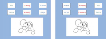
Illustration of color and spatially coded mapping template used to train active and passive sentences in both phases (Errorless Mapping Treatment, Errorful Mapping Treatment) of the intervention.
Time-series graphs of treatment and generalization response for G01 and J02 during the course of the study. BL = Baseline, TXELM = Errorless Mapping Treatment, TXEFM = Errorful Mapping Treatment. Numbers indicate the session.
Change in error distribution for G01 and J02 sentence probes pre- and posttreatment (TX). A ratio of the frequency of occurrence of each error type over the total number of errors made was used to calculate the proportion of each error relative to the total sample.

Mean percent correct of baselines for active and passive sentence production pre- and post-Errorless Mapping Therapy and pre- and post-Errorful Mapping Therapy

Linguistic analyses of narrative discourse (story retelling) prior to and upon completion of mapping for G01 and J02
Volumes (in microliters) of Activity Pre- and Post-Syntactic Mapping Treatment. Note: * indicates areas of significant change between pre- and posttreatment based on the binomial equation (p < .01). Volumes of significant activity in microliters (R2 ≥ .30 for G01 pre- and posttreatment, R2 ≥ .40 for J02 pre- and posttreatment, volume ≥ 100 microliters) are shown for presyntactic mapping treatment fMRI and postsyntactic mapping treatment fMRI. From pre- to posttreatment scans for G01, activity increased in the left inferior frontal gyrus including Broca's area and remained relatively consistent in inferior temporal regions (a). For J02, activity was predominantly frontally mediated both pre- and posttreatment, with a decrease in activity along the inferior frontal sulcus and left middle temporal gyrus from pre- to posttreatment (b). L = left, R = right, IFG = inferior frontal gyrus, IFS = inferior frontal sulcus, MTG = middle temporal gyrus, ITG = inferior temporal gyrus.
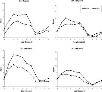
Deconvolved waveforms averaged over voxels within regions of interest in the frontal and temporal lobes. Regions of interest were identified by means of composite images uniting activations observed pre- and posttreatment. For G01, Broca's area and left middle temporal gyrus are depicted. For J02, left inferior frontal sulcus and left middle temporal gyrus are depicted.
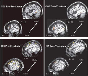
Pre- and Post-Mapping Therapy fMRI Images for G01 and J02 during sentence generation. Frontal activity during naming for group comparison. Pretreatment images are shown for G01 and J02 on the left side of the figure and posttreatment images for both patients are shown on the right side of the figure. Images for G01 show changes in activity in Broca's area, red = R2 ≥ .30; yellow = R2 ≥ .35 for pre- and posttreatment. Prior to the mapping treatment, G01 showed some activity in this region (BA 44, 45). Posttreatment, activity in Broca's area increased substantially to include pars opercularis (BA 44) along the anterior ascending ramus and extended medially within pars triangularis (BA 45) and pars orbitalis (BA 47) along the anterior horizontal ramus of the Sylvian fissure. Images for J01 show the left lateral frontal and temporal cortices, red = R2 ≥ .40; yellow = R2 ≥ .45 for pre- and posttreatment. His pattern of activation remained relatively consistent pre- and posttreatment, with a decrease in activity in the left inferior frontal sulcus and left middle temporal gyrus.
 You have
Access
- 32
- Cited by