Introduction
Integrative taxonomy has been proposed as a powerful approach for species delimitation, combining multiple lines of evidence – such as morphology, molecular data, ecology, and biogeography (Dayrat Reference Dayrat2005). This approach overcomes the limitations inherent in traditional taxonomy, which often relies on a single data source and may neglect, for example, cryptic diversity (Yeates et al. Reference Yeates, Seago, Nelson, Cameron, Joseph and Trueman2011). Although traditional procedures remain useful in many cases, integrative taxonomy provides taxonomists with a robust set of tools to inventory the planet’s biodiversity, which remains regrettably underestimated (Padial et al. Reference Padial, Miralles, De la Riva and Vences2010). One consequence of this trend is the revolution in research on parasitic lineages, in which the combined use of molecular techniques and morphological data has provided more robust insights into their diversity and evolutionary relationships (Osaki-Pereira et al. Reference Osaki-Pereira, Yamada, Ebert and Da Silva2025).
Molecular findings have also led to taxonomic revisions, including the suppression of the traditional class Monogenea, and the elevation of the subclasses Monopisthocotylea and Polyopisthocotylea to class level (Monopisthocotyla and Polyopisthocotyla, respectively) (Brabec et al. Reference Brabec, Salomaki, Kolísko, Scholz and Kuchta2023). Monopisthocotyls are the most diverse helminth parasites of freshwater fish and one of the best-studied taxa in the Neotropical region (Cohen et al. Reference Cohen, Justo and Kohn2013; Luque et al. Reference Luque, Pereira, Alves, Oliva and Timi2017). In South America alone, approximately 650 species have been identified, with the family Dactylogyridae Bychowsky, 1933 being the richest in species diversity (approximately 476 species described to date) (De Aguiar et al. Reference De Aguiar, Mathews, Domingues and Da Silva2025), reported to parasitise various taxa of teleost fish hosts (Kmentová et al. Reference Kmentová, Cruz-Laufer, Pariselle, Smeets, Artois and Vanhove2022). However, despite the great diversity of dactylogyrid parasites of fish in South America, De Aguiar et al. Reference De Aguiar, Mathews, Domingues and Da Silva2025. suggest that fewer than 5% of the species in this family have been formally described.
Dactylogyridae constitutes a monophyletic group formed by nine main lineages, namely Anacanthorinae, Ancylodiscoidinae, Ancyrocephalinae, Dactylogyrinae, Hareocephalinae, Heterotesiinae, Linguadactylinae, Linguadactyloidinae, and Pseudodactylogyrinae (Kritsky and Boeger Reference Kritsky and Boeger1989). However, more recent molecular phylogenetic studies have led to a revision of this classification, resulting in the synonymisation of Heteronchocleidinae and Pseudodactylogyrinae with Dactylogyrinae, and of Anacanthorinae and Ancylodiscoidinae with Ancyrocephalinae (Kmentová et al. Reference Kmentová, Cruz-Laufer, Pariselle, Smeets, Artois and Vanhove2022). Within Ancyrocephalinae, the genus Anacanthorus stands out for presenting a great diversity of species, distributed exclusively among Characiformes fish species of the Neotropical region (Cohen et al. Reference Cohen, Justo and Kohn2013), being represented by 98 valid species, of which 46 parasitise fish of the family Serrasalmidae (including piranhas, pacus, and their relatives), the most common host group of Anacanthorus spp. (Moreira et al. Reference Moreira, Da Silva Carneiro, Ruz and Luque2019a; Morey et al. Reference Morey, Sarmiento, Chu, Cruces and Chero2024, Reference Morey, Sarmiento, Isern and Aguinaga2025a, Reference Morey, Pinedo, Oliveira, Vieira, Chu, De Sousa and Viana2025b; Silva et al. Reference Silva, Cohen, Santos-Clapp, Brasil-Sato, Costa and Justo2024; Yamada et al. Reference Yamada, Diniz, De Sousa, Yamada and Tavares-Dias2024) (see Table 1).
List of species of Anacanthorus parasites of Serrasalmidae from the Neotropical region described to date

Table 1. Long description
The table contains 40 rows of data under six headers.* Column 1: Monopisthocotyla species (e.g., Ameloblastella chavarriai, Anacanthorus species, Boegeriella conica).* Column 2: Host species (e.g., Rhamdia quelen, Serrasalmus maculatus, Pygocentrus nattereri).* Column 3: Host family (e.g., Heptapteridae, Serrasalmidae, Pimelodidae).* Column 4: Locality (primarily Brazil, with some entries for Mexico, Peru, and China).* Column 5: GenBank 28S r D N A (alphanumeric accession codes like KP056251, MH843721).* Column 6: Reference (citations including Mendoza-Palmero et al. 2015, Moreira et al. 2019b, and Zago et al. 2021).A key entry in bold indicates a new species from the present study: Anacanthorus penedensis n. sp., hosted by Pygocentrus nattereri in Brazil, with GenBank accessions Q B 068625 through Q B 068628. The final row lists Murraytrema pricei from China as an outgroup.
* The first column lists species such as Anacanthorus amazonicus, A. anacanthorus, A. brandtii, A. camposbacae, and A. penedensis n. sp.
* The second column identifies host species including Serrasalmus rhombeus, Pygocentrus nattereri, Myloplus schomburgkii, and a hybrid of Piaractus mesopotamicus times Piaractus brachypomus.
* The third column indicates the country of origin, primarily Brazil, with several entries for Peru (A. camposbacae, A. carmenrosae, A. cultro, A. santaclarae).
* The fourth column provides citations ranging from Mizelle and Price (1965) to Morey et al. (2025b).
The final row is highlighted in bold, identifying the new species Anacanthorus penedensis n. sp. hosted by Pygocentrus nattereri in Brazil, documented in the present study.
The new species is shown in bold.
Serrasalmidae is a diverse family of freshwater Characiformes fish widely distributed throughout the river basins of South America (Silva et al. Reference Silva, Cohen, Santos-Clapp, Brasil-Sato, Costa and Justo2024). Among these, the genus Pygocentrus Müller and Troschel, 1844 has three valid species: Pygocentrus cariba (Humboldt, 1821), Pygocentrus piraya (Cuvier, 1819), and Pygocentrus nattereri Kner, 1858 (Raick et al. Reference Raick, Huby, Kurchevski, Godinho and Parmentier2020). To date, nine genera of monopisthocotyls have been reported parasitising P. nattereri, among which Anacanthorus is the most species-rich. The following species of Anacanthorus have been described from this host: Anacanthorus anacanthorus Mizelle and Price, 1965; Anacanthorus brazilensis Mizelle and Price, 1965; Anacanthorus maltai Boeger and Kritsky, 1988; Anacanthorus neotropicalis Mizelle and Price, 1965; Anacanthorus reginae Boeger and Kritsky, 1988; Anacanthorus rondonensis Boeger and Kritsky, 1988; Anacanthorus stachophallus Kritsky, Boeger, and Van Every, 1992; Anacanthorus thatcheri Boeger and Kritsky, 1988 (Boeger and Kritsky Reference Boeger and Kritsky1988; Kritsky et al. Reference Kritsky, Boeger and Van Every1992; Mizelle and Price Reference Mizelle and Price1965).
Molecular markers, such as large subunit ribosomal DNA (LSU rDNA), have been widely used to identify species and reconstruct the phylogenies of monopisthocotyls (Jorge et al. Reference Jorge, Ebert and Da Silva2025; Santos-Neto et al. Reference Santos-Neto, Paixão and Domingues2024; Soares et al. Reference Soares, Adriano, Luque, Balbuena, Domingues and Maia2025; Vale et al. Reference Vale, Soares, Dalmas-Branchi, Sales, Adriano and Domingues2026; Yamada et al. Reference Yamada, De Sousa, Ebert, Diniz, Tavares-Dias, Da Silva and Yamada2025). Although recent studies have used molecular markers to perform phylogenetic and cophylogenetic analyses of Anacanthorus spp. (Cabrera-Soregui et al. Reference Cabrera-Soregui, Torres-Lozano, Garcia-Candela, Mesias-Valle, Martinez-Rojas, Cruz-Neyra and Cala-Delgado2024; Da Graça et al. Reference Da Graça, Fabrin, Gasques, Prioli, Balbuena, Prioli and Takemoto2018; Moreira et al. Reference Moreira, Da Silva Carneiro, Ruz and Luque2019a, Reference Moreira, Luque and Šimková2019b; Müller et al. Reference Müller, Ceccarelli and Ueta2016), many gaps remain in our knowledge, since most descriptions are based on morphological data and do not include the molecular characterisation of the species (Leão et al. Reference Leão, São Clemente and Cohen2015; Morey et al. Reference Morey, Aliano and Grandez2019, Reference Morey, Sol and Cachique2021, Reference Morey, Sarmiento, Chu, Cruces and Chero2024, Reference Morey, Sarmiento, Isern and Aguinaga2025a, Reference Morey, Pinedo, Oliveira, Vieira, Chu, De Sousa and Viana2025b; Pereira et al. Reference Pereira, Mota, Paiva and Tavares2020; Santos-Neto et al. Reference Santos-Neto, Muriel-Cunha and Domingues2019; Silva et al. Reference Silva, Cohen, Santos-Clapp, Brasil-Sato, Costa and Justo2024; Yamada et al. Reference Yamada, Diniz, De Sousa, Yamada and Tavares-Dias2024).
Considering the great diversity of species, host specificity, and the scarcity of phylogenetic studies, efforts are needed to integrate molecular markers and morphological data to provide additional evidence for species delimitation, as well as to achieve a comprehensive and stable phylogeny for Anacanthorus species and related dactylogyrids. To address this gap, we conducted an inventory of monopisthocotyl parasites of serrasalmid fish from the Lower São Francisco River, northeastern Brazil, focusing on an integrative taxonomic approach. As part of this ongoing inventory, a new species of Anacanthorus was described parasitising the gills of P. nattereri, supported by morphological characters and partial 28S rDNA sequences.
Materials and methods
Host collection
Twenty-nine specimens of P. nattereri (20, 8–21 cm in standard length and 171, 42–614 g in weight) were collected by local fishermen using gill nets between February 2023 and November 2024 in the Lower São Francisco River (10°16′52.1″ S, 36°35′31.6″ W), municipality of Penedo, state of Alagoas, northeastern Brazil. The collected fish were individualised in plastic bags and gradually cooled in thermal boxes containing ice and sent to the Parasitology Laboratory of the Institute of Biological and Health Sciences of the Federal University of Alagoas (UFAL), Maceió, state of Alagoas.
Collection and preparation of parasites
During necropsy, gill arches were removed and placed in vials containing tap water. Each vial was shaken vigorously and then the contents of each vial were passed through a 53-micrometre mesh sieve. The contents retained on the sieve, including the parasites, were transferred to Petri dishes and examined under a stereomicroscope. Monopisthocotyl specimens were removed from the gills or sediments with fine needles and micropipettes, and immediately processed for morphological and molecular analyses. To study sclerotised structures, some specimens were mounted on permanent slides in Gray and Wess’s medium (12 specimens) (Kritsky et al. Reference Kritsky, Thatcher and Boeger1986). Other specimens were stained with Gomori’s trichrome and mounted in Canada balsam to visualise the internal soft structures (three specimens) (Kritsky et al. Reference Kritsky, Leiby and Kayton1978). Four specimens were mounted on slides with a drop of water, covered with coverslips, and examined under a microscope for morphological identification. These specimens were then fixed in absolute ethanol for subsequent molecular analysis. The specimens were registered in SISGEN (Sistema Nacional de Gestão do Patrimônio Genético e do Conhecimento Tradicional Associado) under the registration number (XXXX).
Morphological and morphometric analyses were carried out using a Biofocus BLUE1600BA-L-BAT computerized image analysis system and a Leica ICC50W microscope equipped with differential interference contrast (DIC). The illustrations were made using a drawing tube attached to a Nikon E200 microscope with phase-contrast optics and vectorised using Inkscape 1.4 software. Measurements, all in micrometres (μm), represent straight-line distances between extreme points and are expressed as the mean followed by the range and the number (n) of structures measured in parentheses; body length includes the haptor. The specific terminology of the genus Anacanthorus was based on Mizelle and Price (Reference Mizelle and Price1965) and Kritsky et al. (Reference Kritsky, Thatcher and Kayton1979); the numbering and distribution of hook pairs follow the method recommended by Mizelle and Price (Reference Mizelle and Price1963). The definitions of prevalence, mean intensity, and mean abundance were calculated according to Bush et al. (Reference Bush, Lafferty, Lotz and Shostak1997).
Type specimens of monopisthocotyls were deposited in the Collection of the Oswaldo Cruz Institute (CHIOC) (i.e., holotype CHIOC (XXXXX) and paratypes CHIOC (XXXXX)), municipality of Rio de Janeiro, state of Rio de Janeiro, Brazil.
DNA extraction, amplification, and sequencing
Genomic DNA was extracted from four specimens of Anacanthorus penedensis n. sp. using the QIAamp DNA Mini Kit (Qiagen), following the manufacturer’s instructions. A partial fragment of the 28S rDNA gene (large ribosomal subunit) was amplified by polymerase chain reaction (PCR) using the primer pair C1 and D2 (Mollaret et al. Reference Mollaret, Jamieson, Adlard, Hugall, Lecointre, Chombard and Justine1997). PCR reactions were performed using the Promega PCR Master Mix under the conditions described by Ogawa et al. (Reference Ogawa, Mizuochi, Yamaguchi, Shirakashi, Asai and Agawa2021). The resulting amplicons were purified with the QIAquick PCR Purification Kit (Qiagen). Sequencing was carried out using the BigDye Terminator v3.1 Cycle Sequencing Kit on an ABI 3730 DNA Analyzer at the RPT01A Sequencing Platform (Instituto Oswaldo Cruz (IOC), Rio de Janeiro, Brazil). Consensus sequences were assembled and edited in Geneious R9.1 (Kearse et al. Reference Kearse, Moir, Wilson, Stones-Havas, Cheung, Sturrock and Drummond2012) and deposited in GenBank under accession numbers QB068625 – QB068628.
Phylogenetic inferences
Sequence alignment and selection of the most appropriate nucleotide substitution model were performed in MEGA 11 (Tamura et al. Reference Tamura, Stecher and Kumar2021). The ribosomal 28S rDNA gene sequences used for the ingroup, as well as the outgroup, were chosen based on closely related families and genera previously published and obtained from GenBank (Table 2). Maximum likelihood (ML) and Bayesian inference (BI) analyses were conducted in MEGA 11 (Tamura et al. Reference Tamura, Stecher and Kumar2021) and Geneious R9.1 (Kearse et al. Reference Kearse, Moir, Wilson, Stones-Havas, Cheung, Sturrock and Drummond2012), respectively. The evolutionary model used was general time-reversible, with gamma-distributed rates and invariant sites (GTR + G + I), calculated using the ‘Find Best DNA/Protein Model’ tool through the MEGA software. Nodal support was assessed using 1,000 bootstrap replicates for ML analyses and posterior probabilities for BI analyses. MEGA 11 software was also used to estimate pairwise genetic distances among Anacanthorus specimen sequences.
List of monopisthocotyls included in the phylogenetic analyses

Table 2. Long description
The table contains 41 rows and 6 columns. The columns are labeled: Monopisthocotyla species, Host species, Host family, Locality, GenBank (specifically 28S r D N A), and Reference.
Key entries include:
* Anacanthorus penedensis n. sp. (bolded) found on Pygocentrus nattereri (Serrasalmidae) in Brazil, listed as ‘Present study’.
* Multiple Anacanthorus species (amazonicus, jegui, lepyrophallus, maltai, paraxaniophallus, penilabiatus, rondonensis, thatcheri) primarily hosted by Serrasalmidae in Brazil and Peru.
* Other genera listed include Ameloblastella, Aphanoblastella, Boegeriella, Cacatuocotyle, Characithecium, Cosmetocleithrum, Demidospermus, Diaphorocleidus, Heteropriapulus, Jainus, Nanayella, Mymarothecium, Rhinoxenus, Tereancistrum, Trinigyrus, Unibarra, Unilatus, Urocleidoides, and Vancleaveus.
* Host families represented include Heptapteridae, Serrasalmidae, Pimelodidae, Acestrorhamphidae, Characidae, Doradidae, Loricariidae, Anostomidae, Prochilodontidae, Parodontidae, and Gymnotidae.
* Localities are predominantly Brazil, with several from Peru and Mexico.
* The final row lists Murraytrema pricei from the family Diplectanidae, hosted by Nibea albiflora in China, marked as an outgroup.
New sequences obtained in this study are shown in bold.
a indicates sequence used as outgroup.
Results
Taxonomic summary
Class Monopisthocotyla Brabec, Salomaki, Kolísko, Scholz, and Kuchta, 2023
Order Dactylogyridea Bychowsky, 1937
Family Dactylogyridae Bychowsky, 1933
Subfamily Ancyrocephalinae Bychowsky, 1937
Anacanthorus Mizelle and Price, 1965
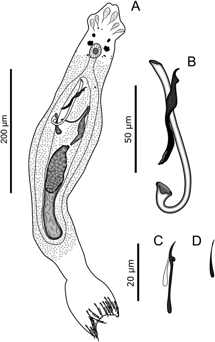
Anacanthorus penedensis n. sp. (Figures 1 and 2)
Anacanthorus penedensis n. sp. parasite of the gills of Pygocentrus nattereri Kner, 1858 (Characiformes: Serrasalmidae), in the Lower São Francisco River, municipality of Penedo, state of Alagoas, Brazil. (a) Whole mount (composite, ventral view); (b) Male copulatory organ (MCO), accessory piece, ventral view; (c) Hooks (d) 4A hooks. Scale bars: (a) = 200 μm; (b–d) = 50 μm.

Figure 1. Long description
Panel A shows a whole mount composite in ventral view of the elongated parasite. It features a cephalic region with four lobes, two pairs of eyespots, a circular pharynx, and internal organs including a granular ovary and testis. The posterior ends in a haptor with small hooks. A vertical scale bar to the left indicates 200 micrometers. Panel B illustrates the M C O and accessory piece, showing a curved tubular structure intertwined with a darker, solid accessory piece. A scale bar to the left indicates 50 micrometers. Panel C depicts hook pairs 1, 5, and 6, which have a long straight shaft and a small curved point. Panel D shows hooks 2, 3, 4, and 7, which are smaller and simpler in structure. Panel E displays the 4 A hooks, which are the smallest and most slender. Panels B through E share a common scale bar of 50 micrometers.
Photomicrographs of Anacanthorus penedensis n. sp. from the gills of Pygocentrus nattereri Kner, 1858 (Characiformes: Serrasalmidae), in the Lower São Francisco River, municipality of Penedo, state of Alagoas, Brazil. (a) Male copulatory organ (MCO), accessory piece (ventral view). (b) Hooks. Scale bars: (a) = 50 μm; (b) = 20 μm.

Type host: Pygocentrus nattereri Kner, 1858 (Characiformes: Serrasalmidae).
Type locality: Lower São Francisco River (10°16′52.1″ S, 36°35′31.6″ W), municipality of Penedo, state of Alagoas, Brazil.
Site of infestation: Gill filaments.
Infestation parameters: prevalence: 48.3% (14 of 29 analysed hosts); total number of parasites: 121; mean intensity and standard error: 8.6 ± 2.7; range of intensity: 1–34.
Specimens deposited: Holotype CHIOC (XXX); Paratypes CHIOC (XXX); Vouchers CHIBB (XXX).
Molecular sequence data: 28S rDNA (818 base pairs (bp) long, GenBank accession number QB068625; 798 bp long, GenBank accession number QB068626; 825 bp long, GenBank accession number QB068627; 805 bp long, GenBank accession number QB068628).
ZooBank registration: urn:lsid:zoobank.org:act:0A1DA352-662B-4457-B469-D7FC923328DF
Etymology: The specific name refers to the municipality of Penedo, Alagoas, Brazil, the type locality of the new species.
Description: (Based on 15 specimens: three stained with Gomori’s trichrome and 12 mounted in Gray and Wess’s medium). Body elongated, fusiform, 606 (531–730; n = 12) long including the haptor, 99 (64–128; n = 14) wide at the height of germarium. Two terminal and two bilateral well-developed cephalic lobes; three bilateral pairs of head organs. Eyes present, with the posterior pair bigger than the anterior one. Accessory granules present in the cephalic area. Pharynx subspherical 22 (18–40; n = 11) long, 25 (18–27; n = 11) wide. Copulatory complex comprising male copulatory organ (MCO) and accessory piece. MCO 92 (83–98; n = 15) long, J-shaped, elongated and slightly curved shaft, gradually tapering in the distal region; moderately wide and gently rounded base, forming a slightly curved proximal region. Accessory piece 44 (41–46; n = 15) long, with hooked acute tip, non-articulated to MCO base; submedial expansion extended to distal region, conspicuous laminar subterminal expansion originating at one of the margins of the shaft. Gonads slightly overlapping; germarium fusiform, 74 (72–84; n = 3) long, 34 (27–47; n = 3) wide; testis elongate, 71 (62–78; n = 3) long, 21 (12–25; n = 3) wide, dorsal to germarium. Prostatic reservoir piriform, seminal vesicle as an elongated sac. Oviduct, ootype, uterus, eggs, and seminal receptacle not observed. Peduncle elongated. Haptor subquadrangular 105 (85–109; n = 9) long, 57 (47–66; n = 12) wide. Seven pairs of hooks, similar in shape; each with truncated and slightly depressed thumb, shank expanded proximally. Pairs 1, 5, 6, 24 (22–26; n = 14) long, proximal expansion about 0.6 shank length. Pairs 2, 3, 4, 7, 28 (24–31; n = 14) long, proximal expansion about 0.4 shank length. Filamentous hook (FH) loop ½ shank length. Similar 4A hooks, 9 (7–12; n = 12) long. Vitelline follicles dense, extending from the level posterior to the pharynx to the peduncle, absent in the regions of the reproductive organs.
Remarks: Anacanthorus penedensis n. sp. can be easily distinguished from all congeners described from serrasalmid hosts by a unique combination of characters of the accessory piece and by having a markedly longer MCO. Anacanthorus penedensis n. sp. most closely resembles Anacanthorus jegui Van Every and Kritsky, Reference Van Every and Kritsky1992 in the general morphology of the accessory piece and hooks. However, it differs from this species in having two clearly distinct expansions of the accessory piece: a submedial expansion extending to the distal region and a conspicuous subterminal laminar expansion originating from one margin, whereas in A. jegui, the subterminal expansion originates from both margins of the accessory piece. Moreover, it differs from this species in having a simple base of the MCO (short basal flap in A. jegui).
The new species also resembles A. reginae and Anacanthorus simpliciphallus Silva, Cohen, Costa, and Justo, 2024 in the morphology of the MCO, particularly in having a simple base. However, it can be distinguished from these species by the configuration of the hooks. In Anacanthorus penedensis n. sp., the hooks have a truncated and slightly depressed thumb, and a shank expanded proximally, whereas in A. reginae, the hooks have a short recurved point and a depressed thumb. In A. simpliciphallus, the hooks have a truncated, slightly depressed thumb, a curved shaft, and a short point. It also differs from these species in the morphology of the accessory piece, which bears two distinct expansions in Anacanthorus penedensis n. sp., vs. a variable subterminal expansion in A. reginae and a midlength expansion extending toward the distal region in A. simpliciphallus.
The new species further differs from A. jegui, A. reginae, and A. simpliciphallus by having a markedly longer MCO, with a mean length of 92 (83–98), compared with 48 (45–51) in A. jegui, 67 (57–76) in A. reginae, and 70 (60–78) in A. simpliciphallus. These species also differ in the relative length of the MCO and accessory piece. In Anacanthorus penedensis n. sp., the accessory piece reaches approximately half the total length of the MCO, whereas in A. jegui and A. reginae this structure extends along almost the entire length of the MCO, and in A. simpliciphallus, it is more than half the length of the MCO.
Molecular data and phylogenetic inference
Four partial sequences of the 28S rDNA gene were obtained from four specimens of Anacanthorus penedensis n. sp. The aligned dataset comprised 47 taxa and 894 characters, of which 204 were conserved, 675 were variable, and 580 were parsimony-informative. BI yielded a mean estimated marginal likelihood of −17,044.244, with a median value of −17,043.88. Effective sample size (ESS) values for all parameters averaged 837.6.
ML and BI analyses recovered different topologies, differing mainly in clade arrangement. In particular, ML recovered the family Dactylogyridae as monophyletic, whereas BI resolved it as polyphyletic. The final topology presented (Figure 3) is based on the ML tree.
Phylogenetic relationships among 28S rDNA sequences of Monopisthocotyla, including Anacanthorus penedensis n. sp. (in bold). The final topology presented was based on the topology obtained from the ML analysis. The evolutionary model used was general time-reversible, with gamma-distributed rates and invariant sites (GTR + G + I). The nodal support is described at the left by bootstrap replicates (ML) and at the right by posterior probability (BI). The scale bar indicates the number of substitutions per site. * indicates a distinct clade topology between ML and BI analyses.

Figure 3. Long description
A phylogenetic tree with a scale bar of 0.10 at the bottom left. The tree branches from a single root on the left toward numerous species on the right.At the top, a large clade is highlighted in light blue, labeled in the legend as True piranha hosts parasites. This group includes species such as Anacanthorus jegui, A. paraxaniophallus, A. maltai, A. thatcheri, A. rondonensis, A. amazonicus, A. lepyrophallus, and four entries for Anacanthorus penedensis n. sp. (Q B 0 6 8 6 2 7, Q B 0 6 8 6 2 5, Q B 0 6 8 6 2 8, and Q B 0 6 8 6 2 6).Below the blue section is a single entry for Anacanthorus sp. 1 (M H 8 4 3 7 2 2) highlighted in yellow.Following this is a second large clade highlighted in yellow, labeled in the legend as Pacu hosts parasites. This group contains seven entries for Anacanthorus penilabiatus with accession numbers M H 8 4 3 7 1 9, P Q 0 4 8 8 3 5, P Q 0 4 8 8 3 3, P Q 0 4 8 8 3 0, P Q 0 4 8 8 3 4, P Q 0 4 8 8 3 2, and P Q 0 4 8 8 3 1.The remainder of the tree consists of non-highlighted species including Aphanoblastella travassosi, Cosmetocleithrum bulbocirrus, Demidospermus spirophallus, Nanayella processusclavis, Boergeriella conica, Demidospermus mortenthaleri, Characithecium paranapanemense, Urocleidoides sinus, Urocleidoides digitabulum, Tereancistrum ancistrum, Diaphorocleidus neotropicalis, Diaphorocleidus magnus, Cacatuocotyle papilionis, Rhinoxenus paranaensis, Rhinoxenus euryenus, Urocleidoides uncinus, Heteropriapulus anchoradiatus, Unilatus unilatus, Trinidgyrus peregrinus, Jainus piava, Urocleidoides indianensis, Urocleidoides tenuis, Mymarothecium viatorum, Unibarra paranoplatensis, Ameloblastella chavarriai, Vancleaveus janauacaensis, and Murraytrema pricei at the base.Numerical values representing bootstrap support and posterior probability (e.g., 100 / 1, 99 / 0.89) are positioned at the branching nodes.
At the top, a large blue-shaded clade labeled True piranha hosts parasites contains several Anacanthorus species including A. jegui, A. paraxaniophallus, and A. rondonensis. Within this blue block, four bolded entries for Anacanthorus penedensis n. sp. form a distinct sub-clade with support values of 100 forward slash 1.
Below the blue block is a single entry for Anacanthorus species 1 highlighted in yellow. Further down, a yellow-shaded clade labeled Pacu hosts parasites contains seven entries for Anacanthorus penilabiatus with nodal support of 100 forward slash 1.
The lower half of the tree consists of various genera including Aphanoblastella, Cosmetocleithrum, Demidospermus, Urocleidoides, and Rhinoxenus. An asterisk is placed on a major node in the lower section to indicate a distinct clade topology between M L and B I analyses. The outgroup at the very bottom is Murraytrema pricei.
Monophyly of Anacanthorus and relationships within Dactylogyridae using 28S rDNA
In both ML and BI analyses, the genus Anacanthorus was recovered as a monophyletic clade with high nodal support in ML (bootstrap value (bv) = 99%) and BI (posterior probability (pp) = 1). In both topologies, Anacanthorus was resolved as sister to a clade comprising representative sequences of the genera Aphanoblastella, Boegeriella, Cosmetocleithrum, Demidospermus, and Nanayella. This relationship was strongly supported in the BI analysis (0.99) but received lower support in the ML analysis (56%). It is interesting to note that, within this sister clade, these species clustered with a parasite lineage from Pimelodidae (Boegeriella conica Mendoza-Palmero and Hsiao, 2020, Demidospermus mortenthaleri Mendoza-Palmero, Scholz, Mendoza-Franco, and Kuchta, 2012, and Nanayella processusclavis Acosta, Mendoza-Palmero, da Silva, and Scholz, 2019), a parasite lineage from Doradidae + Loricariidae (Cosmetocleithrum bulbocirrus Thatcher and Boeger, 1986 and Demidospermus spirophallus Franceschini, Zago, Müller, Francisco, Takemoto, and da Silva, 2017), and, more distantly, a parasite lineage from Heptapteridae (Aphanoblastella travassosi Kritsky, Mendoza-Franco, and Scholz, 2000).
Interspecific relationships and host-associated patterns in Anacanthorus using 28S rDNA
The newly generated sequences clustered together as a distinct lineage with high node support values in both analyses (100% and 1). The Anacanthorus penedensis n. sp. clade grouped, with high support (98% and 1), with a clade composed of A. amazonicus, A. jegui, A. lepyrophallus, A. maltai, A. rondonensis, and A. thatcheri, indicating a close phylogenetic relationship among these taxa. Genetic distances among Anacanthorus species sequences used in the present phylogenetic analyses are shown in Table 3.
Pairwise genetic distances among representative Anacanthorus sequences from the present phylogenetic analyses.

Table 3. Long description
A table of pairwise genetic distances for 19 sequences. The rows and columns are numbered 1 through 19.
* Row 1: P Q 0 4 8 8 3 5 Anacanthorus penilabiatus.
* Row 2: P Q 0 4 8 8 3 4 Anacanthorus penilabiatus; Column 1 is 0.0075.
* Row 3: P Q 0 4 8 8 3 3 Anacanthorus penilabiatus; Column 1 is 0.0025, Column 2 is 0.0050.
* Row 4: P Q 0 4 8 8 3 2 Anacanthorus penilabiatus; Column 1 is 0.0079, Column 2 is 0.0000, Column 3 is 0.0053.
* Row 5: P Q 0 4 8 8 3 1 Anacanthorus penilabiatus; Column 1 is 0.0074, Column 2 is 0.0000, Column 3 is 0.0050, Column 4 is 0.0000.
* Row 6: P Q 0 4 8 8 3 0 Anacanthorus penilabiatus; Column 1 is 0.0025, Column 2 is 0.0050, Column 3 is 0.0000, Column 4 is 0.0053, Column 5 is 0.0050.
* Row 7: M H 8 4 3 7 1 9 Anacanthorus penilabiatus; Column 1 is 0.0099, Column 2 is 0.0050, Column 3 is 0.0050, Column 4 is 0.0053, Column 5 is 0.0194, Column 6 is 0.0050.
* Row 8: M H 8 4 3 7 2 2 Anacanthorus species 2; distances range from 0.5759 to 0.6152 against sequences 1 through 7.
* Row 9: M H 8 4 3 7 2 1 Anacanthorus amazonicus; distance to sequence 8 is 0.1702.
* Row 10: M H 8 4 3 7 2 0 Anacanthorus jegui; distance to sequence 9 is 0.0802.
* Row 11: M H 8 4 3 7 1 8 Anacanthorus lepyrophallus; distance to sequence 9 is 0.0327.
* Row 12: M H 8 4 3 7 1 7 Anacanthorus species 1; distance to sequence 10 is 0.0608.
* Row 13: M H 8 4 3 7 1 6 Anacanthorus maltai; distance to sequence 12 is 0.0508.
* Row 14: M H 8 4 3 7 1 5 Anacanthorus thatcheri; distance to sequence 13 is 0.0261.
* Row 15: M H 8 4 3 7 1 4 Anacanthorus rondonensis; distances to sequences 9 through 14 range from 0.0549 to 0.0885.
* Rows 16 through 19: X X X X Anacanthorus penedensis new species. These rows show high genetic distances (approximately 0.58 to 0.61) compared to sequences 1 through 7, and lower distances (0.0786 to 0.2138) compared to sequences 8 through 15. Intra-species distances for Anacanthorus penedensis (Rows 17 to 19 compared to 16) are very low, ranging from 0.0013 to 0.0086.
Note: The new species is shown in bold
The present study reveals that the phylogenetic relationships of Anacanthorus only partially reflect the phylogeny of its serrasalmid hosts. Specimens parasitising ‘pacu’ hosts are here represented by Anacanthorus penilabiatus Boeger, Husak, and Martins, Reference Boeger, Husak and Martins1995 from Piaractus mesopotamicus (Holmberg, 1887), and Anacanthorus sp. 1 from Myleus setiger Müller and Troschel, 1844, whereas specimens parasitising true ‘piranha’ hosts are represented by the newly generated sequences and the remaining Anacanthorus sequences included in the present study. However, Anacanthorus sp. 1 was nested within the true ‘piranha’ clade, occupying a basal position within this clade, clustering with high support values (99% and 1).
Discussion
Species of Anacanthorus are parasites that exclusively inhabit the gills of freshwater Neotropical Characiformes fish. The genus currently comprises 98 nominal species, of which 46 have been described in serrasalmid fishes (Boeger and Kritsky Reference Boeger and Kritsky1988; Boeger et al. Reference Boeger, Husak and Martins1995; Kritsky, Reference Kritsky1974; Kritsky et al. Reference Kritsky, Thatcher and Kayton1979, Reference Kritsky, Boeger and Van Every1992; Leão et al. Reference Leão, São Clemente and Cohen2015; Mizelle and Kritsky Reference Mizelle and Kritsky1969; Mizelle and Price Reference Mizelle and Price1965; Monteiro et al. Reference Monteiro, Kritsky and Sato2010, Reference Monteiro, Cohen and Brasil-Sato2015; Moreira et al. Reference Moreira, Da Silva Carneiro, Ruz and Luque2019a; Morey et al. Reference Morey, Aliano and Grandez2019, Reference Morey, Sol and Cachique2021, Reference Morey, Sarmiento, Chu, Cruces and Chero2024, Reference Morey, Sarmiento, Isern and Aguinaga2025a, Reference Morey, Pinedo, Oliveira, Vieira, Chu, De Sousa and Viana2025b; Pereira et al. Reference Pereira, Mota, Paiva and Tavares2020; Santos-Neto et al. Reference Santos-Neto, Muriel-Cunha and Domingues2019; Silva et al. Reference Silva, Cohen, Santos-Clapp, Brasil-Sato, Costa and Justo2024; Van Every and Kritsky Reference Van Every and Kritsky1992; Yamada et al. Reference Yamada, Diniz, De Sousa, Yamada and Tavares-Dias2024). Despite the increasing number of described species, molecular data are currently available for only 14 species (Cabrera-Soregui et al. Reference Cabrera-Soregui, Torres-Lozano, Garcia-Candela, Mesias-Valle, Martinez-Rojas, Cruz-Neyra and Cala-Delgado2024; Da Graça et al. Reference Da Graça, Fabrin, Gasques, Prioli, Balbuena, Prioli and Takemoto2018; Moreira et al. Reference Moreira, Da Silva Carneiro, Ruz and Luque2019a, Reference Moreira, Luque and Šimková2019b; Müller et al. Reference Müller, Ceccarelli and Ueta2016). This limited molecular sampling remains a major challenge for a comprehensive understanding of phylogenetic relationships within Anacanthorus. In the present study, a new species of Anacanthorus is described, increasing the number of valid species to 99, and contributing new molecular data for the genus.
Anacanthorus was proposed by Mizelle and Price (Reference Mizelle and Price1965) to accommodate monopisthocotyls found parasitising the gills of P. nattereri obtained from the Steinhart Aquarium in San Francisco, California, United States. Currently, P. nattereri represents the Serrasalmidae host with the highest documented diversity of Anacanthorus, with at least eight species previously reported, including the original record and subsequent records from Brazilian Amazonian systems (Boeger and Kritsky Reference Boeger and Kritsky1988; Kritsky et al. Reference Kritsky, Boeger and Van Every1992; Mizelle and Price Reference Mizelle and Price1965). The description of Anacanthorus penedensis n. sp. from P. nattereri raises the number of Anacanthorus species reported from this host to nine. Notably, the discovery of a new species in the type host of the genus further highlights the importance of P. nattereri as a host for these monopisthocotyls.
The diversity of Anacanthorus associated with serrasalmid fishes in the São Francisco River basin remains poorly documented. Silva et al. (Reference Silva, Cohen, Santos-Clapp, Brasil-Sato, Costa and Justo2024) reported A. brandtii parasitising Serrasalmus brandtii Lütken, 1875, in the Upper São Francisco River, whereas the present study records a new species from a serrasalmid host in the Lower São Francisco River. This finding broadens the known distribution of Anacanthorus and its associations with serrasalmid hosts across different regions of the basin, reinforcing the need for further investigations to better document the diversity and distribution of the genus. Additionally, previously described monopisthocotyls are reported, including A. reginae, Amphithecium falcatum Boeger and Kritsky, 1988, and Enallothecium aegidatum (Boeger and Kritsky, Reference Boeger and Kritsky1988), whereas Rhinoxenus sp. was identified to the genus level and another dactylogyrid remained unidentified.
Species of Anacanthorus are characterised by having a bilobed haptor with seven pairs of hooks and two pairs of reduced hooks (4A); tandem or slightly overlapping gonads; and absence of anchors, bars, and vagina (Kritsky et al. Reference Kritsky, Thatcher and Kayton1979, Reference Kritsky, Boeger and Van Every1992; Mizelle and Price Reference Mizelle and Price1965). The morphological characteristics observed in the new species are consistent with the diagnostic characters of Anacanthorus, supporting its placement within the genus.
Recent studies show that the sclerotised structures of Anacanthorus spp., such as the copulatory complex and hooks, appear to have a high degree of morphological specificity in relation to the host family level (Neto et al. Reference Neto, Muriel-Cunha and Domingues2019; Silva et al. Reference Silva, Cohen, Santos-Clapp, Brasil-Sato, Costa and Justo2024). The Anacanthorus species that parasitise members of the Serrasalmidae are characterised by having a J-shaped MCO, an accessory piece that is not articulated to the MCO, and hooks with a truncated thumb and a shaft with proximal dilation (Silva et al. Reference Silva, Cohen, Santos-Clapp, Brasil-Sato, Costa and Justo2024). The discovery of Anacanthorus penedensis n. sp. in P. nattereri reinforces a previously observed trend among Anacanthorus species parasitising serrasalmid hosts, which tend to exhibit these morphological characteristics (Santos et al. Reference Santos-Neto, Muriel-Cunha and Domingues2019; Silva et al. Reference Silva, Cohen, Santos-Clapp, Brasil-Sato, Costa and Justo2024).
Although Anacanthorus parasites of serrasalmids present these characteristics, species such as Anacanthorus cultro Morey, Sarmiento, Chu, Cruces, and Chero, 2024, Anacanthorus maranhensis Morey, Pinedo, Oliveira, Vieira, Chu, De Sousa, and Viana, 2025, Anacanthorus paraspathulatus Kritsky, Boeger, and Van Every, 1992, Anacanthorus santaclarae Morey, Sarmiento, Isern, and Aguinaga, 2025, and Anacanthorus toledoensis Leão, São Clemente, and Cohen, 2015 exhibit a flattened tube-shaped MCO (Kritsky et al. Reference Kritsky, Boeger and Van Every1992; Morey et al. Reference Morey, Sarmiento, Chu, Cruces and Chero2024, Reference Morey, Sarmiento, Isern and Aguinaga2025a, Reference Morey, Pinedo, Oliveira, Vieira, Chu, De Sousa and Viana2025b). This morphological variation confirms that there are still many gaps to be filled regarding the evolutionary history of this group. Therefore, further phylogenetic analyses are needed to clarify its evolutionary significance.
Variations in the morphology of accessory pieces, including conspicuous expansions, have been repeatedly documented in Anacanthorus and are considered important characters for distinguishing closely related species within the genus (Boeger and Kritsky Reference Boeger and Kritsky1988). According to Morey et al. (Reference Morey, Ramírez, Rojas, Anable, Quiroz, Oliveira and Aguinaga2026), variations in this structure may represent reproductive adaptations associated with mechanisms of reproductive isolation, potentially contributing to speciation within the group. Nevertheless, further investigations are needed to assess this hypothesis. Anacanthorus penedensis n. sp. presents an accessory piece with two distinct expansions: a submedial expansion extending to the distal region, and a conspicuous laminar subterminal expansion originating from one of the margins of the shaft, with a hooked acute tip. This configuration, combined with differences in the relative lengths of the MCO and accessory piece, supports the distinction of Anacanthorus penedensis n. sp. from its congeners.
Our results reinforce the monophyly of Anacanthorus, supporting previous hypotheses based on morphological and molecular data (Cabrera-Soregui et al. Reference Cabrera-Soregui, Torres-Lozano, Garcia-Candela, Mesias-Valle, Martinez-Rojas, Cruz-Neyra and Cala-Delgado2024; Kritsky and Boeger Reference Kritsky and Boeger1989; Moreira et al. Reference Moreira, Da Silva Carneiro, Ruz and Luque2019a, Reference Moreira, Luque and Šimková2019b; Van Every and Kritsky Reference Van Every and Kritsky1992). Phylogenetic inference based on partial 28S rDNA sequences placed Anacanthorus penedensis n. sp. as the sister taxon to a clade composed of A. amazonicus, A. lepyrophallus, A. maltai, A. rondonensis, and A. thatcheri. These species share general morphological characteristics, such as a J-shaped MCO (except for A. rondonensis, which has a sickle-shaped MCO), an accessory piece that is not articulated to the MCO, hooks with a truncated thumb, and a shaft with proximal dilation. Thus, to some extent, the phylogenetic topology and the evaluated genetic distances reflected this degree of similarity, with the molecular analysis corroborating the morphological comparisons, supporting a close evolutionary relationship between them. However, they differ markedly from Anacanthorus penedensis n. sp.: A. amazonicus (MCO with submedial ‘feather’); A. lepyrophallus (MCO shaped like a ‘feather’ with a submedial cirral ‘feather’ and a base with a small flap); A. maltai (MCO with a conspicuous posterior projection shaped like a heel); A. thatcheri (MCO with a base with an anterior pointed process, a cirral apex with a subterminal aperture, and a spatulate distal projection).
The presence of sequences identified as Anacanthorus sp. in the phylogenetic analysis indicates the existence of additional lineages, not yet formally described, within the genus. For example, the specimens identified by Moreira et al. (Reference Moreira, Luque and Šimková2019b) as Anacanthorus sp. 1 represent an undescribed parasitic species of M. setiger. These findings suggest that the currently recognised diversity of Anacanthorus is still underestimated, possibly due to taxonomic limitations and sampling gaps. According to Karbstein et al. (Reference Karbstein, Kösters, Hodač, Hofmann, Hörandl, Tomasello and Wäldchen2024), current challenges in taxonomy, such as resolving hundreds of synonyms or dubious names and describing global biodiversity, require greater attention from evolutionary biology. Therefore, we reinforce the need for future integrative taxonomic studies to improve species delimitation.
Molecular phylogenetic analyses based on 28S rDNA sequences recovered Anacanthorus as sister to a clade formed by Aphanoblastella, Boegeriella, Cosmetocleithrum, Demidospermus, and Nanayella. All of these genera are currently included in Ancyrocephalinae following the classification of Kmentová et al. (Reference Kmentová, Cruz-Laufer, Pariselle, Smeets, Artois and Vanhove2022). Our result is consistent with the study by Moreira et al. (Reference Moreira, Luque and Šimková2019b), which placed Anacanthorus species in a clade comprising freshwater representatives of Ancyrocephalinae and species previously assigned to Ancylodiscoidinae. Notably, Anacanthorus is predominantly associated with characiform fishes, whereas the genera forming its sister clade typically parasitise siluriform fishes. The evolutionary significance of this pattern should be interpreted with caution, particularly given the limited phylogenetic resolution of the 28S rDNA marker. Therefore, broader taxon sampling, additional molecular markers, and cophylogenetic analyses will be important for clarifying the evolutionary relationships and patterns of host association within Ancyrocephalinae.
In this study, the phylogenetic patterns among Anacanthorus species correspond to the patterns among Serrasalmidae, corroborating Moreira et al. (Reference Moreira, Luque and Šimková2019b). Currently, Serrasalmidae is composed of two subfamilies, Colossomatinae and Serrasalminae, with Serrasalminae divided into two tribes: Myleini, which includes most pacu species, and Serrasalmini, represented by Metynnis, Catoprion, Pygocentrus, and other piranha genera (Mateussi et al. Reference Mateussi, Melo, Ota, Roxo, Ochoa, Foresti and Oliveira2020; Patel Reference Patel2024). Our results show that Anacanthorus penedensis n. sp. and its sister taxon were recovered from the same host lineage (Serrasalmini). However, to investigate the phylogenetic congruence between Anacanthorus spp. and their serrasalmid hosts, cophylogenetic analyses are necessary. Additionally, similarly to previous studies, our phylogenetic analysis recovered a ‘pacu’ parasite specimen positioned basally within the ‘true piranha’ clade (Cabrera-Soregui et al. Reference Cabrera-Soregui, Torres-Lozano, Garcia-Candela, Mesias-Valle, Martinez-Rojas, Cruz-Neyra and Cala-Delgado2024; Moreira et al. Reference Moreira, Luque and Šimková2019b).
According to Da Graça et al. (Reference Da Graça, Fabrin, Gasques, Prioli, Balbuena, Prioli and Takemoto2018), duplications were the most frequent historical association events in the speciation of Anacanthorus parasitising species of the same family, followed by cospeciations. Moreira et al. (Reference Moreira, Luque and Šimková2019b), when investigating the phylogenetic position of Anacanthorus spp. within the Dactylogyridae family in serrasalmids from two Brazilian river basins, suggested that cospeciation processes play an important role in this Anacanthorus–Serrasalmidae system. Despite advances in research on the genus, the pattern of host specificity in Anacanthorus species may also reflect differences in the morphology of the copulatory complex, potentially containing useful phylogenetic information, a possibility that could be explicitly tested in future studies.
The findings presented in this study, which include the description of Anacanthorus penedensis n. sp., advance our knowledge of the taxonomic and phylogenetic diversity of Anacanthorus. Although molecular analyses were incorporated, the study is limited by the scarcity of available sequences, as only a small proportion of the currently known diversity of Anacanthorus has molecular data available. The low support values observed for some nodes in our phylogenetic analyses, as well as the divergence between certain topological configurations recovered by ML and BI, reflect the existence of molecular gaps within Dactylogyridae. To address these limitations, we suggest that future studies expand the molecular dataset, including previously described and newly discovered species. Moreover, the use of mitochondrial markers may help fill this gap and improve species delimitation within the genus, and the use of integrative taxonomic approaches could provide deeper insights into the evolutionary history of the Anacanthorus.
Acknowledgements
The authors are grateful to the Laboratório de Parasitologia – LABPAR (Parasitology Laboratory) and to the Laboratório de Ecologia Parasitária – LABEP (Parasitic Ecology Laboratory) for their support and infrastructure throughout this study.
Author contribution
Writing the manuscript, methodology, and morphometrical analysis, M.N.M.C. and A.G.C.F.; visualisation (camera lucida drawings and vectorised illustrations), M.N.M.C. and W.B.B.S.; molecular data and phylogenetic inferences, A.B.M. and J.L.L.; review-editing, R.K.A., J.L.L., F.H.Y., and V.D.A. All authors have read and approved the published manuscript version.
Financial support
We would like to thank the Conselho Nacional de Desenvolvimento Científico e Tecnológico (CNPq, Brazil) for the research productivity grant awarded to V.D.A. (304498/2022-0) and the Fundação de Amparo à Pesquisa do Estado de Alagoas (FAPEAL, Brazil) for the research project grant granted to V.D.A. (E:60030.0000000161/2022); CNPq (grant numbers – J.L.L.: 310761/2025-5) and (grant numbers – F.H.Y.: 304502/2022-7 and 174814/2023-2); M.N.M.C. and A.B.M. were supported by a doctoral scholarship from the Coordenação de Aperfeiçoamento de Pessoal de Nível Superior (CAPES, Brazil) (grant# 88887.904638/2023-00) and (grant# 88887.958070/2024-00); A.G.C.F. was supported by a doctoral scholarship from the Fundação de Amparo à Pesquisa do Estado de Alagoas (FAPEAL, Brazil) (grant# E:60030.0000001107/2025); W.B.B.S. was supported by a doctoral scholarship from the Fundação de Apoio à Pesquisa do Estado da Paraíba (FAPESQ, Brazil) (grant# 427/2025).
Competing interests
The authors declare that they have no conflict of interest.
Consent for publication
All authors have read and consent to the publication of the manuscript.
Ethical standards
There was no need to submit the project to the Animal Use Ethics Committee (CEUA) of the Federal University of Alagoas, since the fish were acquired from artisanal fishermen and considered as consumable material.