Introduction
Paralichthyidae, included in Pleuronectiformes, are represented by a distinct group of fish with a very peculiar morphology. Members of this family live on the sea bed, burrowed in the sand, and are extremely mimetic. They are of great ecological importance and are represented by a large number of species with wide geographical distribution. Paralichthys isosceles Jordan, 1890 occurs from central Argentina to north-eastern Brazil, whereas Paralichthys patagonicus Jordan, 1889 occurs from Argentinean Patagonia to Rio de Janeiro; both host species can be found in waters that reach up to 200 m in depth (Carvalho-Filho, Reference Carvalho-Filho1999; Figueiredo & Menezes, Reference Figueiredo and Menezes2000).
The presence of fish parasites is a cause for great concern, considering the huge economic losses affecting the commercialization of fish worldwide (Balbuena et al., Reference Balbuena, Karisbakk, Kvonseth, Saksvik and Nylund2000) and due to the zoonotic potential of some helminths that occasionally become harmful to humans (Roepstorff et al., Reference Roepstorff, Karl, Bloemsma and Huss1993).
Trematodes of the family Didymozoidae Monticelli, 1888 are a very peculiar group, which parasitize, either as larvae or adults, the tissues and organs of fish, mostly those of marine species (Kohn & Justo, Reference Kohn and Justo2008). As the complete life cycle of didymozoids has not yet been described and the taxonomic position of this group of trematodes continues to be poorly understood, the proper identification of juveniles certainly adds new data to the current body of knowledge about these parasites.
This investigation describes juvenile didymozoids recovered from specimens of P. isosceles and P. patagonicus in the littoral of the State of Rio de Janeiro, Brazil, and includes data on taxonomy, parameters of infection and infected sites.
Materials and methods
From December 2006 to March 2008, 60 specimens of Paralichthys isosceles (25.0–39.5 cm total length, 164–680 g) and 25 specimens of P. patagonicus (31.0–59.0 cm total length, 305–2530 g) from the municipalities of Angra dos Reis, Rio de Janeiro, Maricá, Saquarema, Cabo Frio, Arraial do Cabo, Macaé and Campos, State of Rio de Janeiro, Brazil (21°15′S–23°23′S; 40°29′W–44°28′W), were investigated for helminths. Identification of fish followed the methodology of Figueiredo & Menezes (Reference Figueiredo and Menezes2000). Juvenile didymozoids were collected and processed for study according to usual helminthological procedures (Eiras et al., Reference Eiras, Takemoto and Pavanelli2000); adopted classification of juveniles was that proposed by Pozdnyakov & Gibson (Reference Pozdnyakov, Gibson, Bray, Gibson and Jones2008). Slices of the dorsal musculature were examined through a negatoscope; sections of the abdominal musculature were sliced and examined under a stereomicroscope. Measurements are in micrometres (μm), unless otherwise indicated. Drawings were made with the aid of a drawing tube connected to a bright field Nikon Eclipse E 800 microscope and confocal laser scanning micrographs with a ZEISS LSM 510 microscope. Analysis of the parameters of infection, related to prevalence (P), mean intensity (MI), mean abundance (MA) and range of infection (RI) were based on Bush et al. (Reference Bush, Lafferty, Lotz and Shostak1997). Representative specimens were deposited in the Helminthological Collection of the Oswaldo Cruz Institute (CHIOC), Rio de Janeiro, Brazil.
Results
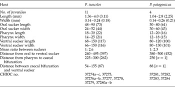
Forty-eight of 60 specimens of P. isosceles were infected with 137 juveniles from the abdominal cavity, abdominal musculature, stomach, intestine, gonad serosa, gonad, kidney and liver. Parameters of infection were: P = 80%, MI = 2.85 ± 2.26; MA = 2.28 ± 2.06 and RI = 1–8. Ten of 25 specimens of P. patagonicus were infected with 32 juveniles distributed in the same sites of infection as observed in P. isosceles and with parameters of infection of P = 40%, MI = 3.2 ± 1.94; MA = 1.28 ± 1.74 and RI = 1–4. Didymozoids were identified as Torticaecum and Neotorticaecum types (Pozdnyakov & Gibson, Reference Pozdnyakov, Gibson, Bray, Gibson and Jones2008).
Torticaecum Yamaguti (Reference Yamaguti1942)
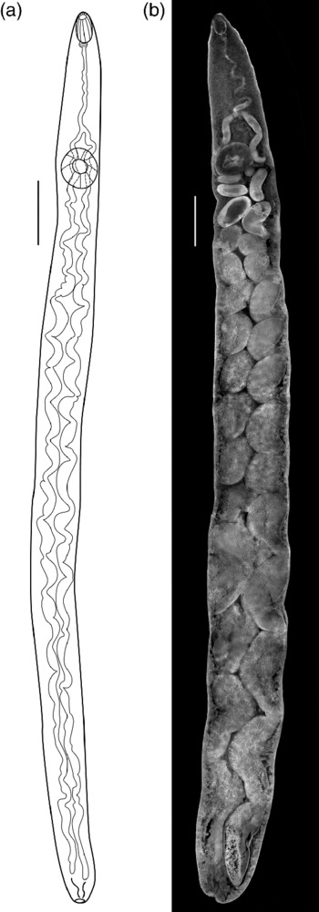
Torticaecum Yamaguti (Reference Yamaguti1942) from Paralichthys isosceles: (a) entire worm using a bright field microscope; (b) entire worm using a confocal laser scanning microscope. Scale bars = 0.25 mm (a), 0.20 mm (b).

Measurements of didymozoids of the type, Torticaecum Yamaguti (Reference Yamaguti1942) recovered from Paralichthys isosceles and P. patagonicus from the littoral of the State of Rio de Janeiro, Brazil, deposited in the Helminthological Collection of Instituto Oswaldo Cruz (CHIOC).

Measurements in micrometres unless indicated; means in parentheses. n, Number of specimens.
General description. Body elongate, relatively narrow, with rounded extremities, tegument smooth, oral sucker terminal, muscular, followed by a small muscular pharynx, well-developed ventral sucker, larger than oral sucker. Oesophagus narrow, long and sinuous, bifurcating anterior to ventral sucker without glandular cells. ‘Drüsenmagen’ absent. Caeca tubular at bifurcation level, enlarging in the region below the ventral sucker, forming 13–19 chambers on each side, occupying almost the entire body and becoming progressively elongated as they approach the end of the body. Excretory vesicle saccular, posterior to caeca.
Neotorticaecum Kurochkin & Nikolaeva (Reference Kurochkin and Nikolaeva1978)
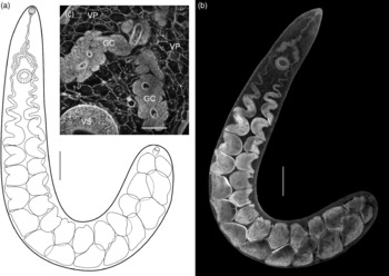
Neotorticaecum Kurochkin & Nikolaeva (Reference Kurochkin and Nikolaeva1978) from Paralichthys patagonicus: (a) entire worm using a bright field microscope; (b) entire worm using a confocal laser scanning microscope; (c) detail by a confocal laser scanning microscope, showing vesicular parenchyma (VP), glandular cells (GC) and ventral sucker (VS). Scale bars = 0.50 mm (a, b), 0.10 mm (c).

Measurements of didymozoids type Neotorticaecum Kurochkin & Nikolaeva (Reference Kurochkin and Nikolaeva1978) recovered from Paralichthys isosceles and P. patagonicus from the littoral of the State of Rio de Janeiro, Brazil, deposited in the Helminthological Collection of Instituto Oswaldo Cruz (CHIOC).
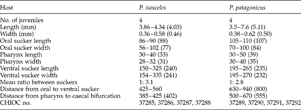
Measurements in micrometres unless indicated; means in parentheses.
General description. Body elongate, tegument smooth, parenchyma vesicular, oral sucker subterminal, muscular, followed by a muscular pharynx, well-developed ventral sucker, larger than oral sucker. Oesophagus, narrow and sinuous, bifurcating anterior to the ventral sucker and encircled by glandular cells that extend up to the middle portion of the oesophagus. ‘Drüsenmagen’ absent. Caeca chambers posterior to ventral sucker increasing in size as they reach the end of body. Excretory vesicle saccular, posterior to caeca.
Discussion
Yamaguti (Reference Yamaguti1942) described the genera Monilicaecum and Torticaecum as including some immature fish trematodes from Japan, unaware that they belonged to juvenile Didymozoidae, and considered Distomum fenestratum Linton, 1907, as a synonym for Torticaecum. Further, these genera were allocated to the Didymozoidae, and Yamaguti (Reference Yamaguti1970, Reference Yamaguti1971) proposed that the generic nominations should be applied to larval collective groups. According to Pozdnyakov & Gibson (Reference Pozdnyakov, Gibson, Bray, Gibson and Jones2008), the classification of the didymozoids is still controversial and some authors have used either numeric nomenclatures (Nikolaeva, Reference Nikolaeva1964, Reference Nikolaeva1965, Reference Nikolaeva1970; Gaevskaya & Nigmatullin, Reference Gaevskaya and Nigmatullin1976; Kurochkin & Nikolaeva, Reference Kurochkin and Nikolaeva1978; Køie & Lester, Reference Køie and Lester1985), or alphabetical organization (Fischthal & Kuntz, Reference Fischthal and Kuntz1964; Fischthal & Thomas, Reference Fischthal and Thomas1968; Gaevskaya, Reference Gaevskaya1977) to properly identify larval didymozoids presenting small structural differences. Kurochkin & Nikolaeva (Reference Kurochkin and Nikolaeva1978) suggested a nomenclatural system based on the presence or absence of the ventral sucker, pharynx, ‘Drüsenmagen’ and gland cells encircling the oesophagus and/or the anterior portion of the caeca. In 1985, Køie & Lester reported that the disappearance of the ‘stomach’ (‘Drüsenmagen’) is related to the worms' aging and observed that the development of younger stages reveals the presence of the pharynx and ventral sucker; thus, these characters are devoid of taxonomic value; it was also suggested that Latin names ought not to be used and that numerical arrangements should be adopted for identification. Pozdnyakov & Gibson (Reference Pozdnyakov, Gibson, Bray, Gibson and Jones2008) agree with Køie & Lester (Reference Køie and Lester1985) that morphological characteristics of young didymozoids may change with age, although numerical and alphabetical trials could lead to misinterpretations. The former authors recommend, in agreement with Yamaguti (Reference Yamaguti1970, Reference Yamaguti1975), that structural types should be considered for identification and hence, in the present study, this is based on Pozdnyakov & Gibson (Reference Pozdnyakov, Gibson, Bray, Gibson and Jones2008). Two juvenile didymozoids were identified as the types, Torticaecum and Neotorticaecum. The main differences observed were the absence (Torticaecum) and the presence (Neotorticaecum) of gland cells in the oesophageal region and in the caecal bifurcation.
The first juvenile didymozoids mentioned in Brazil were reported by Amato (Reference Amato1983), parasitizing specimens of Parona signata (Jenyns, 1841). The trematode was designated as ‘Didymozoid larvae I’ after being compared to species Torticaecum Yamaguti, Reference Yamaguti1942, considering body size, aspect and location of ventral sucker. The Torticaecum type, described here, although larger, can be associated with Torticaecum nipponicum, described by Yamaguti (Reference Yamaguti1942), differing from Didymozoid larvae I (Amato, Reference Amato1983) by the absence of an equatorial ring in the oral sucker. Likewise, specimens described by Køie & Lester (Reference Køie and Lester1985) as species 9, showed similarities in body shape and location of ventral sucker to the Torticaecum type; and species 5, also described by Køie & Lester (Reference Køie and Lester1985), is similar to the Neotorticaecum type, mainly considering the presence of gland cells around the oesophagus and caecal bifurcation. The second reference to juvenile didymozoids in Brazil was that of Kohn & Justo (Reference Kohn and Justo2008), reporting on the finding of all larval stages associated with adult Didymocystis lamotheargumedoi, infecting three different species of tuna fish.
Although other helminth surveys of marine fish from the littoral of Rio de Janeiro refer to the presence of juveniles, the parasites are designated only as immature didymozoids (Ribeiro et al., Reference Ribeiro, Luque and Alves2002; Luque et al., Reference Luque, Alves and Ribeiro2003; Bicudo et al., Reference Bicudo, Tavares and Luque2005; Cordeiro & Luque, Reference Cordeiro and Luque2005; Luque et al., Reference Luque, Felizardo and Tavares2008). Juveniles presently described were found free or attached to the infection sites, and never associated with cysts, suggesting the possibility of parasite migration to different organs of infected hosts, thus explaining the presence of the trematodes at several sites of infection in the same host specimen. Cable & Nahhas (Reference Cable and Nahhas1962) considered that this migration probably promotes the maturation of parasites.
Although there are no reports of the zoonotic potential of these juveniles, the prevalence of 22.3% in the musculature indicates the need for further studies, taking into account that high adult worm burdens are responsible for the distasteful appearance of infected fish and their subsequent devaluation. Some authors have referred to the presence of didymozoid eggs in human faeces (Kamegai, Reference Kamegai1971; Chung & Cross, Reference Chung and Cross1975; Carney et al., Reference Carney, Nocun, Vergel, Pagaran, Mercado and Cross1987; Cross et al., Reference Cross, Zaraspe, Alquizia and Ranoa1989; Traub et al., Reference Traub, Macaranas, Mungthin, Leelayoova, Cribb, Murrell and Thompson2009); yet, according to Traub et al. (Reference Traub, Macaranas, Mungthin, Leelayoova, Cribb, Murrell and Thompson2009), presumably these eggs were passed mechanically in the faeces.
Prado-Rosas et al. (Reference Prado-Rosas, Alvarez-Cadena, Segura-Puertas and Lamothe-Argumedo1999) reported the occurrence of Torticaecum sp. metacercaria parasitizing the chaetognath, Serratosagitta serratodentata, in Caribbean waters. The present data enlarge the geographical distribution of Torticaecum and Neotorticaecum types in South America, in the Atlantic Ocean; Paralichthys isosceles and P. patagonicus are referred to as new hosts for the studied didymozoids.
Acknowledgements
To Pedro Paulo de Abreu Manso and Bernardo Pascarelli, Laboratório de Patologia, Instituto Oswaldo Cruz for access to the confocal laser scanning microscope; to Heloisa Maria Nogueira Diniz, Serviço de Produção e Tratamento de Imagens (IOC/Fiocruz) and to Flávia Garcia de Carvalho, Serviço de Comunicação (ICICT/Fiocruz) for technical assistance with the figures, and to the Conselho Nacional de Desenvolvimento Científico e Tecnológico (CNPq), Brazil.