Anomalous origin of the left pulmonary artery from the aorta is extremely rare. Reference Kutsche and Van Mierop1 In most cases, the anomalous origin of the left pulmonary artery originates from the ascending aorta and is strongly associated with tetralogy of Fallot, right aortic arch and anomalous subclavian artery. Reference Kutsche and Van Mierop1,Reference Aru, English, Gaymes and Heath2 To the best of our knowledge, only 3 cases of anomalous origin of the left pulmonary artery arising from the descending aorta have been previously reported. Reference Gnanappa, Laohachai, Orr and Ayer3–Reference Rajanbabu and Chigullapally5 None of them had associated cardiac anomalies. We report here such a case in association with heterotaxy syndrome.
Case report
A full-term male newborn who had prenatal suspicion of VACTERL syndrome (oesophageal atresia, anal atresia, right kidney agenesis, ectopic pelvic left kidney) without cardiac anomalies and normal CGH array was referred for systematic cardiac assessment at day 1. Anal and oesophageal atresia was not confirmed at birth. Clinical examination was normal. The transthoracic echocardiogram showed a normal-sized right pulmonary artery following the pulmonary trunk, but the left pulmonary artery was not found. A large arterial duct was clearly patent between the pulmonary trunk and the left-sided aortic arch. Intra-cardiac anatomy was normal.
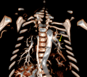
A CT angiogram confirmed the abnormal origin of the left pulmonary artery from the lateral wall of the left-sided descending aorta, far below the origin of the left subclavian artery. This vessel supplied all the left pulmonary segments. Three-dimensional reconstruction image from CT angiogram helped to identify anatomic relationships and to confirm vessels anatomy (Fig 1).
CT imaging showing the left pulmonary artery (LPA) arising from descending aorta. Three-dimensional frontal CT angiogram with LPA (*) arising from the lateral surface of the descending aorta (DAo)

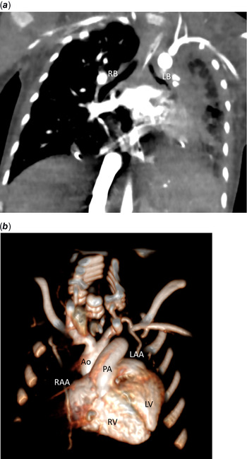
Moreover, neonatal comprehensive screening demonstrated several laterality defects including a left bronchial isomerism (Fig 2a), a midline liver, an asplenia and an intestinal malrotation. In addition, right kidney agenesis and ectopic pelvic left kidney were confirmed. There were also a micro-gastria and a post-axial thumb duplication. Intra-cardiac anatomy was normal on echocardiography, except for bilateral superior caval veins with a left superior caval vein draining into the coronary sinus. The electrocardiogram was in sinus rhythm with normal P wave axis. Atrial appendages on CT scan had a similar external shape, triangular with multiple indentations (Fig 2b). However, the resolution of the CT scan did not allow us to assess the morphology of the pectinate muscles inside the atria. Taken together, these findings were in favour of a heterotaxy syndrome, according to Lin’s criteria. Reference Lin, Krikov and Riehle-Colarusso6
CT imaging showing intra-thoracic laterality defects. ( a ) Two-dimensional frontal CT angiogram with bilateral morphologically left bronchi (late first branching of both bronchi), RB = right bronchus, LB = left bronchus. ( b ) Three-dimensional CT imaging showing similar external shape of the two atrial appendages; RAA = right atrial appendage; LAA = left atrial appendage; Ao = aorta; PA = pulmonary artery; RV = right ventricle; LV = left ventricle.

At one-month-old, the patient underwent surgical re-implantation of the left pulmonary artery by direct anastomosis to the pulmonary trunk. At the time of surgery, transthoracic echocardiography recorded systemic pulmonary hypertension. The right ventricle was dilated but had a preserved systolic function. The surgery was performed by median sternotomy under cardiopulmonary bypass with deep systemic hypothermia (25°C). Operative findings confirmed imaging description including an arterial ligament between the roof of pulmonary trunk and the left-sided aortic arch, a left superior caval vein and a 3.5 mm- left pulmonary artery arising from the lateral surface of the descending aorta. The arterial duct and the left superior caval vein were ligated. Then, the left pulmonary artery was carefully dissected and mobilised to enable a tension-free direct anastomosis without interposition of a synthetic graft. Post-operative course was complicated by severe left pulmonary atelectasis related to an extrinsic compression of the left bronchus by local oedema. Post-operative CT imaging confirmed a small-calibre, but patent re-implanted left pulmonary artery far away from the left main bronchus. Despite selective right intubation and optimal medical treatment, the patient presented a severe infection and necrosis of the left lung and died 20 days after the surgery.
Discussion
Anomalous left pulmonary artery from the descending aorta is exceedingly rare. The three cases reported so far share common characteristics: left-sided aortic arch, anomalous origin of the left pulmonary artery origin from the ipsilateral wall of the descending aorta at a significant distance from the left subclavian artery, patent arterial duct connecting the pulmonary trunk to the under surface of the left aortic arch in two cases and no associated intra-cardiac lesions. Reference Gnanappa, Laohachai, Orr and Ayer3,Reference Rajanbabu and Chigullapally5 Our case is very similar, with a patent arterial duct, and in addition, a left superior caval vein to coronary sinus. These cases are very different from the more frequent, albeit rare, anomalous origin of the left pulmonary artery from the ascending aorta. Various embryologic hypotheses have been reported about the constitution of anomalous origin of branch pulmonary arteries from either right or left aortic arch. Reference Aru, English, Gaymes and Heath2,Reference Prifti, Bonacchi and Murzi4
Developmentally, central pulmonary arteries have a dual origin: the pulmonary trunk originates from the embryonic outflow tract, while the right and left pulmonary arteries originate from the ventral pharyngeal mesenchyme (splanchnic plexus) connecting within the aortic sac to the pulmonary arch, Reference Waldo and Kirby7 formerly named sixth aortic arch. Reference Graham, Poopalasundaram, Shone and Kiecker8 Intra-pulmonary arteries develop within the lung buds and connect to the dorsal aorta through the intersegmental arteries, which disappear when the connection with the central pulmonary arteries occurs. On the left side, the left pulmonary artery derives from the proximal part of the left pulmonary arch, while the distal part gives rise to the arterial duct. Various hypotheses have been raised for anomalous origin of one pulmonary artery from the ascending aorta, the most prevalent being an asymmetric development of the aortic sac. This hypothesis is not applicable for the anomalous origin of left pulmonary artery from descending aorta. This rare anomaly could be due to a lack of development of the left pulmonary arch, total when no duct is present, Reference Prifti, Bonacchi and Murzi4 or partial, involving only the proximal part of the arch, like in our case and others. Reference Gnanappa, Laohachai, Orr and Ayer3,Reference Rajanbabu and Chigullapally5 Another subject of interrogation is the nature of the vessel connecting the descending aorta and the intra-parenchymal left pulmonary artery. If this anomaly results from lack of development of the left pulmonary arch, this vessel should be of another origin, like in tetralogy of Fallot with pulmonary atresia. In this setting, major aortopulmonary collateral arteries originating from the descending aorta are considered persistent intersegmental arteries. In our case like in other cases described, the argument against this anomalous vessel being a major aortopulmonary collateral artery is that it does supply the entire left pulmonary arterial tree, while major aortopulmonary collateral arteries, if they also enter in the hilum, usually supply a lobe or a segment and not the entire lung. Reference Gnanappa, Laohachai, Orr and Ayer3,Reference Rajanbabu and Chigullapally5 However, some cases with tetralogy of Fallot with pulmonary atresia with one lung supplied by a single collateral artery arising from the descending aorta have been published. Reference Jefferson, Rees and Somerville9
The anatomical differences regarding associated anomalies in anomalous origin of the left pulmonary artery from the ascending aorta and anomalous origin of the left pulmonary artery from the descending aorta may reflect a different embryological background. Anomalous origin of the left pulmonary artery from the ascending aorta is commonly associated with tetralogy of Fallot and right aortic arch, which could be related to disturbances in neural crest cells migration. Indeed, anomalous origin of the left pulmonary artery from the ascending aorta was described in patients with DiGeorge syndrome and Waardenburg syndrome. Reference Dodo, Alejos and Perloff10 On the contrary, the rare cases reported of anomalous origin of the left pulmonary artery from the descending aorta display no associated intra-cardiac lesions and have a left aortic arch, despite a similar alleged mechanism, namely the absence of proximal pulmonary arch.
The diagnosis of heterotaxy in our case was made according to Lin’s criteria, based on the association between: abdominal situs abnormality (midline liver), spleen abnormality (asplenia), left bronchial isomerism, bilateral superior vena cava and intestinal malrotation, despite the absence of intra-cardiac defects. Reference Lin, Krikov and Riehle-Colarusso6 The definition of heterotaxy and the very term “heterotaxy” itself are still subject to many controversies. According to the IPCCC ICD-11 nomenclature, heterotaxy is defined as “a congenital malformation in which the internal thoraco-abdominal organs demonstrate abnormal arrangement across the left-right axis of the body”, right and left isomerisms are defined as variants of heterotaxy, and “heterotaxy does not include normal and mirror-imaged arrangements of the internal organs (situs solitus or inversus)”. Reference Franklin, Béland and Colan11 According to Van Praagh, heterotaxy is defined as “an abnormal symmetry of certain viscera and veins (lungs, liver, caval veins) and situs discordance between various organ systems and between the various segments of the heart”. Reference Van Praagh12 Although some anatomical studies claimed that atrial appendages were uniformly isomeric in the setting of heterotaxy, Reference Uemura, Ho and Devine13,Reference Tremblay, Loomba and Frommelt14 several other reports demonstrate that this is not always the case. Reference Loomba, Pelech, Shah and Anderson15,Reference Yim, Nagata and Lam16 We cannot affirm in our case that there was isomerism of the pectinate muscles, even if their external appearance looked similar on the CT scan, but the presence of lateralised atrial appendages would not preclude heterotaxy anyway. Reference Loomba, Pelech, Shah and Anderson15,Reference Yim, Nagata and Lam16 Moreover, the absence of intra-cardiac defects does not rule out the diagnosis of heterotaxy, as it is now recognised that 10–16% of heterotaxy patients have no CHD. Reference Lin, Krikov and Riehle-Colarusso6,Reference Cohen, Anderson and Cohen17 The fact that the electrocardiogram was normal with a normal P wave axis does not rule out heterotaxy neither, as assessed by Wren et al. Reference Wren, Macartney and Deanfield18 Some authors have defined a CHD in the setting of heterotaxy as “any intracardiac lesion or an abnormality of venous returns to the heart”. Reference Loomba, Pelech, Shah and Anderson15 For all these reasons, the diagnosis of heterotaxy, made on the association of left bronchial isomerism, abdominal organs laterality defects and bilateral superior caval vein, is unequivocal in our patient. Disharmony between the arrangement of the bronchi and the abdominal organs is relatively frequent (20% in the series by Yim Reference Yim, Nagata and Lam16 ), but the association of left bronchial isomerism and asplenia is rare. All these considerations are in favour of an analytic approach based on accurate description of bronchi, abdominal organs and cardiac features independently, including the venous and arterial connections. Reference Loomba, Pelech, Shah and Anderson15,Reference Sanders and Geva19 We have no embryologic explanation to propose for the unusual association between anomalous origin of the left pulmonary artery from the descending aorta and heterotaxy syndrome found in our patient.
Complete diagnosis and anomalous origin of the left pulmonary artery origin analysis are not always easy by echocardiography alone. CT angiogram and three-dimensional reconstruction were very helpful in our case to detail the lesions and guide the surgeon.
Overall, the surgical repair strategies for the re-implantation of the anomalous origin of the left pulmonary artery to the pulmonary trunk show good long-term results despite the late high risk of post-operative restenosis across the anastomotic site. When the distance between anomalous origin of the left pulmonary artery and pulmonary trunk is large, cardiopulmonary bypass is required during the procedure. Although early repair avoids pulmonary hypertension and irreversible pulmonary vascular disease, outcome of our patient was complicated by ventilation issues related to post-operative oedema compressing the left bronchus and severe bronchiectasis.
In conclusion, we present a rare case of anomalous origin of the left pulmonary artery from the descending aorta, associated with a particular form of heterotaxy, with disharmonious patterns of bronchi and abdominal organs, bilateral superior caval veins and no intra-cardiac lesions. This complex and unusual case reinforces the growing consensus that all the still pending controversies about heterotaxy should be resolved by the accurate description of each thoracic and abdominal organ independently, including the segments of the heart and the venous and arterial connections. Reference Loomba, Pelech, Shah and Anderson15,Reference Sanders and Geva19,Reference Loomba, Tretter and Anderson20
Financial support
This research received no specific grant from any funding agency, commercial or not-for-profit sectors.
Conflict of interest
None.
Ethical standards
This article does not contain any studies with animals performed by any of the authors.