Introduction
Peripheral vascular malformation is an unusual and sometimes life-threatening anomaly, which has been successfully managed by surgery and vascular occlusive embolic therapy or radiotherapy in the intracranial cavity,Reference Baskaya, Jea and Heros1–Reference Starke, Komotar and Hwang5 and the relative roles are well defined. Surgery and embolisation have been used in the extra-cranial setting with usually excellent results.Reference Seccia, Salgarello and Farallo6,Reference Kohout, Hansen and Pribaz7 However, those patients who do not respond to these traditional therapies have few options. Radiotherapy has been used in a variety of benign diseases with success and usually acceptable morbidity.Reference Donaldson, McDougall, Alberti and Sagerman8–Reference Tripuraneni11 However, few data exist in the published literature regarding the use of radiotherapy for vascular malformations in the extra-cranial setting, and no standard dose-schedule has been determined.Reference Hida, Shirato and Seki12–Reference Rudman and Clark15 To our review, no data exist in the literature describing the use of radiotherapy for peripheral vascular malformations of the extremities. In our patient, we have shown that high dose per fraction external beam radiotherapy can be effectively used to treat arterio-venous malformations (AVMs) even in large fields in the extremities.
Case history
A 50-year-old black male originally presented in 1994 with a tender mass lesion of the right lateral thigh measuring ∼2.0 cm. The patient manifested no other symptomatology at that time, and the remainder of his physical exam was unremarkable. His past medical history was significant for coronary artery disease, pulmonary hypertension, multiple myocardial infarctions, mild chronic renal failure, non-insulin dependent diabetes mellitus and a colonic resection to remove benign non-adenomatous polyps. The mass in the thigh was felt to be a solitary AVM and was surgically resected in an extensive procedure. Pathologic review demonstrated benign vascular findings consistent with an AVM.
In 2003, a painful mass re-presented at the same site. However, this mass demonstrated serosanguineous drainage for ∼1 week before evaluation. At that time he had no other abnormal physical findings. He underwent a magnetic resonance arteriography and venography, which demonstrated a large feeder artery to an area of increased vascularity on the lateral right thigh in the area of previous resection. A percutaneous embolisation of this feeder artery, the associated vascular abnormality and the deep perforating branches of the right profunda femoris artery was performed. In mid-2004, the patient developed a non-healing ulcer of the right lateral thigh in the prior site of the recurrent AVM. The ulcer was conservatively managed for nearly 2 years; however, chronic blood loss increased and the patient required multiple transfusions. Computed tomographic (CT) studies and angiography of the right lower extremity in early 2006 revealed multiple globular vascular lesions superficial to the quadriceps muscle again involving the deep perforating branches of the profunda femoris. Plastic and reconstructive surgery and interventional radiology were consulted at that time; however, the patient developed dyspnoea requiring emergency inpatient admission for anaemia and congestive heart failure. Repeat transfusions were once again performed secondary to substantive blood loss from the ulcerative lesion. Cardiac evaluation revealed an ejection fraction of 17% secondary to multiple myocardial infarctions causing perfusion defects in the anterior wall, septum, and inferior wall of the myocardium as demonstrated by a chemical stress test. Because of his poor cardiac functionality, surgical management was not recommended. Additionally, the patient had recurrent episodes of ventricular arrhythmias, which were treated medically but also required the placement of an implantable defibrillator.
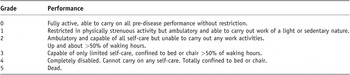
Despite these concurrent medical issues, the patient continued to maintain an active lifestyle with an Eastern Cooperative Oncology Group performance status of two (Table 1). A repeat percutaneous embolisation procedure was attempted in February of 2006; however, the arteriography demonstrated vascular malformations of the end arteries not amenable to embolisation. The patient was discharged from the hospital with oral ferrous sulphate and subcutaneous epoetin-α. The ulcerated area of the thigh continued to exude blood with associated haemoglobin level as low as 4.5 g/dl requiring multiple unit transfusions on numerous occasions. Repeated superficial wound infections also developed over this period, which were successfully treated with intravenous and oral antibiotics. In December of 2007, the patient was admitted to the Allegheny General Hospital with more profuse bleeding from the wound. Admission haemoglobin was 7.9 g/dl. Interventional radiology and surgical consultations were once again obtained and no further intervention was recommended. Finally, consultation with radiation oncology was obtained.
ECOG Performance status

At the time of our review the patient was normotensive with a blood pressure of 130/74 and a regular pulse rate of 68 beats per minute, and a respiratory rate of 13 breaths per minute. He was ambulatory with crutches for only short distances. A large pressure dressing over the right thigh covered a 10 × 10 cm ulcerated non-healing lesion actively oozing dark red blood. There were multiple areas of superficial venous dilatations distal to the lesion and generalized +3 pitting oedema of the entire lower extremity caudad to the lesion. The opposite extremity was normal on examination. Following multidisciplinary consultation with vascular surgery and interventional radiology, the only available management option was amputation of the right lower extremity. After careful and complete discussion with the patient and family a course of high dose per fraction external radiotherapy was recommended.
Management with Radiotherapy
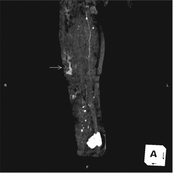
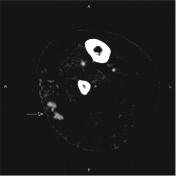
The patient was informed that only limited data supported such an approach and that possible sequelae might be tissue slough or even loss of the extremity. After careful consideration, the patient and his family voiced understanding and agreed to treatment. Magnetic resonance angiography could not be performed because of the patient's implanted defibrillator and low volume contrast CT was used to localize the lesion in view of the patient's history of mild chronic renal failure. At the time of the localization no single lesion was evident and the area of malformation was described as a “field effect” of widespread small AVMs (Figures 1 and 2) extending nearly the entire length of the lateral thigh (∼15 cm). The patient's lower extremity was immobilized in a vacuum bag. The fields used were opposed tangential obliques delivered with 6 MV photons without a bolus. Three-dimensional treatment planning was performed with a planning treatment volume which covered the radiographically identifiable lesions plus a 2-cm margin. A radiation dose of 3,000 cGy (centiGray) was delivered to the volume in six fractions of 500 cGy over an 11-day elapsed time. The patient experienced no untoward side effects during the course of therapy.
A coronal image from three-dimensional contrast-enhanced CT angiography study performed seven days prior to initiation of radiation therapy. The arrow points to serpiginous vascular structures located mainly in superficial soft tissues of the right leg, which are part of patient's known vascular malformation.

This is an axial image from the same study. The arrow again points to abnormal vascular structures containing contrast.

Results
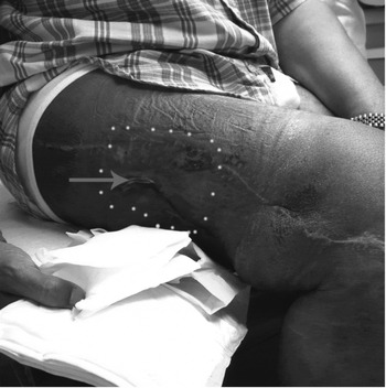
Three weeks following completion of therapy the ulcer size was unchanged; however, the amount of venous oozing had decreased by ∼50% as evidenced by the number of dressing changes that were required daily. Three months after the completion of therapy venous oozing had essentially ceased. At the 6-month follow-up the ulcer had nearly completely regressed with a residual 2 cm × 3 mm slit-like crease as the only evidence of the previous wound (Figure 3). The patient has required no blood transfusions since the completion of radiotherapy and is ambulatory with a cane for support. His haemoglobin has increased to 13.0 g/dl, and he has stopped his recombinant erythropoietin and ferrous sulphate. At his 1-year follow up, there has been complete healing of the wound and no chronic sequelae have developed. He continues to ambulate without an assistance device and has had no further blood loss.
Oblique view of Patient's lower extremity. The dotted line demarcates the pretreatment extent of the ulcerated malformation. The residual wound is demarcated by the arrow.

Discussion
AVMs can be managed successfully by surgery and radiotherapy when they occur intracranially. The mechanism of action of radiation for vascular malformations is poorly understood; however, it is thought to act on the endothelial cells of the vessels, which tend to multiply following treatment. However, the management of an extra- cranial vascular malformation refractory to surgery and embolisation therapy can be complicated. Little has been published on the role of radiotherapy in this challenging disease, and nothing has been published to date describing the use of radiotherapy in vascular malformations of the extremities. Intracranial anomalies have been managed with single high-dose fractions,Reference Hartmann, Mast and Choi4–Reference Starke, Komotar and Hwang5 which would be inappropriate for use in large fields due to the massive volume of treatment and the likely development of significant and devastating acute and chronic sequelae. However, our case history demonstrates a novel way to approach this challenge with minimal morbidity. No clear dose fractionation schedule has been developed as schedules varied from 32 Gy in 2 fractions to 40 Gy in 20 fractions.Reference Hida, Shirato and Seki12–Reference Rudman and Clark15 Additionally, no clear margins of treatment have been established.Reference Seccia, Salgarello and Farallo6 It is important to note that this approach, while successful to date in this case requires further investigation through clinical trials to establish efficacy. However, it seems reasonable that this approach be considered in patients with malformations refractory to conventional treatment in lieu of amputation. As this technique involves the use of ionising radiation to treat a benign disease, patients should be cautioned about the rare potential development of radiation induced malignancies.
Conclusion
Radiotherapy for peripheral vascular malformations is feasible and can yield excellent results in selected cases. Further investigation is warranted to determine appropriate dose-fraction size and effective total dose.