Introduction
Meat quality is a major issue both for the process meat industry and the consumers. Pale, soft and exudative (PSE) meat, which is seen in pig meat production, has seldom been analysed in poultry meat, apart from turkey (Alvarado and Sams, Reference Alvarado and Sams2002). So-called PSE meats show low water-holding capacity, bad textural properties and reduced protein extractability (Tankson et al., Reference Tankson, VizzierThaxton, Thaxton, May and Cameron2001). A relationship also exists between colour, pH and water-holding capacity (Woelfel et al., Reference Woelfel, Owens, Hirschler and Sams2002; Campo et al., Reference Campo, Gil, Dávila and Muñoz2005). PSE meat from pigs loses more water during cooking compared with normal meat (Tankson et al., Reference Tankson, VizzierThaxton, Thaxton, May and Cameron2001). In a study on boneless and skinless fillets from broilers, Barbut et al. (Reference Barbut, Zhang and Marcone2005) also reported dark, firm and dry (DFD) meat. In poultry, stress has severe consequences on the quality of the final product, with effects on pH, pigmentation, water-holding or fat percentage (Fletcher, Reference Fletcher1999; Tankson et al., Reference Tankson, VizzierThaxton, Thaxton, May and Cameron2001; Campo et al., Reference Campo, Gil, Dávila and Muñoz2005). In poultry, secretions from the preen (or uropygial) gland have a role in physiology and behaviour as their composition is affected by age and season, as well as by whether or not a bird has been feather pecked (Sandilands et al., Reference Sandilands, Powell, Keeling and Savory2004). It is also known that hens raising their chicks produce specific hormonal secretions from this gland (Richard-Yris et al., Reference Richard-Yris, Garnier and Le boucher1983; Bohnet et al., Reference Bohnet, Rogers, Sasaki and Kolattukudy1991). Their lack may be related to the level of stress observed in broiler husbandries, as observed by Madec et al. (2005) and Madec et al. (Reference Madec, Pageat, Bougrat, Saffray, Falewee, Gervasoni, Bollart and Gabarrou2006). In order to test the effect on the quality of broilers’ production, the secretion from the uropygial gland of hens was isolated under natural mothering conditions. A synthetic analogue was prepared according to the analysis of the secretion. The purpose of the present study was to investigate the effect of this synthetic analogue on several meat quality indicators in heavy broilers, along with carcass performances.
Material and methods
Animals and breeding conditions
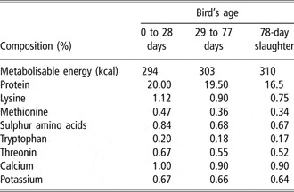
The experiment was conducted in two similar buildings, both with a stocking density of 11 birds per square metre (i.e. approximately 25 kg/m2 when birds are due for slaughter). A heavy broiler strain (Sasso T56N) was used, and a total of 4400 chicks were kept in each building from 1 day of age and then slaughtered at day 80. The floor in the buildings was composed of soil covered with wood shavings and birds had free access to fresh water and food (Table 1). From 4 weeks of age until slaughter, the birds also had day-time free-range access outside the buildings through trapdoors operated by the stockperson. In the buildings, all broilers shared floor and air space. Health status of the birds was examined upon arrival (D0) and then on a weekly basis until completion of the study. Ethical aspects of the experiment were in accordance with European Convention 95/29/CE.
Composition of the diet, depending on birds’ age

Values are per 100 g diet.
Treatment
In order to construct the Mother Hen Uropygial Secretion Analogue (MHUSA), samples of the natural secretions were obtained from hens by squeezing their preen glands once a day for 14 days after hatching of their chicks. When the pattern was analysed, we observed a stabilisation of the uropygial secretion from 12 days post hatching. Thus, samples from 12 days post hatching were analysed using gas chromatography – mass spectometry (GC/MS, Turbo Mass; Perkin-Elmer, Boston, MA, USA). A synthetic analogue was then created (MHUSA, Pat. PCT/EP03/007144). This consisted of a synthetic reconstruction of a fraction of the natural secretion, composed of 12- to 18-carbon fatty acid methyl esters (FAME). The treatment was incorporated into a commercially available gelatin matrix block (Nicols S.A., Bertry, France) weighing 150 g and composed of water (135 g), non-ionic surfactant (7 g) and a gelling gum (5 g), plus either 3 g of water (control) or 3 g of MHUSA. The gelatin matrix block (control or MHUSA) was held in specially manufactured perforated plastic container suspended 120 cm above the ground, out of reach of the birds. The components of MHUSA are heavier than air, and this delivery arrangement allowed the treatment to diffuse into the air around the birds. Treatment was blinded since it was impossible to tell the difference between MHUSA and control matrixes (their odour and appearance were identical), and the persons involved in the trial were unaware of which group was given which treatment. Treatment was installed in each building on the day before the arrival of the chicks. One block was used for every 50 m2 of floorspace, and the blocks were replaced every 4 weeks, giving a total of 24 blocks used during the 80-day trial. After this first experiment, the buildings were cleaned and prepared for a new batch of chicks. The whole experiment was then repeated precisely as before, but with the building treatments reversed in order to control against building effect.
Data collection
For all measurements, a randomly selected group of 40 males and 40 females were used per building treatment, meaning that a total of 160 samples were analysed (80 per treatment). At 24 h post mortem, eviscerated carcass weight (CW) was measured (Bird Weighing System-1050, Weltech Int., Cambridgeshire, UK) before excision of body parts subject to measurements. Then both pectoralis major (fillet weight or FW) and abdominal fat were weighed. All measurements were performed inside a refrigerated room at constant temperature (2°C), using the same procedure for all animals, in the same order and by a certified technician, following a method described by Fletcher et al. (Reference Fletcher, Qiao and Smith2000). After excision and weighing, muscle pH was recorded using a pH meter (CG 843; SCHOTT Instruments, Woburn MA, USA). The electrode was inserted into the anterior area of the ventral part of right pectoralis major to a depth of 100 mm. After each pH measurement, the electrode was rinsed with distilled water and dried with soft tissue paper. Each pectoralis major was then stored at 4°C in polyethylene bags for further weight, pH and colour measurements. Colour measurements were taken from the posterior area of the ventral side of these samples. CIE (International Commission on Illumination) Laboratory colour coordinates (L* = lightness, a* = degree of redness, b* = degree of yellowness) were measured using a colorimeter (CR-10, Minolta France S.A., France). The a* and b* coordinates were subsequently used to calculate the hue (H* = tan−1b*/a*, angle that the metric chroma line makes with the a* axis) and saturation (or chroma C* = (a*2 + b*2)−1). The colour of the samples was compared to a standardised white ceramic (L* = 94.3, a* = −2.86 and b* = 1.92). At 6 days post mortem, excess liquid was drained from the bag containing each pectoralis major muscle, before recording the weight, pH and colour of the tissue. For meat-surface colour measurements, readings were taken from the selected areas that were free from obvious defects (bruises or haemorrhages) that might have affected the uniformity of the colour reading. A 35-g sample from the left anterior side of the left pectoralis major was then analysed for weight, pH and colour variations between before and after cooking. The cooking procedure (CP) consisted of placing each 35 g sample in a microwave oven for 60 s at 850 W power, after which it was allowed to cool at room temperature (22°C) for 90 min before measurements were taken. Together with sample weight loss during cooking, FW loss relative to CW was recorded at both 24 h and 6 days post mortem. Finally, because we wanted to imitate industrial conditions in which meat quality is not differentiated on the basis of sex, we choose to pool male and female results.
Statistical treatment of results
Data were analysed using Statistica version 5 Software (StatSoft Inc., Tulsa OK, USA). Means were compared using Student’s t-test. The relationship between different variables was determined using Pearson’s correlation coefficient, to look for differences among treatments. Results are expressed as mean ± s.e. Results were considered significant if P < 0.05. Differences between the two replications of the experiment were investigated as treatment effect.
Results
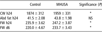
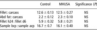
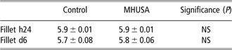
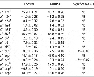
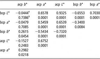
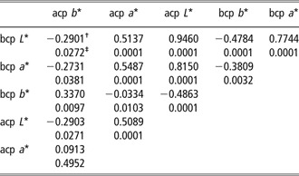
CW at 24 h post mortem, FW at 24 h and at 6 days post mortem were all significantly higher (P < 0.05) for broilers within the MHUSA-treatment group (Table 2), while abdominal fat did not show a significant difference between the groups. There was no significant effect on yield (Table 3) or fillet pH (Table 4). Colorimetry results (Table 5) showed that meat from birds that received treatment with MHUSA had a significantly lower degree of yellowness (b*, P < 0.05) and almost significantly lower colour saturation (C*, P = 0.06) at 6 days after slaughter, as well as reduced redness after cooking (a*, P = 0.07). The correlation coefficients between colour values are presented in Table 6 (for control) and Table 7 (for MHUSA). For both treatment types, we observed strong positive correlations (0.70 or higher) between lightness before and after the CP, and between lightness before CP and redness before CP. In the control group, lightness after CP was also highly negatively correlated with yellowness after CP, while it was positively correlated with redness before CP in the MHUSA group.
Treatment effect on mean weights (g) of carcass, abdominal fat and fillet at different times post slaughter
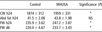
CW = carcass weight; h24 = 24 h post mortem; Abd fat = abdominal fat; FW = fillet weight; d6 = 6 days post mortem; MHUSA = Mother Hen Uropygial Secretion Analogue; NS = not significant.
Values are mean ± s.e.
*P < 0.05.
Treatment effect on means of fillet and abdominal fat relative to carcass weight, fillet weight loss from 24 h to 6 days post mortem and sample weight loss during cooking

h24 = 24 h post mortem; d6 = 6 days post mortem; bcp = before cooking procedure; acp = after cooking procedure; MHUSA = Mother Hen Uropygial Secretion Analogue; NS = not significant.
Values are mean ± s.e.
Treatment effect on means of fillet pH
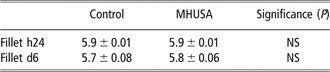
Fillet h24 = pH of the fillet 24 h post mortem; Fillet d6 = pH of the fillet 6 days post mortem; MHUSA = Mother Hen Uropygial Secretion Analogue; NS = not significant.
Values are mean ± s.e.
Treatment effect on mean colour measurements at 24 h and 6 days post mortem and after the cooking procedure

†Colour measurements on fillet samples 24 h post mortem.
‡Colour measurements on fillet samples 6 days post mortem.
§Colour measurements on fillet samples after the cooking procedure.
L* = lightness; a* = degree of redness; b* = degree of yellowness; H* = Hue = tan−1b*/a*; C* = saturation = (a*2 + b*2)−1; MHUSA = Mother Hen Uropygial Secretion Analogue; NS = non-significant.
Values are mean ± s.e.
*P < 0.05.
Pearson’s correlation coefficients and probabilities for lightness, redness, yellowness, hue and saturation of the 35 g fillet samples before and after the cooking procedure – control group

bcp = before cooking procedure; acp = after cooking procedure. L* = lightness; a* = degree of redness; b* = degree of yellowness; H* = Hue = tan−1b*/a*; C* = saturation = (a*2 + b*2)−1.
†Pearson’s correlation coefficient.
‡Probability.
Pearson’s correlation coefficients and probabilities for pH, lightness, redness, yellowness, hue and saturation of the fillet 35 g samples before and after the cooking procedure – MHUSA group

bcp = before cooking procedure; acp = after cooking procedure. L* = lightness; a* = degree of redness; b* = degree of yellowness; H* = Hue = tan−1b*/a*; C* = saturation = (a*2 + b*2)−1; MHUSA = Mother Hen Uropygial Secretion Analogue.
†Pearson’s correlation coefficient.
‡Probability.
Discussion
In the present experiment, we found that treatment with MHUSA had a positive effect on measured weight, with birds having higher CW and FW. The general performance of birds in the present study was in accordance with results obtained for the same broiler type by Berri et al. (Reference Berri, Le Bihan-Duval, Baeza, Chartrin, Picgirard, Jehl, Quentin, Picard and Duclos2005), except for abdominal fat weight, which was lower in our study. Computed yields are similar to those reported by Kannan et al. (Reference Kannan, Heath, Wabeck, Souza, Howe and Mench1997a) for cooking loss, and to those by Lin et al. (Reference Lin, Sui, Jiao, Buyse and Decuypere2006) for both abdominal fat and fillet. Cooking losses in the present study differed from the results of Lin et al. and those of other authors (such as Bianchi et al., Reference Bianchi, Fletcher and Smith2005; Barbut et al., Reference Barbut, Zhang and Marcone2005 or Mehaffey et al., Reference Mehaffey, Pradhan, Meullenet, Emmert, McKee and Owens2006). This is probably due to differences in the CP, which was performed using either a conventional oven or a warm water bath by those cited authors. In the study by Lin et al. (Reference Lin, Sui, Jiao, Buyse and Decuypere2006), stress mimicked by the administration of corticosterone produced a significant decrease in fillet yield and an increase in abdominal fat percentage. As stated earlier, apart from a lack of significant differences for abdominal fat values (fat weight and fat yield), we have the same tendency in our results. Normally, stress is expected to increase body fat content (Siegel, Reference Siegel1995), but our results are supported by a study in laying hens, which showed that a stress-like physiological state induced by ACTH injection did not affect body fat content. Our results showed that broilers from both treatment and control groups had comparable fillet yield and fat percentage, but that both CW and FW were significantly higher in case of MHUSA-treatment group. Zerehdaran et al. (Reference Zerehdaran, Vereijken, van Arendonk and van der Waaij2005) have shown that abdominal fat and CW are strongly correlated, and that live weight is correlated with both CW and abdominal fat. Moreover, Mehaffey et al. (Reference Mehaffey, Pradhan, Meullenet, Emmert, McKee and Owens2006) have shown that overall meat quality is not correlated with fillet yield. We can thus hypothesise that continuous exposure to MHUSA during the growing period of broilers significantly reduces the percentage of body fat. Moreover, our results show that MHUSA treatment has no effect on fillet water loss or cooking loss, which indicates that this form of treatment has no effect on meat quality with regard to water content. Barbut et al. (Reference Barbut, Zhang and Marcone2005) studied meat quality in various strains of broilers that had a similar range of body weight to those in the present study. According to their results, meat samples from birds in our study would have been categorised as PSE with respect to pH value. However, the pH values for the meat in the present study are in accordance with those from a study by El Rammouz et al. (Reference El Rammouz, Berri, Le Bihan-Duval, Babilé and Fernandez2004), which investigated the comparative impact of different rearing conditions (no stress, handling and heat stress) in slow-growing broilers. In the cited study, meat pH varied from 5.79 to 5.89. When comparing textural differences among broiler breast meat ranging from PSE to DFD in their fresh forms, Zhang and Barbut (Reference Zhang and Barbut2005) showed that PSE meat had significantly lower pH and water-holding capacity values compared with normal meat. During cooking, PSE meat lost significantly more liquid than normal meat, and PSE meat proteins were generally more severely affected. PSE meat also had significantly higher lightness value. From our results, MHUSA treatment had no effect on meat pH value compared with the control group, and none of the other results indicated that the meat should be categorised as PSE. Knowing that the overall quality of the meat depends upon several indicators and taking into account the results from other studies, none of the meat produced in the present study should be considered to be PSE, but as normal. Considering colour measurements, we observed that meat from the control-group birds was more yellow compared with that from the MHUSA group. This could be explained by a higher percentage of meat-surface fat or by some other change in fillet colour, for example, due to stress-related denaturing of muscle proteins, although this was not measured in our study. Comparing colour values between broilers stressed by shackling and control birds, Kannan et al. (Reference Kannan, Heath, Wabeck and Mench1997b) did not find a difference in lightness or redness, but they did find a difference in yellowness, saturation and hue angle, all three of which were higher in the stressed birds. These results are similar to ours for both saturation and yellowness but not for hue angle. As a general comment on colour values, it appears difficult to make accurate comparisons due to the difference in white reference used in other studies. Nevertheless, it is possible to compare correlations between colour values, as done in a previous study of broiler meat obtained from a commercial plant (Qiao et al., Reference Qiao, Fletcher, Northcutt and Smith2002). The authors observed the same general relationship between raw fillet, cooked fillet and colour, as we did. The main difference concerned a* values, which were negatively correlated with L* values before CP in the study by Qiao et al. (Reference Qiao, Fletcher, Northcutt and Smith2002), as this was the opposite of our findings for both MHUSA and control. Nevertheless, our results for colour values seem to be strong enough to argue in favour of a lack of difference between treatment groups. Our results also show that MHUSA increases meat quantity without any significant effect on meat quality. This might suggest that chicks, and growing birds, possess olfactory systems that are able to detect and respond to MHUSA in the air, without actual contact with it. This mechanism is the same as what would be expected in a natural response to uropygial secretions. Porter et al. (Reference Porter, Picard, Arnould and Tallet2002) have shown that chicks develop olfactory abilities, which supports the idea that they may be able to detect MHUSA. Nevertheless, this has not yet been ascertained. It would be interesting to know more about the precise effects of MHUSA, such as its effect on stress or anxiety, or its ability to attract chicks. This will be of great interest in further studies. What we can say is that MHUSA has a positive effect on growth rate similar to the one that has been observed in piglets exposed to teat secretions from lactating sows (McGlone and Anderson, Reference McGlone and Anderson2002). The findings from that study are comparable to ours since we observed that MHUSA has an influence on CW and FW. Since constant exposure to MHUSA appears to enhance meat yield, use of this treatment during routine husbandry or during stressful situations such as transportation (to be investigated) could be of great interest.