INTRODUCTION
Sponges are an integral part of coral reefs and play an important role in these ecosystems as suspension feeders (Wilkinson, Reference Wilkinson1983, Reference Wilkinson1987; Wilkinson & Cheshire, Reference Wilkinson and Cheshire1990) whereby they contribute to nutrient cycling (de Goeij & van Duyl, Reference de Goeij and van Duyl2007; de Goeij et al., Reference de Goeij, van den Berg, van Oostveen, Epping and van Duyl2008, Reference de Goeij, van Oevelen, Vermeij, Osinga, Middelburg, Goeij and Admiraal2013). They also provide habitat for several marine species (e.g. fish, brittle stars and shrimps) (Diaz & Rützler, Reference Diaz and Rützler2001). The ecological success of sponges may be due to their close association with highly diverse symbiont microbial communities, which contribute to sponge nutrition, health (e.g. production of antibiotics and degradation of toxic substances) and nutrient cycling (Sipkema et al., Reference Sipkema, Franssen, Osinga, Tramper and Wijffels2005; de Goeij et al., Reference de Goeij, van Oevelen, Vermeij, Osinga, Middelburg, Goeij and Admiraal2013; Colman, Reference Colman2015). Although many sponge species have been shown to host diverse and dense microbial communities, microbial abundance can vary greatly among different sponge species. Depending on the magnitude of the number of microbial cells per gram of tissue, sponges have been classified as high microbial abundance (HMA) or low microbial abundance (LMA) species. HMA sponges harbour more abundant and more diverse microbial communities than LMA sponges (Hentschel et al., Reference Hentschel, Fieseler, Wehrl, Gernert, Steinert, Hacker and Horn2003; Ribes et al., Reference Ribes, Jimenez, Yahel, Lopez-Sendino, Diez, Massana, Sharp and Coma2012).
The sponge Xestospongia testudinaria (Lamarck, 1815) is a well-known HMA sponge, which has been studied due to its ecological importance and potential for pharmacological use (Vogel, Reference Vogel2008; Lee et al., Reference Lee, Wang, Yang, Lafi, Al-Suwailem and Qian2011). Previous studies have showed that X. testudinaria tends to harbour similar bacterial communities dominated by the phyla Acidobacteria, Actinobacteria, Chloroflexi and Proteobacteria (Lee et al., Reference Lee, Wang, Yang, Lafi, Al-Suwailem and Qian2011; Montalvo & Hill, Reference Montalvo and Hill2011; Montalvo et al., Reference Montalvo, Davis, Vicente, Pittiglio, Ravel and Hill2014; Cleary et al., Reference Cleary, de Voogd, Polónia, Freitas and Gomes2015; de Voogd et al., Reference de Voogd, Cleary, Polónia and Gomes2015). Previous studies have assessed the bacterial community of X. testudinaria in a number of locations including the Red Sea, north and south Sulawesi, NW Java and Indonesia (Lee et al., Reference Lee, Wang, Yang, Lafi, Al-Suwailem and Qian2011; Montalvo & Hill, Reference Montalvo and Hill2011; Bayer et al., Reference Bayer, Moitinho-Silva, Brümmer, Cannistraci, Ravasi and Hentschel2014; Cleary et al., Reference Cleary, de Voogd, Polónia, Freitas and Gomes2015; de Voogd et al., Reference de Voogd, Cleary, Polónia and Gomes2015). In the present study, we assess the bacterial communities of X. testudinaria, sediment and seawater in the coral reefs of Singapore. According to Corlett (Reference Corlett1992), the marine flora and fauna of Singapore has been studied since the 19th century. The first comprehensive study of Singaporean sponges was, however, only in 2009 (de Voogd & Cleary, Reference de Voogd and Cleary2009). de Voogd & Cleary (Reference de Voogd and Cleary2009) showed that the Singaporean sponge communities appeared to be composed by low-light resistant species and were quite diverse, despite the pronounced disturbance to Singaporean coral reefs caused by anthropogenic activities. This study also revealed that X. testudinaria was one of the most common species in Singaporean coral reefs. In line with these findings, Lim et al. (Reference Lim, de Voogd and Tan2012) reported that X. testudinaria was one of 13 sponge species commonly found and widespread in Singapore. In the present study, our main goals were to (1) characterize and compare the bacterial communities from different biotopes (X. testudinaria, seawater and sediment) and (2) identify dominant bacterial symbionts of X. testudinaria in a Singaporean coral reef ecosystem.
MATERIALS AND METHODS
Study site and sampling
According to de Voogd & Cleary (Reference de Voogd and Cleary2009), extensive land reclamation and busy port activities have had a great impact on the Singaporean coral reef ecosystem. The Republic of Singapore, at the southern tip of the Malayan Peninsula, consists of a major island (separated from Malaysia by the narrow Johor Strait) and some 50 islets to the south (separated from the Riau Islands Province in Indonesia by the Singapore Strait). In this study, all samples were collected in waters surrounding these small islets.
Sediment, seawater and X. testudinaria were collected from reefs surrounding the islets using scuba in August 2013. Three samples of X. testudinaria (from different sponge individuals, collected at 4–7 m depth) and sediment and one seawater sample were taken. The samples were collected from the Singapore Southern Islands of Semakau (01°12.744′N 103°45.378′E) and Kusu (01°13.566′N 103°51.531′E) – one sample of each biotope was collected in the area of Kusu island and two samples of X. testudinaria and sediment were taken in the area of Semakau island. Plastic disposable syringes with the end cut were used to sample minicores of sediment (top 5 cm were collected; ~5 g) (Capone et al., Reference Capone, Dunham, Horrigan and Duguay1992; Polónia et al., Reference Polónia, Cleary, Duarte, de Voogd and Gomes2014; Cleary et al., Reference Cleary, de Voogd, Polónia, Freitas and Gomes2015). One litre of seawater was filtered using a Millipore® White Isopore Membrane Filter (0.22 µm pore size) as previously described (Sogin et al., Reference Sogin, Morrison, Huber, Welch, Huse, Neal, Arrieta and Herndl2006; Bowen et al., Reference Bowen, Morrison, Hobbie and Sogin2012). About 1 g of segments from surface to interior containing pinacoderm and choanoderm tissue of X. testudinaria were sampled as previously described (Polónia et al., Reference Polónia, Cleary, Duarte, de Voogd and Gomes2014). After sampling, all samples were stored in 96% EtOH and placed at 4°C. Once in the laboratory all the samples were stored at −20°C until DNA extraction.
DNA extraction and pyrosequencing
Total community DNA was extracted from seawater (material retained in membrane filter) and subsamples of sediment (0.5 g) and sponge tissue (0.5 g; comprising approximately equal amounts of both pinacoderm and choanoderm layers). DNA extraction was performed using the FastDNA® SPIN Kit for soil (MP Biomedicals), following the manufacturer's instructions. Bacterial cell lysis was performed using the FastPrep® Instrument (Q Biogene) for 40 s at speed 6.0 m s−1 twice. This method has frequently been used for DNA extraction from sponge samples (Costa et al., Reference Costa, Keller-Costa, Gomes, da Rocha, van Overbeek and van Elsas2013; Polónia et al., Reference Polónia, Cleary, Duarte, de Voogd and Gomes2014). DNA obtained was stored at −20°C until use.
Pyrosequencing was performed as previously described (Cleary et al., Reference Cleary, de Voogd, Polónia, Freitas and Gomes2015). Briefly, before pyrosequencing, a nested PCR (25 cycles) approach was employed to increase the yield of PCR fragments of 16S rRNA genes and to standardize 16S rRNA gene amplification from all DNA samples. For this goal, bacterial specific primers 27F and 1494R were used (Gomes et al., Reference Gomes, Heuer, Schönfeld, Costa, Mendonça-Hagler and Smalla2001). A previous study from Fan et al. (Reference Fan, Li, Mao, Zhu, Wang and Quan2009) showed that nested PCR produces congruent results with those obtained with reconditioning PCR for molecular analysis of microbial communities. Cleary et al. (Reference Cleary, Smalla, Mendonça-Hagler and Gomes2012) also showed that a range of nested PCR-DGGE approaches were highly congruent with barcoded pyrosequencing analysis. For pyrosequencing, samples were amplified individually using barcoded fusion primers specific for the target region (V3/V4). The primers contained the Roche-454 A and B Titanium sequencing adapters, an eight-based barcode sequence in adaptor A and specific sequences for the ribosomal region, forward V3 primer 5′-ACTCCTACGGGAGGCAG-3′ (Yu et al., Reference Yu, Lee, Kim and Hwang2005) and V4 reverse degenerate primer 5′ -TANVRRGTHTCTAATYC-3′ (Vaz-Moreira et al., Reference Vaz-Moreira, Egas, Nunes and Manaia2011). The resulting amplicons were quantified and reunited in equimolar pools to perform the emulsion-PCR (30 cycles) needed for the pyrosequencing.
Barcoded pyrosequencing libraries were analysed using Quantitative Insights Into Microbial Ecology (QIIME; Caporaso et al., Reference Caporaso, Kuczynski, Stombaugh, Bittinger, Bushman, Costello, Fierer, Pena, Goodrich, Gordon, Huttley, Kelley, Knights, Koenig, Ley, Lozupone, McDonald, Muegge, Pirrung, Reeder, Sevinsky, Tumbaugh, Walters, Widmann, Yatsunenko, Zaneveld and Knight2010) software package (http://www.qiime.org/) using fasta and qual files as input for the split_libraries.py script. Default arguments were used except for the minimum sequence length, which was set at 218 bp after removal of forward primers and barcodes, backward primers were removed using the ‘truncate only’ argument, and a sliding window test of quality scores was enabled with a value of 50 as suggested in the QIIME description for the script (http://qiime.org/scripts/split_libraries.html). The resulting sequences had an average length of 420 bp.
OTUs were selected using UPARSE with USEARCH7 (Edgar, Reference Edgar2013) and the UCHIME algorithm was used for chimera checking, which is the fastest and most sensitive chimera-checking algorithm currently available (Edgar et al., Reference Edgar, Haas, Clemente, Quince and Knight2011). With a cut-off threshold at 97%, the – cluster_otus command was used to perform OTU clustering. For the OTU picking and taxonomic assignment, we used the most recent Greengenes database (http://greengenes.secondgenome.com/downloads/database/13_5). The relative abundance of the bacterial groups in each biotope and the representative sequences of the most abundant OTUs (≥100 sequences) were determined using the RDP method (Wang et al., Reference Wang, Garrity, Tiedje and Cole2007). Closely related organisms to numerically abundant OTUs (≥100 sequences) were identified using the NCBI Basic Local Alignment Search Tool (BLAST) command line ‘blastn’ tool with the -db argument set to nt (Zhang et al., Reference Zhang, Schwartz, Wagner and Miller2000).
The DNA sequences generated in this study were submitted to the NCBI SRA: accession number SRP078745.
Statistical analysis
A square matrix containing the abundance of all OTUs per sample was imported into R (R Core Team, 2013) using the read.table() function. After importing into R, sequences not classified as Bacteria, or classified as mitochondria or chloroplast were removed prior to statistical analysis and total rarefied OTU richness (estimated number of OTUs for a given number of sequences) for all the samples was estimated using a self-written function (Gomes et al., Reference Gomes, Cleary, Pinto, Egas, Almeida, Cunha, Mendonca-Hagler and Smalla2010). After the imported matrix was log10(x + 1) transformed (to normalize the distribution of the data), we used the Bray–Curtis index with the vegdist() function in the vegan package (Oksanen et al., Reference Oksanen, Kindt, Legendre, O'Hara, Simpson, Solymos, Stevens and Wagner2009) to construct a distance matrix. This index is one of the most applied (dis)similarity indices used in ecology (Legendre & Gallagher, Reference Legendre and Gallagher2001; Cleary, Reference Cleary2003; Cleary & Genner, Reference Cleary and Genner2004; Becking et al., Reference Becking, Cleary, de Voogd, Renema, de Beer, van Soest and Hoeksema2006; de Voogd et al., Reference de Voogd, Becking and Cleary2009; Cleary et al., Reference Cleary, Becking, de Voogd, Pires, Polónia, Egas and Gomes2013; Polónia et al., Reference Polónia, Cleary, Duarte, de Voogd and Gomes2014, Reference Polónia, Cleary, Freitas, de Voogd and Gomes2015).
With the Bray–Curtis distance as input and using the cmdscale() function in R (R Core Team, 2013), a principal coordinates analysis (PCO) was used to assess the variation in bacterial composition among biotopes. Weighted averages scores were computed for OTUs on the first two PCO axes using the wascores() function in the vegan package. A heatmap was constructed to visualize the distribution of the dominant OTUs (≥100 sequence reads). The heatmap was generated using the function heatmap2() in the R package gplots (http://www.cran.r-project.org/).
RESULTS
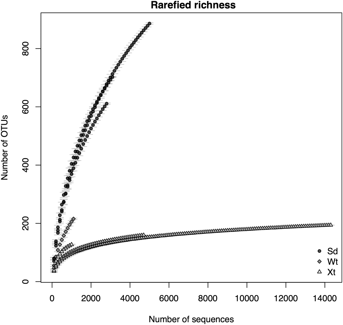
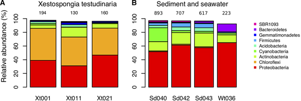
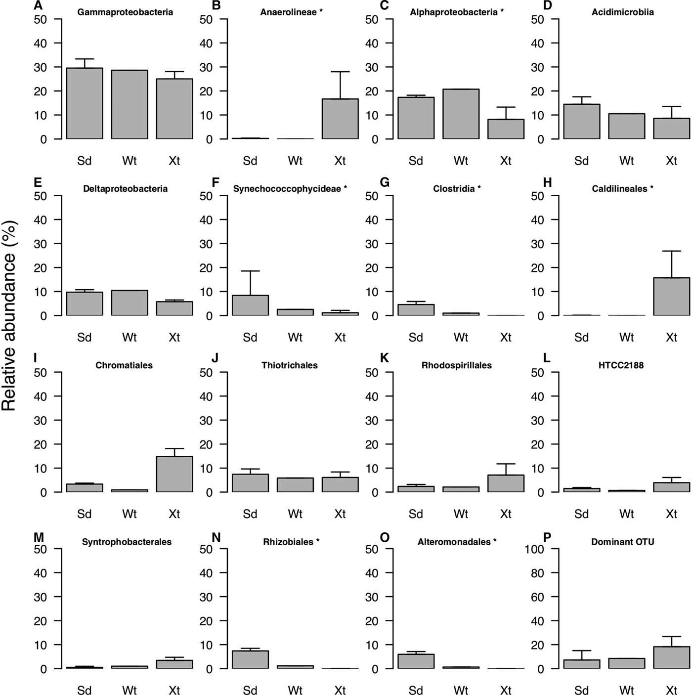
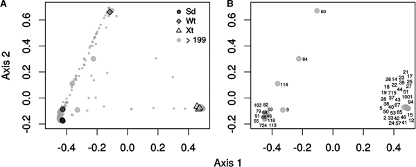
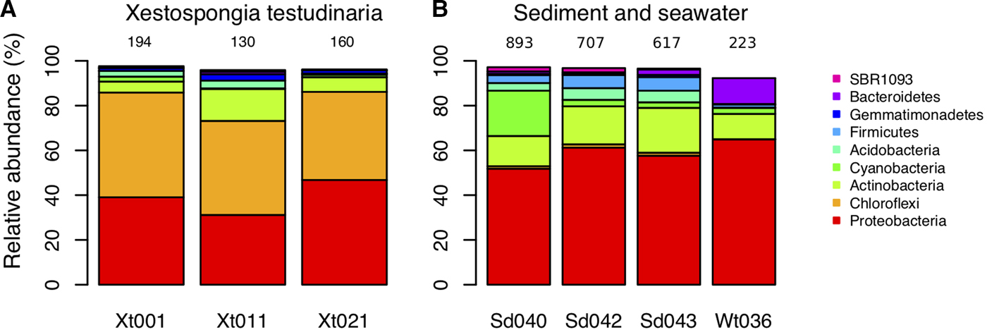
Barcoded pyrosequencing analysis yielded 32,473 sequences assigned to 1884 OTUs after quality control, OTU picking and removal of chimera, chloroplasts and mitochondria. OTUs were assigned to 37 phyla, 88 classes and 117 orders. Richness was highest in sediment and lowest in X. testudinaria samples (Figure 1). In order to compare samples of unequal abundances we calculated the rarefied richness for a fixed number of sequences (N = 1000; based on the number of sequences presented by the least abundant sample Xt011), OTU richness was 205.66 ± 3.58 in seawater, and varied from 95.86 ± 4.69 to 127.19 ± 1.55 in X. testudinaria and from 308.10 ± 10.68 to 404.65 ± 9.6 in sediment. Qualitatively, the Chao1 richness estimators revealed a pattern similar to that for rarefied richness, with the highest expected number of OTUs occurring in sediment, followed by seawater and X. testudinaria (Supplementary Table S1). However, the estimated Chao 1 richness was substantially higher than the observed OTU richness. The taxa composition analysis showed that at phylum level, Proteobacteria was by far the most abundant phylum (Figure 2). The phylum Chloroflexi was markedly more abundant in X. testudinaria than in other biotopes. Proteobacteria was the most abundant phylum in sediment and seawater (Figure 2). The composition analysis of lower taxa (Figure 3) showed that, at the class level, Gammaproteobacteria was the most abundant across all biotopes, and that the class Anaerolineae was most abundant in X. testudinaria. At the order level (Figure 3), the Caldilineales, Chromatiales, Rhodospirillales, HTCC2188 and Syntrophobacterales were more abundant in X. testudinaria while the orders Rhizobiales and Alteromonadales were more abundant in sediment. In line with the richness and taxa analysis, the PCO ordination (Figure 4) showed compositional differences among bacterial communities from different biotopes. While the first PCO axis separated samples of X. testudinaria from samples of sediment and seawater, the second PCO axis separated seawater samples from X. testudinaria and sediment samples. However, the use of only one seawater sample precludes the use of F-test to evaluate the statistical significance of the differences observed between this biotope, X. testudinaria and sediment samples. A PCO ordination analysis comprising only X. testudinaria and sediment samples (Supplementary Figure S1) showed that the compositional differences among these two biotopes were statistically significant (F 1,4 = 39.411, P = 0.107, R 2 = 0.91) and explained 91% of the variation in OTU composition.
Rarefaction curve of bacterial communities as a function of the number of 16S rRNA gene sequences obtained from Xestospongia testudinaria (Xt), sediment (Sd) and seawater (Wt) samples.

Relative abundance of the most abundant bacterial phyla in Xestospongia testudinaria (Xt), sediment (Sd) and seawater (Wt) biotopes. Total number of OTUs per sample is indicated on the top of each column.
Relative abundance of the most abundant bacterial classes (a–g) and orders (h–o) and the most abundant OTU (p) in Xestospongia testudinaria (Xt), sediment (Sd) and seawater (Wt) biotopes. The bars represent a single standard deviation. The dominant OTU represents the mean abundance for the single most dominant OTU in each biotope, thus not necessarily the same OTU. *indicate classes/orders with significant differences among sediment and X. testudinaria samples.

First two axes of the principal coordinates analysis (PCO) showing the variation of bacterial community composition in different biotopes. (a) Symbols represent samples from Xestospongia testudinaria (Xt), sediment (Sd) and seawater (Wt). Each grey dot represents a single OTU, where big dots represent OTUs ≥100 sequence reads and smaller dots represent OTUs <100 sequence reads. (b) Numbers represent the code of dominant OTUs referred to in Table 1; each dot represents a dominant OTU (≥100 sequence reads).

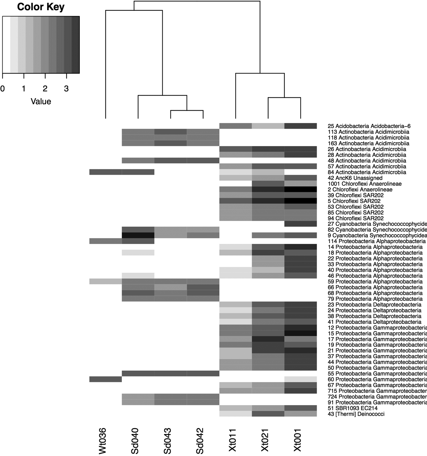
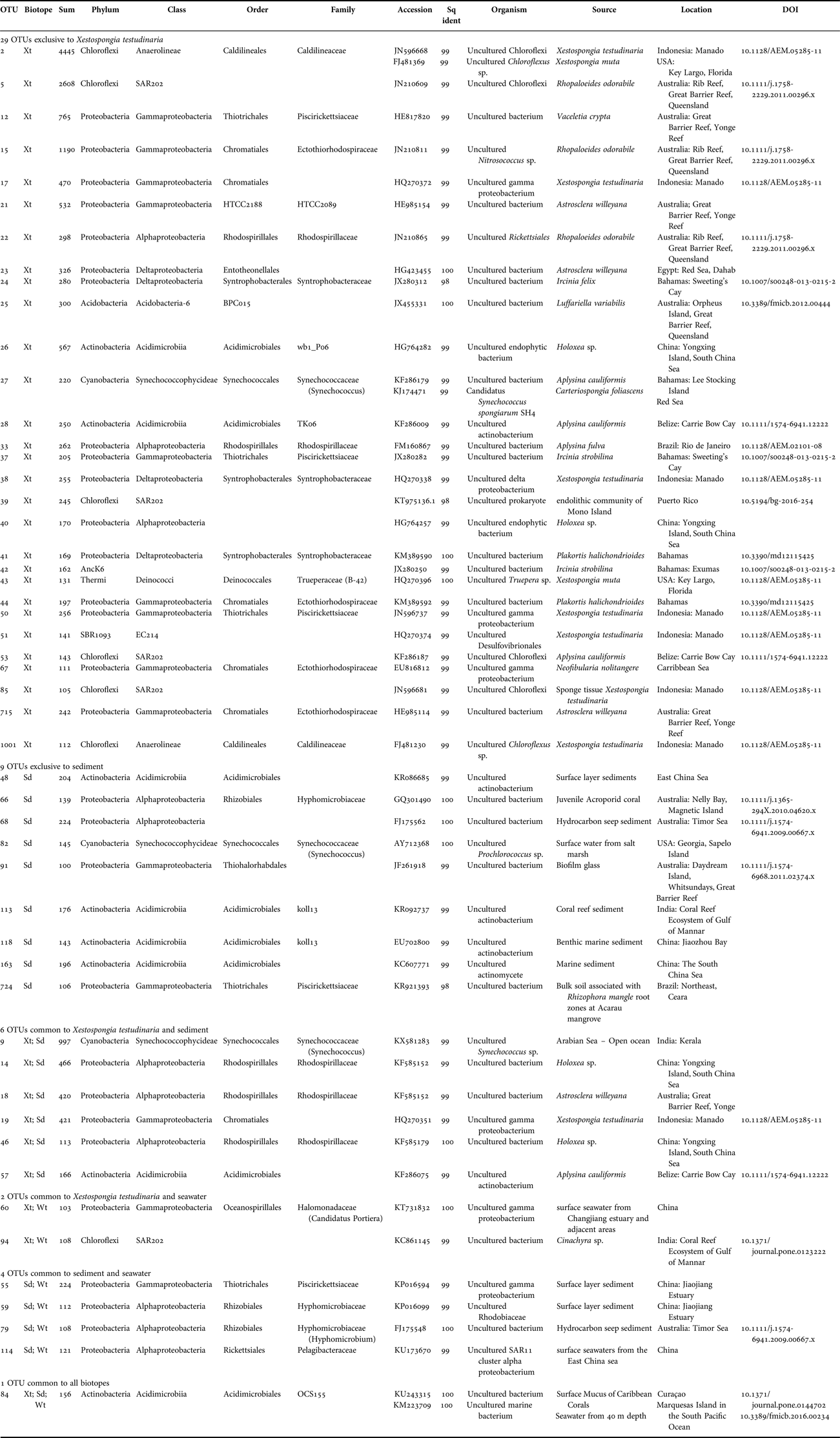
The in-depth bacterial composition analysis detected a total of 51 abundant OTUs (≥100 sequence reads) (Table 1). Of these, 29 OTUs were enriched (i.e. exclusive and highly abundant) in X. testudinaria and 9 in sediment biotopes. None of the most abundant OTUs were restricted to the seawater biotope (Figure 5, Table 1). OTU-84 was the only OTU detected in all three biotopes but was more abundant in sediment and seawater (Figure 5, Table 1). Closely related organisms to the most abundant OTUs were identified with BLAST (Table 1). OTU-84 was assigned to the OCS155 family (order Acidimicrobiales) and was closely related to two uncultured marine bacteria previously detected in coral mucous in Curaçao (accession number – Acc KU243315) and in seawater in the Marquesas Islands (Acc KM223709; French Polynesia). The two most abundant OTUs overall were OTUs 2 and 5 (4445 and 2608 sequences, respectively), both assigned to the Chloroflexi phylum and restricted to X. testudinaria (Table 1). OTU-15 was the third most abundant OTU (1190 sequences) and was also found exclusively in X. testudinaria and was assigned to the family Ectothiorhodospiraceae (order Chromatiales) (Figure 5, Table 1). Eight abundant OTUs were restricted to the sediment biotope (OTUs 48, 66, 68, 82, 91, 113, 118, 163 and 724; Table 1); most of these were closely related to organisms found in sediment samples (Table 1). The heatmap (Figure 5) showed a clear taxa restriction to X. testudinaria and suggests an evident clustering in the distribution of dominant OTUs.
Heatmap showing the abundance of 16S rRNA gene sequence reads of dominant OTUs (≥100 sequence reads). Samples are indicated on x-axis: Xt – Xestospongia testudinaria, Sd – sediment and Wt – seawater. Abundant OTUs are indicated on y-axis by OTU numbers, phyla and class assignment. The abundance of each OTU is indicated in greyscale ranging from white (low abundance or absent) to black (high abundance). The scale bar indicates abundance using a logarithmic base 10 scale.

List of the most abundant OTUs (≥100 sequence reads) and closely related organisms identified using BLAST search.

OTU: OTU number; Biotope: biotope or biotopes where the OTUs were found; Sum: number of sequence reads; Accession: accession number of closely related organisms identified using BLAST; Sq ident: sequence similarity of these organisms with our representative OTU sequences; Source: isolation source of organisms identified using BLAST; Location: sampling location of organisms identified using BLAST.
DISCUSSION
Overall, the richness analysis indicated a relatively low number of OTUs observed in X. testudinaria samples when compared with most recent results obtained with Illumina (e.g. Thomas et al., Reference Thomas, Moitinho-Silva, Lurgi, Björk, Easson, Astudillo-García, Olson, Erwin, López-Legentil, Luter, Chaves-Fonnegra, Costa, Schupp, Steindler, Erpenbeck, Gilbert, Knight, Ackermann, Lopez, Taylor, Thacker, Montoya, Hentsche and Webster2016). Probably, such a difference may be explained due to the higher yield of sequence reads obtained with this technology when compared with pyrosequencing. Luo et al. (Reference Luo, Tsementzi, Kyrpides, Read and Konstantinidis2012) compared Roche 454 and Illumina sequencing technologies on the same DNA sample and concluded that the higher number of reads (sequencing effort) obtained with Illumina resulted in a significantly higher number of OTUs. Nevertheless, in agreement with our results, a previous study showed that sponges (Stylissa carteri, Aaptos suberitoides and X. testudinaria) from the Misool coral reef system (Indonesia) had a much lower OTU richness than sediment. With a sequencing effort of 50,223 sequences (assigned to 3797 OTUs, for all biotopes), sponge richness was in general less than 250 OTUs (Cleary et al., Reference Cleary, Polónia, Becking, de Voogd, Purwanto and Gomes2018). Bayer et al. (Reference Bayer, Moitinho-Silva, Brümmer, Cannistraci, Ravasi and Hentschel2014) also showed that X. testudinaria from the Red Sea, at the minimum sampling effort (8925 sequences per sample), had less than 250 OTUs.
In the present study, bacterial communities associated with sediment and seawater biotopes were compositionally distinct from bacterial communities found in X. testudinaria. Proteobacteria was the most abundant phylum in sediment and seawater whereas Chloroflexi was most abundant in X. testudinaria. Previous studies showed that Chloroflexi is one of the most common and diverse bacterial phyla in sponges with numerous sponge-specific lineages (Brück et al., Reference Brück, Brück, Self, Reed, Nitecki and McCarthy2010; Schmitt et al., Reference Schmitt, Deines, Behnam, Wagner and Taylor2011). Some other studies suggested that members of this phylum may provide synergistic survival advantages to the host in nutrient-poor environments (Brock et al., Reference Brock, Madigen, Martinko and Parker1984; Brück et al., Reference Brück, Brück, Self, Reed, Nitecki and McCarthy2010). For example, Brück et al. (Reference Brück, Brück, Self, Reed, Nitecki and McCarthy2010) suggested that Chloroflexi might be able to provide organic photosynthates (e.g. glycerol) to sponge hosts in shallow waters, due to the ability of these bacteria to fix atmospheric CO2 in these environments. Members of the Chloroflexi phylum may also be able to synthesize polyketides (bioactive compounds) in marine sponges (Siegl & Hentschel, Reference Siegl and Hentschel2010), indicating that they might also play a role in sponge defence against predators.
At lower taxonomic levels, our results showed a specific enrichment of Anaerolineae (Chloroflexi) populations in X. testudinaria. Members of this family have been previously detected in a range of environments such as marine and freshwater sediments, marine sponges and human microbiota and are commonly described as versatile carbohydrate fermenters (Yamada et al., Reference Yamada, Sekiguchi, Hanada, Imachi, Ohashi, Harada and Kamagata2006; Cleary et al., Reference Cleary, Coelho, Oliveira, Gomes and Polónia2017; Campbell et al., Reference Campbell, Schwientek, Vishnivetskaya, Woyke, Levy, Beall, Griffen, Leys and Podar2014; de Voogd et al., Reference de Voogd, Cleary, Polónia and Gomes2015; Xia et al., Reference Xia, Wang, Wang, Chin and Zhang2016). In addition to Anareolineae, other members of the Chloroflexi cluster ‘subphylum IV’ (SAR202) were specifically enriched in X. testudinaria. Representatives of the SAR202 cluster have been found in a variety of habitats, including deep terrestrial subsurface, soil, marine sponges, seawater and freshwater environments (Dunbar et al., Reference Dunbar, Barns, Ticknor and Kuske2002; Hentschel et al., Reference Hentschel, Hopke, Horn, Friedrich, Wagner, Hacker and Moore2002; Morris et al., Reference Morris, Rappé, Urbach, Connon and Giovannoni2004). However, so far, no cultured representatives have been isolated and no information about their ecophysiology is known (Morris et al., Reference Morris, Rappé, Urbach, Connon and Giovannoni2004; Thrash et al., Reference Thrash, Seitz, Baker, Temperton, Gillies, Rabalais, Henrissat and Mason2016). Our results also showed that the order Caldilineales (Chloroflexi) was almost exclusively found in X. testudinaria. While Anaerolineae members are obligately anaerobic, Caldilineae members are facultative aerobes (Yamada et al., Reference Yamada, Sekiguchi, Hanada, Imachi, Ohashi, Harada and Kamagata2006). In general, despite the relatively high abundance and putative functional importance of members of Chloroflexi in sponge biotopes, there is limited information available on cultured representative strains belonging to this phylum (Brück et al., Reference Brück, Brück, Self, Reed, Nitecki and McCarthy2010; Schmitt et al., Reference Schmitt, Deines, Behnam, Wagner and Taylor2011).
The in-depth bacterial community analysis showed that OTU-84, assigned to family OCS155, was the only abundant OTU common to all three biotopes with high dominance in seawater and sediment samples. According to Fan et al. (Reference Fan, Reynolds, Liu, Stark, Kjelleberg, Webster and Thomas2012) and references therein, members of this family are frequently detected in seawater worldwide. The family OCS155 belongs to the order Acidimicrobiales, a group of planktonic photo-heterotrophic free-living microorganisms commonly inhabiting tropical and temperate areas (Ghai et al., Reference Ghai, Mizuno, Picazo, Camacho and Rodriguez-Valera2013; Mizuno et al., Reference Mizuno, Rodriguez-Valera and Ghai2015). Members of the Acidimicrobiales order include obligatory acidophilic organisms capable of ferrous iron, sulphur oxidation and ferric iron reduction (Hardoim et al., Reference Hardoim, Esteves, Pires, Gonçalves, Cox, Xavier and Costa2012). OTU-15 was specifically enriched in X. testudinaria and was assigned to the order Chromatiales (family Ectothiorhodospiraceae) and showed high sequence similarity with an uncultured clone of Nitrosococcus sp. (Acc JN210811) previously detected in the sponge Rhopaloeides odorabile in Rib Reef (Great Barrier Reef, Queensland; Pollock et al., Reference Pollock, Wilson, Johnson, Morris, Willis and Bourne2010). Members of this order, known as purple sulphur bacteria, can perform photosynthesis under anoxic conditions using hydrogen sulphide as electron donor and are able to fix molecular nitrogen (Proctor, Reference Proctor1997; Imhoff, Reference Imhoff, Brenner, Krieg, Staley and Garrity2005). Such functions might contribute to the sponge nitrogen metabolism and may explain why members of this group are consistently found in marine sponges (Pollock et al., Reference Pollock, Wilson, Johnson, Morris, Willis and Bourne2010; Montalvo & Hill, Reference Montalvo and Hill2011; Hardoim et al., Reference Hardoim, Esteves, Pires, Gonçalves, Cox, Xavier and Costa2012; Pita et al., Reference Pita, López-Legentil and Erwin2013; Della Sala et al., Reference Della Sala, Hochmuth, Teta, Costantino and Mangoni2014). Other dominant OTUs, belonging to the orders Entotheonellales (OTU-23) and Syntrophobacterales (OTUs 24, 38 and 41), were also specifically enriched in X. testudinaria. The known bacterial lineages related to the Entotheonellales are widely distributed in marine sponges and have a diverse metabolism; they are able to produce chemically diverse bioactive products of medical interest (Wilson et al., Reference Wilson, Mori, Rückert, Uria, Helf, Takada, Gernert, Steffens, Heycke, Schmitt, Rinke, Helfrich, Brachmann, Gurgui, Wakimoto, Kracht, Crüsemann, Hentschel, Abe, Matsunaga, Kalinowski, Takeyama and Piel2014). All OTUs assigned to the Syntrophobacterales were classified within the Syntrophobacteraceae family (Table 1). Members of this family are strictly anaerobic sulphate-reducing bacteria, mainly found in freshwater, sewage sludge and marine habitats (Kuever, Reference Kuever, Rosenberg, DeLong, Lory, Stackendt and Thompson2014). However, they have also previously been detected in sponges (Fiore et al., Reference Fiore, Baker and Lesser2013; Cleary et al., Reference Cleary, de Voogd, Polónia, Freitas and Gomes2015; Wang & Wu, Reference Wang and Wu2017).
Overall, dominant OTUs belonging to the Alphaproteobacteria class were detected in all three biotopes, however, OTUs belonging to the Rhodospirillaceae family were mainly found in X. testudinaria and sediment. All the dominant OTUs assigned to this family (OTUs 14, 18, 22, 33 and 46) showed high similarity with uncultured bacteria previously detected in a diverse range of sponge species (Rhopaloeides odorabile, Aplysina fulva, Holoxea sp. and Astrosclera willeyana) located across a broad geographic range (Table 1). OTUs 22 and 33 were exclusively detected in X. testudinaria (Table 1). Members of the Rhodospirillaceae are purple non-sulphur bacteria with diverse metabolic and nutritional properties, comprising lineages able to grow heterotrophically, photoheterotrophically under anoxic conditions in the light and chemoheterotrophically in the dark (Baldani et al., Reference Baldani, Videira, Teixeira, Reis, Oliveira, Schwab, Souza, Pedraza, Baldani, Hartmann, Rosenberg, DeLong, Lory, Stackendt and Thompson2014). A number of OTUs (OTUs 12, 37 and 50) assigned to the order Thiotrichales were also enriched in X. testudinaria. These OTUs were assigned to the Piscirickettsiaceae family and were similar (99%) to organisms previously detected in different sponge species including Vaceletia crypta, Ircinia strobilina and X. testudinaria across a broad geographic range (Table 1). The Piscirickettsiaceae family comprises metabolically diverse organisms, including chemoorganotrophs, methylotrophs and chemolithotrophic sulphur-oxidizers (Garrity et al., Reference Garrity, Bell, Lilburn, Brenner, Krieg, Staley and Garrity2005).
OTUs assigned to the order Rhizobiales were exclusively or predominantly found in sediment and were not detected in X. testudinaria. Members of this order can be found in several environments such as freshwater, soils, seawater, brackish water and sediment (Gliesche et al., Reference Gliesche, Fesefeldt, Hirsch, Brenner, Krieg, Staley and Garrity2005; Oren & Xu, Reference Oren, Xu, Rosenberg, DeLong, Lory, Stackendt and Thompson2014). Only one abundant OTU (OTU-114) was assigned to the order Rickettsiales (family Pelagibacteraceae) and was dominant in both sediment and seawater biotopes. Its closest relative was an uncultured bacterium previously detected in surface seawaters of the East China sea (Acc KU173670). Previous studies have reported that members of this family are adapted to low nutrient environments and may be one of the most abundant Alphaproteobacteria in seawater (Campbell et al., Reference Campbell, Fleisher, Sinigalliano, White and Lopez2015; Angly et al., Reference Angly, Heath, Morgan, Tonin, Rich, Schaffelke, Bourne and Tyson2016; Frank et al., Reference Frank, Garcia, Herndl and Reinthaler2016).
In general, our results showed that the structure of the bacterial communities associated with X. testudinaria in the Singaporean coral reef ecosystem followed a profile similar to other communities associated with X. testudinaria from the Red Sea, north and south Sulawesi, NW Java and Misool Indonesia (Lee et al., Reference Lee, Wang, Yang, Lafi, Al-Suwailem and Qian2011; Montalvo & Hill, Reference Montalvo and Hill2011; Bayer et al., Reference Bayer, Moitinho-Silva, Brümmer, Cannistraci, Ravasi and Hentschel2014; Cleary et al., Reference Cleary, de Voogd, Polónia, Freitas and Gomes2015, Reference Cleary, Polónia, Becking, de Voogd, Purwanto and Gomes2018; de Voogd et al., Reference de Voogd, Cleary, Polónia and Gomes2015). These studies revealed that X. testudinaria bacterial communities were also dominated by the phyla Chloroflexi, Proteobacteria and Actinobacteria.
CONCLUSION
In this study, we have provided an in-depth compositional analysis of bacterial communities inhabiting distinct biotopes in a Singaporean coral reef ecosystem. Our data revealed that biotope affects the richness, composition, abundance and putative function of bacterial communities. Overall, bacterial communities of X. testudinaria diverged greatly from the surrounding sediment and seawater bacterial communities. Our results also showed that Proteobacteria was the most abundant phylum in sediment and seawater while Chloroflexi was more abundant in X. testudinaria. Members of the order Caldilineales (fermentation of organic substrates), Chromatiales (purple sulphur bacteria), Rhodospirillales (purple non-sulphur bacteria) and Syntrophobacterales (sulphate-reducing bacteria) were enriched in X. testudinaria samples.
SUPPLEMENTARY MATERIAL
The supplementary material for this article can be found at https://doi.org/10.1017/S0025315418000188
FINANCIAL SUPPORT
This research was supported by Ecotech-Sponge (PTDC/BIA-MIC/6473/2014 – POCI-01-0145-FEDER-016531); LESS CORAL (PTDC/AAC-AMB/115304/2009). Thanks are also due to the financial support to CESAM (UID/AMB/50017 – POCI-01-0145-FEDER-007638), to FCT/MCTES through national funds (PIDDAC), and the co-funding by the FEDER, within the PT2020 Partnership Agreement and Compete 2020. V.O. (Post-Doc grant SFRH/BPD/101919/2014), A.R.M.P. (Post-Doc grant SFRH/BPD/117563/2016) and A.C.C.P. (PhD grant SFRH/BD/90340/2012) were financed by the Portuguese Foundation for Science and Technology (FCT).