Ectopia cordis is a rare congenital cardiac malformation defined as a complete or partial protrusion of the heart through a ventral defect. Reference Gabriel, Donnelly and Kuc1 Despite advances in neonatal cardiac surgery, complete ectopia cordis remains a surgical challenge with few long-term survivors. Here, we present a case report of successful balloon valvuloplasty using direct puncture of the heart for severe pulmonary stenosis in a premature infant with single ventricle and ectopia cordis.
Case report
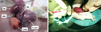
Fetal echocardiogram revealed ectopia cordis, single ventricle, severe pulmonary stenosis and small ductus arteriosus at 29 gestational weeks. At 31 weeks’ gestation, an emergency caesarean section was performed due to fetal distress. His birth weight was 1756 g, Apgar score was 3/6 and oxygen saturation (SpO2) was 80% on 100% O2 under respirator support. The sternum was entirely deficient and the heart was completely extruded out of the thoracic cavity without pericardial protection. The upper abdominal wall and anterior diaphragm were both absent, and part of the liver was also outside his body (Fig 1a). Epicardial echocardiogram showed a double inlet right ventricle without rudimentary left ventricle, pulmonary stenosis (severe valvular and mild infundibular) and malposition of the great arteries, and the ductus arteriosus was already closed. The echocardiographic findings prompted us to start a continuous intravenous infusion of lipo-prostaglandin E1 at 5.0 ng/kg/min.
(a) The sternum was entirely deficient and the heart was completely extruded out of the thoracic cavity without pericardial protection. The upper abdominal wall and anterior diaphragm were both absent, and a part of liver was also outside his body. RV = right ventricle; RA = right atrium; aAo = ascending aorta. (b) We punctured apex of the heart using 16-gauge intravenous catheter toward pulmonary valve under echo-guidance.

Soon after birth, the patient was kept in systematic treatment and supportive care, and a staged silo closure was performed using a polyurethane sheet to cover his heart. After this procedure, the patient showed increased hypoxaemia, and echocardiogram showed no flow of the ductus arteriosus. We explained to his parents that his prognosis was not optimistic; furthermore, he would require multiple surgeries in the future. We then discussed the therapeutic strategy with them, and they requested aggressive therapy to prolong his life. The risk of surgical systemic to pulmonary shunt was very high, and it seemed impossible to insert the catheter into the pulmonary artery using a percutaneous transvenous approach due to a very tortuous catheter course. Therefore, we decided to perform balloon valvuloplasty by direct puncture of the ventricular wall at 7 days of age. Informed consent was obtained from the parents.
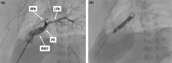
We punctured the apex of the heart using a 16-gauge intravenous catheter (internal diameter 1.30 mm; SURFLO Terumo, Tokyo, Japan) towards the pulmonary valve under echo guidance (Fig 1b). The backflow prevention valve was attached to the proximal end of the catheter to create the handmade ultrashort sheath. Hand injection angiography from the sheath revealed a small and thickened pulmonary valve with a 3.5 mm annular diameter and hypoplastic pulmonary arteries (Fig 2a). We carefully advanced a 0.012-inch double angle GT wire (Terumo, Tokyo, Japan) into the right pulmonary artery with reference to the angiogram, then inserted a 4 mm ARMADA 14 PTA catheter (Abbott Inc., United States of America) to the main pulmonary trunk through the handmade sheath and dilated balloon up to 10 atm (Fig 2b). Soon after inflation, the patient’s SpO2 improved from 70 to 85%. During the procedure, the patient showed no signs of arrhythmia or severe hypoxia.
(a) Hand injection angiography from the catheter. RPA = right pulmonary artery; LPA = left pulmonary artery; PV = pulmonary valve; RVOT = right ventricular outflow tract. (b) Inflated balloon up to 10 atm to dilate pulmonary valve.

At 14 weeks of age, the patient died of low output syndrome caused by kinking of the inferior vena cava after repeated silo closure.
Discussion
Pentalogy of Cantrell was first described in 1958 and consists of five major malformations; a defect within the lower part of the sternum, the anterior portion of the diaphragmatic portion of the pericardium, supra-abdominal wall defect, absence of the anterior diaphragm and various intracardiac malformations. This is an extremely rare syndrome with an incidence of 1 in 65,000 live births. Reference Cantrell, Haller and Ravitch2 Additionally, some severe cases have been associated with herniation of the heart through the defect of the diaphragm, resulting in ectopia cordis. Reference Gabriel, Donnelly and Kuc1
Patients with both pentalogy of Cantrell and ectopia cordis have an even worse prognosis. Reference Tongsong, Wanapirak, Sirivatanapa and Wongtrangan3 Intracardiac defects worsen the prognosis of patients with pentalogy of Cantrell and previous reports have emphasised high mortality in this group of patients requiring surgical intervention. Reference O’Gorman, Tortoriello and McMahon4
Only a few reports of transcatheter treatment for these cases have been reported to date. McMahon reported successful palliative right ventricular outflow tract stenting in a patient with pentalogy of Cantrell, double outlet right ventricle and pulmonary stenosis Reference McMahon and Walsh5 , while Galeczka reported successful atrial septal defect transcatheter closure in a patient with pentalogy of Cantrell and partial ectopia cordis. Reference Galeczka, Fiszer and Knop6 In these papers, the patients’ hearts were covered by skin, and procedures could be performed percutaneously. However, the percutaneous approach can be challenging due to low weight, limited vascular access and a very tortuous catheter course in certain patients. In such cases, combining operative and interventional approaches with direct puncture of the heart may facilitate the procedures. Reference Schmitz, Esmailzadeh and Herberg7 In our case, the whole heart was positioned outside the thoracic cavity completely, and it seemed impossible to insert the catheter into the pulmonary artery by percutaneous transvenous approach due to a very tortuous catheter course. Therefore, direct puncture of the heart was very well suited and we could accomplish the procedure quickly and safely.
This report represents the first transcatheter procedure using direct puncture of the heart in a patient with complete ectopia cordis and severe pulmonary stenosis. This particular procedure is a safe and useful palliation for complete ectopia cordis with cyanotic congenital heart disease.
Acknowledgements
We would like to thank Dr Hironori Matsuhisa (Department of Cardiovascular Surgery, Kobe Children’s Hospital) for help in performing this procedure and reporting this case and Editage (www.editage.com) for English language editing.
Financial support
This research received no specific grant from any funding agency or from commercial or not-for-profit sectors.
Conflicts of interest
The authors have indicated they have no potential conflicts of interest to disclose.
Ethical standards
This case report does not involve human and/or animal experimentation.