Unilateral absence of the pulmonary artery is a rare anomaly. Without any congenital heart defects it is called isolated unilateral absence of the pulmonary artery. Many case reports of isolated unilateral absence of the pulmonary artery in adults have been reported with atypical symptoms. However, there have been few reports of neonatal cases. We herein report two cases of unilateral absence of the pulmonary artery that were diagnosed just after birth, but that suffered complications from the treatment.
Case reports
Case 1
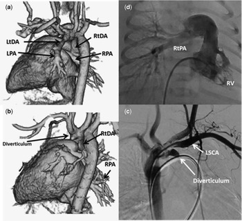
A 32-year-old woman was referred for foetal echocardiography at 29 weeks’ gestation because of a suspected double aortic arch in the foetus. The foetus was delivered vaginally at 41 weeks’ gestation. A postnatal echocardiogram showed a right aortic arch and undetected pulmonary artery branch. A computed tomography (CT) scan of the thorax with contrast revealed that the left pulmonary artery arose from the left subclavian artery through a left-sided patent ductus arteriosus (Fig 1). We considered a right aortic arch and the left-sided patent ductus arteriosus as double aortic arch. Then we diagnosed isolated unilateral absence of the pulmonary artery. We administered intravenous prostaglandin E1 (0.01 μg/kg/minute) to maintain the blood flow through the ductus arteriosus. A CT scan taken on the 3rd day after birth showed occlusion of the left-sided ductus arteriosus and did not visualise the left pulmonary artery. Right heart catheterisation revealed a mean pulmonary pressure of 46 mmHg, the pulmonary vascular resistance was calculated to be 5.7 Wood units, and the cardiac index was calculated to be 4.3 L/minute/m2, and suggested pulmonary hypertension. An angiogram revealed an absent proximal left-sided patent ductus arteriosus, but descending aortography showed a left-sided ductal diverticulum. To reopen the left-sided ductus arteriosus, prostaglandin E1 was administered up to 0.05 μg/kg/minute. The left-sided ductus arteriosus did not reopen. On the contrary, the right-sided ductus arteriosus opened widely. Heart failure was induced. After that, we stopped intravenous prostaglandin E1 and also checked that the right-sided ductus arteriosus was closed. The patient currently has no symptoms, and the pulmonary blood pressure was normal in the echocardiographic study.
( a and b ) Computed tomography (CT) images with three-dimensional (3D) volume-rendered reconstruction. ( a ) The left pulmonary artery (LPA) arising from the left subclavian artery through the left-sided patent ductus arteriosus (LtDA). ( b ). Occlusion of the left-sided patent ductus arteriosus and the absence of the left pulmonary artery. ( c and d ) Angiograms. ( c ) The right pulmonary artery from the right ventricle (RV). ( d ) The left-sided ductal diverticulum originating from the base of the left subclavian artery (LSCA) is noted.

Case 2
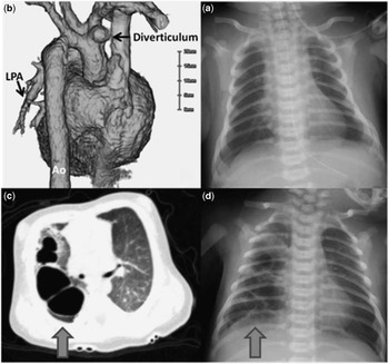
A female infant was delivered vaginally at 40 weeks’ gestation. She needed oxygen therapy from birth. A routine echocardiogram after birth showed undetected pulmonary artery branch, a dilated right atrium, and a flattening of the interventricular septum; suggested pulmonary hypertension without congenital heart defects; and suspected isolated unilateral absence of the pulmonary artery. A CT scan was performed on the 2nd day after birth, and it showed a normal bronchus and lung, an absent right pulmonary artery, and a right-sided ductal diverticulum originating from the base of the right common carotid artery (Fig 2). The echocardiogram and the CT confirmed the presence of isolated unilateral absence of the pulmonary artery. The infant had been started on prostaglandin E1 infusion (0.1 μg/kg/minute) to open the right-sided ductus arteriosus. Her breathing was, however, suppressed by the treatment and she had to be intubated for recurrent apnoea attacks. Therefore, the medical therapy was deemed unsuccessful and was discontinued. After 5 days, she was extubated and we checked the chest X-rays. There were already fibrotic and cystic changes in the right hemithorax. The CT of the chest in a lung window revealed multiple bullous emphysema, which were getting bigger and worse. For a while, she had suffered from a pneumothorax and had been treated with thoracic cavity drainage many times.
( a ) The radiographic findings of the chest after birth. There were no remarkable findings. ( b ) Computed tomography (CT) images with three-dimensional (3D) volume-rendered reconstruction showing an absent right pulmonary artery and a right-sided ductal diverticulum originating from the base of the right common carotid artery. ( c ) CT of the chest in a lung window showing multiple bullous emphysema. ( d ). Radiography of the chest after extubation showing fibrotic and cystic changes in the visualised right hemithorax.

Discussion
An abnormal sixth branch arch morphology causes isolated unilateral absence of the pulmonary artery. In such cases, if the ductus arteriosus closes after birth, the ipsilateral pulmonary artery would lose a source of blood supply without aortopulmonary collateral to that pulmonary artery. Unilateral absence of the pulmonary artery was first described by Frentzel.Reference Frentzel 1 However, there is no consensus on treatment of isolated unilateral absence of the pulmonary artery. Recently, Euloge et al described the therapeutic strategies used for children with an isolated unilateral absence of the pulmonary artery. Reference Krammoh 2 He showed that a “two-stage approach” is required to maintain the blood flow by ductus arteriosus stenting. This transcatheter approach is an alternative to systemic pulmonary shunting for such patients who require a subsequent end-to-end anastomosis. Several years later, their patients underwent anastomosis for the absence of the pulmonary artery to the main pulmonary artery with a graft (Dacron tube). He reported four patients who were all referred for tachypnoea at 1 day to 3.5 months (mean: 2 months) after birth. They underwent interventional therapy. Of the four patients, one patient who was born prematurely at 29 weeks’ gestation had suffered from heart failure with pulmonary hypertension after the procedure and died of severe sepsis. Peter Kruzliak et alReference Kruzliak 3 described similar strategies. They stated that, if the intrapulmonary arteries are well developed, a primary anastomosis with the central pulmonary artery can be made. If the intrapulmonary branches are small, a modified Blalock–Taussing shunt can be used. Welch et al reported that if it would be possible to perform a direct anastomosis in the neonatal period or early infancy, it should be recommended.Reference Welch and Hanley 4
In our cases, we chose a similar strategy to maintain the blood flow through the ductus arteriosus. We used a prostaglandin E1 for both cases, but it could not reopen the ductus arteriosus connecting the ipsilateral pulmonary artery. Moreover, case 1 developed congestive heart failure due to the ductus arteriosus connecting the pulmonary artery on the opposite side. Case 2 suffered from an unidentified pleural effusion caused by ventilator therapy for a side effect of prostaglandin E1. In this case, we think that the intervention to open the ductus arteriosus had been already closed and was associated with the adverse effect. The reperfusion due to the sudden pulmonary blood flow was associated with a risk of pulmonary haemorrhage and pulmonary emphysematous changes.Reference Nabo 6
Although they had not developed cardiac or respiratory symptom, we started medical therapy in our cases. It is difficult to decide whether or not treatment is necessary for unilateral absence of the pulmonary artery without symptoms. It is necessary to carefully choose the way to open a closed ductus arteriosus, because it may adversely affect the pulmonary tissue. In a paediatric case, especially neonate or infant, we consider that we should not use prostaglandin E1. We recommend intervention way such as ductus arteriosus stenting or direct anastomosis for term neonates or infants. In a premature or preterm neonate case, the patient has high risk of persistent pulmonary hypertension or suprasystemic pulmonary hypertension caused by bronchopulmonary displasia. Therefore, to maintain the blood flow through the ductus arteriosus promotes pulmonary hypertension and heart failure. That is why we should not choose an invasive way for them and should observe them carefully.
Acknowledgement
None.
Financial Support
This research received no specific grant from any funding agency, commercial, or not-for-profit sectors.
Conflicts of Interest
None.
Ethical Standards
This work complies with the ethical standards of the relevant national guidelines on human experimentation and with the Helsinki Declaration of 1975, as revised in 2008, and has been approved by the Institutional Committees, Osaka Medical Centre, and Research Institute for Maternal and Child Health.