Introduction
Cranial fasciitis of childhood was first described as a subtype of nodular fasciitis in a series of nine cases by Lauer and Enzinger in 1980.Reference Lauer and Enzinger1 The condition arises from the deep fascia, the periosteum or the fibromembranous layer covering the skull fontanelles and sutures. Although it has been reported in at least one adult, presentation is typically between birth and 11 years of age, with the average age of onset being three years. There is a 2:1 male predominance. Prior trauma and a familial predisposition have been postulated as predisposing factors.Reference Adler and Wong2 Approximately 40 cases have been described in the literature.Reference Keyserling, Castillo and Smith3
Case report
A 10-week-old male infant presented to our department with a three-day history of a right peri-auricular swelling. He had a one-week history of coryzal prodrome symptoms. He was otherwise systemically well, with no associated ear discharge or history of trauma. He had been delivered at full term via a normal vaginal delivery and was up to date with his immunisations.
Clinical examination revealed a non-tender peri-auricular swelling anterior, superior and posterior to the pinna. The swelling measured 3.0 × 4.0 cm and displaced the pinna laterally, with no signs of inflammation. The tympanic membrane was obscured by wax. There were no palpable lymph nodes in the neck and no hepatosplenomegaly.
The total white cell count was at the upper limit of normal, at 11.9 × 10⁹/l, the C-reactive protein was 22 mg/l and the erythrocyte sedimentation rate was 46 mm/hour. The child had a hypochromic anaemia with a haemoglobin level of 8.8 g/l. His absolute reticulocyte count was elevated, at 119.3 × 10⁹/l. All other blood tests were normal.
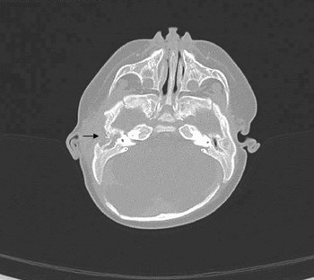
A contrast computed tomography (CT) scan revealed opacification of the right middle-ear cavity and mastoid air cells, with bony destruction of the lateral mastoid cortex. There was marked thinning and irregularity of the tegmen tympani. A soft tissue swelling, with a central area of low attenuation measuring 3 × 1.5 cm was visible lateral to the petrous temporal and mastoid process (Figure 1). This was consistent with a subperiosteal abscess secondary to mastoiditis, although an underlying neoplastic process could not be excluded. There was no evidence of any intracranial abnormality.
Axial computed tomography scan showing a soft tissue swelling lateral to the right petrous temporal and mastoid process, with bony destruction at the lateral mastoid cortex (arrow).

The child was initially treated with intravenous cefotaxime and metronidazole. After 24 hours, in view of the lack of clinical improvement and the concern about a possible underlying malignant process, surgical exploration was scheduled.
At the time of surgery, a dull tympanic membrane was seen and thick, non-infected glue was obtained at myringotomy. The temporalis muscle was exposed via a post-aural approach. Its lower border superior to the external auditory meatus was thickened and white. An incision was made in this area, releasing several millilitres of pus from an apparent abscess cavity. A sample was sent for microbiological analysis. A biopsy was taken from the thickened temporalis muscle surrounding the abscess cavity. The underlying bone was exposed and noted to appear ‘moth-eaten’, with a small defect in the cortex. A limited cortical mastoidectomy was performed, and no further collection was identified. A drain was placed and the wound closed.
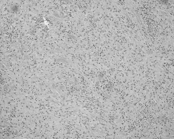
There was no growth on microbiological culture of the pus specimen. Histological examination of the biopsy showed fragments of adipose and connective tissue with small fragments of skeletal muscle, containing an area of spindle proliferation (Figure 2). The spindle cells showed plump, slightly irregular cytoplasm, but no evidence of necrosis, mitotic activity or nuclear atypia. The spindle cells were strongly positive for smooth muscle myosin and CD68, confirming their myofibroblastic–fibroblastic lineage. Staining for desmin, factor 13a, CD34, CAM5.2, S100 and langerin was negative. The histopathological features were consistent with a diagnosis of cranial fasciitis of childhood. There was no evidence of malignancy or necrosis, and no features of acute or chronic inflammation were seen in the biopsy material.
Photomicrograph of the lesion showing bundles of spindle cells with scattered occasional lymphocytes (H&E; ×10).

The post-operative period was uncomplicated, and the drain was removed after 72 hours. After discharge, the swelling slowly settled.
At review six weeks post-operatively, the child remained well, and the swelling had resolved completely.
Discussion
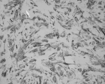
Cranial fasciitis of childhood represents a pseudosarcomatous, self-limiting, reactive proliferation of fibroblasts and myofibroblasts. Although benign, it may infiltrate the outer (and less frequently the inner) skull table. As a sub-set of nodular fasciitis, it shares identical microscopic features. Macroscopically, lesions are grey-white, rubbery, relatively well circumscribed and non-encapsulated. Microscopically, they consist of spindle-shaped fibroblasts in a myxoid matrix. Reactive bone formation and areas of hyalinisation are common. Immunohistochemical staining in previous cases has shown positive reactions with antibodies to α1-antichymotrypsin and smooth muscle actin (Figure 3). Positive staining for alcian blue indicates that hyaluronic acid is the main component of the myxoid background. The α1-antichymotrypsin reaction identifies fibroblasts with phagocytic activity and scattered macrophages, and the reaction to smooth muscle actin indicates the presence of myofibroblasts. These findings have been confirmed on electron microscopy by Patterson et al. Reference Patterson, Moran and Konerding4 It can be difficult to distinguish clinically between cranial fasciitis of childhood and rhabdomyosarcoma, and an incisional biopsy may be needed to confirm the histological nature of the lesion.
Immunocytochemical analysis for smooth muscle actin; photomicrograph shows positive staining in the spindle and stellate-shaped cytoplasm of the cells (×40).

• Cranial fasciitis of childhood is a benign condition and a rare variant of nodular fasciitis
• This paper describes the case of a 10-week-old infant with symptoms and signs consistent with a subperiosteal abscess complicating acute mastoiditis
• The distinctive histopathological features of cranial fasciitis, coupled with an awareness of the condition, are crucial to establishing a definitive diagnosis and, in turn, to instituting appropriate management
Cranial fasciitis of childhood typically presents as a firm, rapidly growing, subcutaneous mass ranging from 1 to 3 cm in size.Reference Lee, Kim and Shin5, Reference Larralde, Boggio, Schroh and Cusumano6 It is usually single, although a case of two synchronous lesions has been reported.Reference Adler and Wong2 Following initial rapid growth, the lesion stabilises after approximately two months. It occurs predominantly in the temporoparietal region.Reference Lauer and Enzinger1 Other associated symptoms can occur due to a mass effect, such as proptosis, diplopia, facial nerve palsies, deafness and hemiparesis.Reference Keyserling, Castillo and Smith3, Reference Clap, Dodson, Pickett and Lambert7
Radiographically, the lesion can be identified on CT or magnetic resonance imaging (MRI) as a well defined soft tissue mass in association with a lytic bone defect, with surrounding sclerotic edges.Reference Keyserling, Castillo and Smith3, Reference Clap, Dodson, Pickett and Lambert7 The lesion enhances with contrast and may contain areas of calcification.Reference Keyserling, Castillo and Smith3 It should be noted that intracranial extension and dural involvement of cranial fasciitis of childhood have been reported in approximately one-third of cases,Reference Keyserling, Castillo and Smith3 supporting a role for CT or MR imaging.
In a child, the differential diagnosis includes the following conditions: Langerhan's cell histiocytosis, fibromatosis, primary bone neoplasms, haemangioma and rhabdomyosarcoma.
The current treatment of choice is surgical excision of the mass, with or without local resection or curettage of the underlying bone.Reference Keyserling, Castillo and Smith3, Reference Lee, Kim and Shin5, Reference Clap, Dodson, Pickett and Lambert7 Results have been equally effective with an incisional biopsy.Reference Clap, Dodson, Pickett and Lambert7 Recent reports have shown systemic and intra-lesional steroid treatment to be equally as effective, particularly with multiple lesions.Reference Lee, Kim and Shin5 Recurrence of the lesion is rare, even with incomplete excision, probably due to spontaneous regression, which has been exhibited by at least one case.Reference Lee, Kim and Shin5