I. INTRODUCTION
Rutile has been found to be potential material for photocatalytic antibacterial activity, Reference Senna, Myers, Aimable, Laporte, Pulgarin, Baghriche and Bowen1 gas sensors, Reference Wu2,Reference Karim, Yeum, Lee and Lim3 batteries, Reference Xu, Jia, Cao and Zhang4,Reference Baudrin, Cassaignon, Koelsch, Jolivet, Dupont and Tarascon5 solar cells, Reference Kim, Jang, Vittal, Lee and Kim6,Reference Byun, Vittal, Kim and Kim7 capacitors, Reference Karim, Lim, Lee, Kim and Yeum8,Reference Karim, Lee, Cheong, Park, Oh and Yeum9 high-k gate insulator, Reference Kadoshima, Hiratani, Shimamoto, Torii, Miki, Kimura and Nabatame10 and so on due to low cost production, thermal stability, and most importantly for its semiconductive, dielectric behavior. Potential photoluminescence Reference Abazovic, Comor, Dramicanin, Jovanovic, Ahrenkiel and Nedeljkovic11,Reference Wu, Shih and Wu12 and chemiluminescence Reference Zhu, Shi, Zhang, Zhang and Zhang13,Reference Corrales, Allen, Edge, Sandoval and Catalina14 properties of rutile provide the additional advantage toward its diversified applications. Rutile is the most stable one among the TiO2 polymorphs and also the most abundant one. Rutile nanoparticle finds more attractive applications for controlled morphology and high specific surface area. Among the synthesis methods of rutile nanoparticles, low-temperature synthesis method is very much attractive to the research community due to low production cost, high purity, soft particle aggregation, higher specific surface area, and uniform shape of the nanoparticles. Several methods have been developed using varying precursors and strategies at reaction temperatures ranging from 20 to 100 °C. Reference Yin, Li, He and Sato15–Reference Park, Cho, Kim and Kim17 Previously, all methods were high-temperature methods at the temperature ranging from 130 to 1400 °C. Reference Ovenstone18,Reference Navrotsky and Kleppa19
Different methods produce rutile nanoparticles of different morphologies ranging from spherical, oval, rod shape, needle shape, shuttle-like shape to even uneven shape. Reference Yin, Li, He and Sato15 But highly organized crystalline nanoparticles with uniform porosity at mesoporous to micro level are highly desired materials for various applications. Here, the organized crystalline microporous metal-oxide semiconductor (rutile) nanoparticles synthesized by the low-temperature dissolution-reprecipitation process (LTDRP) has been reported.
II. EXPERIMENTAL SECTION
A. Reagents
All the chemicals used in this experiment are of analytical grade. Titanium tetraisopropoxide (TTIP) and hydrochloric acid have been purchased from Sigma Aldrich.
B. Synthesis of nanoparticles
The organized crystalline microporous metal-oxide semiconductor (rutile) nanoparticles have been synthesized by a modified method of Yin et al. Reference Yin, Li, He and Sato15 TTIP of 0.005 mol has been added slowly to 18.36 mL water at 24 °C under stirring. White slurry has been produced instantly. HCl up to 1M acidic concentration has been added to that freshly prepared slurry. Stirring has been continued for 4 h at the same temperature and a clear solution obtained. It has been subjected to aging up to 3 days at 24 °C and white rutile nanoparticles have been obtained.
C. Instruments used
Instruments that have been used here are: a) high-resolution transmission electron microscope (HR-TEM; Philips model CM 200, FEI Company, Eindhoven, Netherlands), b) x-ray diffraction (XRD; Philips model X'Pert APD with a Cu Kα source), c) BET surface area and pore size analyzer (Quantachrome Autosorb-3a; at 77 K by N2 adsorption after pretreating them at over 300 °C for 1 h to remove adsorbed water), d) Raman spectrometer (Almega X/Thermo), e) elemental analyzer (CE Instruments, model FISON EA-1110), and f) impedance analyzer (HP4291B RF impedance/material analyzer; 1–1.8 GHz).
III. RESULTS AND DISCUSSION
A. X-ray diffraction data analysis
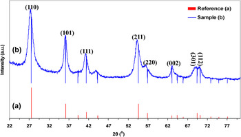
The XRD data (Fig. 1) show all the characteristic peaks of rutile completely matched with the standard reference (JCPDS—International Centre for Diffraction Data). 20 This implies that the synthesized TiO2 nanoparticles are very pure and consist of only rutile polymorph of titania.
XRD pattern of rutile nanoparticles: (a) standard reference 20 and (b) synthesized in this work.

The crystallite size of the rutile nanoparticles is calculated by Scherrer's relation Reference Klug and Alexander21 (using the 110 face):
 $$D = {{K\lambda } \mathord{\left/ {\vphantom {{K\lambda } {\beta \cos \theta }}} \right. \kern-\nulldelimiterspace} {\beta \cos \theta }}\quad ,$$
where K = 0.89, D represents the crystallite size (nm), λ is the wave length of Cu Kα radiation, and β is the corrected value at half width of the diffraction peak. This can be attributed to the fact that “crystallite size” is not synonymous with “particle size” while XRD is sensitive to the crystallite size. Reference Danilchenko, Kukharenko, Moseke, Protsenko, Sukhodub and Sulkio-Cleff22 The characteristic peak of rutile at 2θ = 27.316° has been chosen to calculate the average grain size of rutile nanoparticles and obtained as 11.3 nm.
B. Transmission electron microscopy data analysis
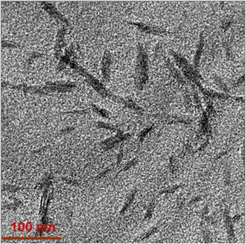
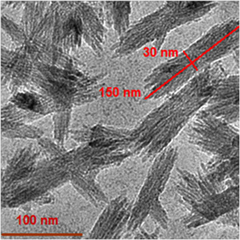
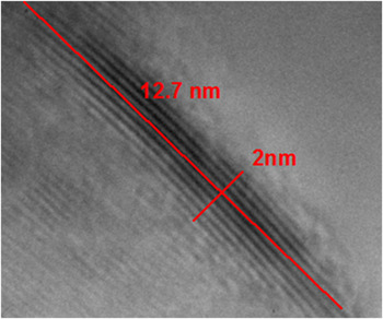
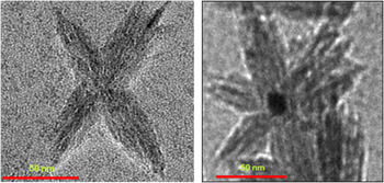
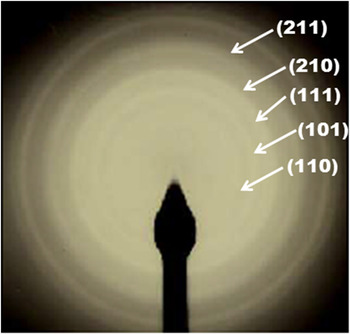
Transmission electron micrographs (TEM) of the rutile nanoparticles show that at the initial stage of aging (e.g., 4 h aged), the particles are not uniform (Fig. 2); but at the longer aging period (e.g., 72 h aged), they are quite uniform with dimensions 25–30 nm in diameter and 130–150 nm in length (Fig. 3). HR-TEM of the rutile nanoparticles (Figs. 4 and 5) shows that the rutile nanoparticles are composed of crystallites in a highly organized fashion. The crystallites of the rutile nanoparticles are of dimensions 1.5–2.5 nm in diameter and 10–13 nm in length. Selected area electron diffraction (SAED) image (Fig. 6) of rutile nanoparticles (72 h aged) confirms the crystalline structure of the particles.
TEM image of rutile nanoparticles obtained after 4 h of aging.

TEM image of rutile nanoparticles obtained after 72 h of aging.

HR-TEM image of a rutile crystallite after 24 h of aging.

HR-TEM image of aggregated rutile nanoparticles after 48 h of aging.

SAED image of synthesized rutile nanoparticles.

C. Surface area and pore size analyzer
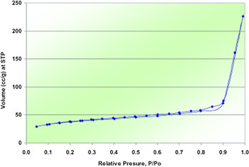
The surface area of the rutile nanoparticles (72 h aged) has been determined from the corresponding nitrogen adsorption/desorption isotherms (Fig. 7). The BET surface area of rutile nanopowder is 132.2 m2/g.
N2 adsorption desorption isotherms for rutile nanoparticles after 72 h of aging.

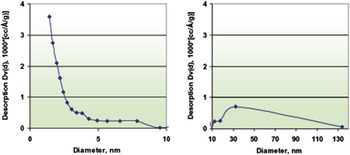
The pore volume and pore size distribution (Fig. 8) are 0.34 cm3/g and 1.4 nm, respectively, determined using the Barrett–Joyner–Halenda (BJH) model from the data of N2 isotherms. The pore size distribution analysis of both the samples shows that pore diameters concentrate in the range of 1–10 nm with a high trend in the microporous range which is in good agreement with the HR-TEM observations. The specific surface area and porosity of the rutile nanoparticles have been summarized in Table I.
Pore size distribution of rutile nanoparticles (72 h aged) determined from the N2 desorption isotherm using the BJH method.

Specific surface area and porosity of rutile nanoparticles after 72 h of aging.

a BET surface area (S BET)
b Pore volume (V p) and average pore diameter (D p) [BJH desorption].
D. Raman spectra
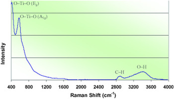
From Fig. 9, the O–Ti–O network peaks over 400–700 cm−1 are characteristics of TiO2 (72 h aged) in correspondence to the Raman active phonons of rutile 441 cm−1 (E g) and 579 cm−1 (A 1g). Reference Watson, Beydoun, Scott and Amal23,Reference Porto, Fleury and Damen24 The C–H stretching peaks that are clearly evident at around 2900 cm−1 confirm the presence of the alkyl groups (–C3H7) on the surface of rutile nanoparticles. The O–H stretch peak (3000–3700 cm−1) attributes to the large amount of water molecules adsorbed on the surface of the particles and condensed in the pores which is very consistent with the Raman data. This also indicates that the nanoparticles are highly porous.
Raman spectra of rutile nanoparticles after 72 h of aging.

E. Elemental analysis
The elemental analysis is also proving the presence of –C3H7 and –OH groups on the rutile nanoparticle surface (72 h aged). Approximately, only 0.08% isopropyl group is present in the sample. Remaining 0.0729% hydrogen quite certainly comes from bound –OH groups and adsorbed water molecules. The elemental analysis data of rutile nanoparticles are given in Table II.
Elemental analysis of rutile nanoparticles after 72 h of aging.

F. Postulations on the mechanism of crystal growth of rutile nanoparticles
When TTIP was added in distilled water, hydrolysis reaction proceeded fast leading to the formation of amorphous Ti(OH)4 by the following reaction Reference Wang, Yuan and Egerton16,Reference Hanaor and Sorrell25 :
 $${\rm{Ti}}{\left( {{{\rm{C}}_3}{{\rm{H}}_7}{\rm{O}}} \right)_4} + 4{{\rm{H}}_2}{\rm{O}} \to {\rm{Ti}}{\left( {{\rm{OH}}} \right)_4} + 4{{\rm{C}}_3}{{\rm{H}}_7}{\rm{OH}}\quad {\rm{Hydrolysis}}\quad {\rm{,}}$$
 $${\rm{Ti}}{\left( {{\rm{OH}}} \right)_4} \to {\rm{Ti}}{{\rm{O}}_2} + 2{{\rm{H}}_2}{\rm{O}}\quad {\rm{Condensation}}\quad {\rm{.}}$$
In the present study, from the observation it is assumed that the amorphous titania is dissolved to a transparent titania gel in stirred acidic solutions, and reprecipitated during the aging time over 72 h at room temperature. The reprecipitated particles are identified as crystalline particles by XRD and TEM.
According to Yin et al., Reference Yin, Li, He and Sato15 the crystallization of titania proceeds from the reprecipitation from transparent solution of hydrated ion [Ti(H2O)6]4+. Before reprecipitation, titanium does not exist in the form of Ti4+ cation but exist as a sixfold coordinated hydrated ion [Ti(H2O)6]4+. These hydrated ions are dehydrated and polymerized to [(H2O)4Ti–(OH)2–Ti(H2O)4]6+, [(H2O)4TiO2 Ti(H2O)4]4+, and finally TiO6 octahedra. It might be suggested that thermodynamically stable rutile nucleus with compact texture is formed by very slow precipitation rate at low temperatures. In this case, the rearrangement of TiO6 octahedra was realized slowly during a long period of time, and finally, the rutile phase with a compact texture was formed. Wang et al. Reference Wang, Yuan and Egerton16 observed that the longer addition time not only increases the fraction of rutile crystals but also alters the particle morphology. Differing from the above-mentioned mechanism, we have found that the rutile phase starts to form from the beginning (within 4 h) of the reprecipitation starts (Fig. 2). At 72 h of aging, the nanoparticles are found to be as quite matured crystalline rutile nanoparticles (Fig. 3).
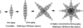
The XRD data are quite consistent with that one obtained from HR-TEM data studies. The average grain size of rutile nanoparticles has been obtained as 11.3 nm by XRD. Again the rutile crystallite dimensions are 1.5–2.5 nm in diameter and 10–13 nm in length, whereas the dimensions of rutile nanoparticles are 25–30 nm in diameter and 130–150 nm in length. So it indicates that the crystallites are the building blocks for the formation of the self-organized nanoparticles. The crystal growth is not continuous, i.e., the crystallites are arranged one after another and interconnected. Thereby the nanoparticles are microporous in nature by dint of intercrystallite voids. This postulation is well consistent with the HR-TEM and BJH porosity measurement data for the nanoparticles. The HR-TEM images also evaluate that some of the nanoparticles aggregate in an orderly fashion to give star-shaped or flower-shaped particles. In Fig. 10, the formation of self-organized rutile nanoparticles and ordered aggregation has been presented.
Schematic representation of the formation of rutile nanoparticles.

G. Electrical properties
Table III has been shown that the dielectric constant of rutile nanoparticles (72 h aged) has been found very high as 57 at 10 MHz AC frequency and DC conductance as 2.3 × 10⁻⁶ S/cm. This highly organized crystalline microporous metal-oxide semiconductor with high dielectric properties might suggest using for gas sensor applications.
Resistance (ρ), conductance (σ), and real part of relative permittivity (ε′) of the rutile nanoparticles.

IV. CONCLUSION
To the best of our knowledge, the LTDRP described here in harmony with Yin et al. Reference Yin, Li, He and Sato15 is the most economic route to synthesis of pure rutile nanoparticles. High yield (>80% at 3 days aging) and high specific surface area (>130 m2/g) are the additional attractive features. Here, we have reported another very attractive feature of the rutile nanoparticles synthesized following that method: these nanoparticles are of highly self-organized crystalline microporous structure. We postulate that in conjunction with the luminescence property, this semiconductive (2.3 × 10⁻⁶ S/cm) and high dielectric constant (57 at 10 MHz) material with the large volume microporosity (average pore diameter, D p = 1.4 nm; BJH desorption) and organized crystalline structure holds very good prospects for different applications.
ACKNOWLEDGMENT
The authors gratefully acknowledge the support from the Deanship of Graduate Research (DSR) program by King Saud University.