Introduction
Nitric oxide (NO) is a highly reactive unstable free radical metabolite. It is generated through oxidation of the amino acid l-arginine to l-citrulline, a reaction catalysed by NO synthase (NOS). This enzyme is found in vertebrates as two main types: constitutive (cNOS) and inducible (iNOS) (Alderton et al., Reference Alderton, Cooper and Knowles2001). In some insects NO is inducibly synthesized by the iNOS in response to infections, as in Anopheles gambiae (Hillyer and Estévez-Lao, Reference Hillyer and Estévez-Lao2009), Anopheles stephensi (Luckhart et al., Reference Luckhart, Vodovotz, Cui and Rosenberg1998) and Rhodnius prolixus (Whitten et al., Reference Whitten, Mello, Gomes, Nigam, Azambuja, Garcia and Ratcliffe2001). In immune-reactive insects, NO is frequently produced by activated cells in association with the reactive oxygen intermediates (ROI) •O − 2 and H2O2. These ROI, particularly H2O2, are activators or secondary messengers of NF-κB (Nappi et al., Reference Nappi, Vass, Frey and Carton2000). To initiate cytotoxic activity, NO reacts with both •O − 2 to form peroxynitrite (ONOO−) (Halliwell and Gutteridge, Reference Halliwell, Gutteridge, Halliwell and Gutteridge1999) and H2O2 to form the highly reactive hydroxyl radical (•OH) (Hogg et al., Reference Hogg, Darley-Usmar, Wilson and Moncada1992). At physiological pH, both NO and H2O2 can readily diffuse from their tissues of origin to damage lipids, proteins and DNA (Nappi et al., Reference Nappi, Vass, Frey and Carton2000). Therefore, NO is toxic to a wide-spectrum of pathogens, including bacteria, fungi, protozoa and viruses (Colasanti et al., Reference Colasanti, Gradoni, Mattu, Persichini, Salvati, Venturini and Ascenzi2002; Faraldo et al., Reference Faraldo, Sá-Nunes, Del Bel, Faccioli and Lello2005). Also, NO performs a signalling function, as indicated in Drosophila melanogaster, in the induction of host defences to Gram-negative bacteria (Foley and O'Farrell, Reference Foley and O'Farrell2003), in neuroinflammatory responses (Ajjuri and O'Donnell, Reference Ajjuri and O'Donnell2013) and in chemosensory transduction and functioning of the odorant receptor cells in the flesh fly Neobellieria bullata (Wasserman and Itagaki, Reference Wasserman and Itagaki2003).
Lysozymes (E.C. 3.2.1.17), 1,4-N-acetylmuramidases, catalyse cleavage of peptidoglycan's β-1,4-glycosidic bond between the alternating linked residues of N-acetylmuramic acid (MurNAc) and N-acetylglucosamine (GlcNAc) that often comprise a principal component of bacterial cell wall, resulting in its depolymerization (Mohamed et al., Reference Mohamed, Elmogy, Dorrah, Yousef and Bassal2013). Lysozymes are one of the components of the insect humoral immune system that respond to bacterial challenge (Hillyer, Reference Hillyer2016) and are upregulated upon infection (Mohamed et al., Reference Mohamed, Zhang, Dorrah, Elmogy, Yousef, Bassal and Duvic2016). This upregulation was reported to take place through injection of the bacterial cell wall components, peptidoglycan (PGN) and lipopolysaccharides (LPS) or fungal β-1,3-glucans such as laminarin (Morishima et al., Reference Morishima, Horiba, Iketami, Nishioka and Yamano1995; Mohamed et al., Reference Mohamed, Elmogy, Dorrah, Yousef and Bassal2013).
Immune mediators of insects play critical roles in relaying immune signals from non-self-recognition to immune effectors, such as fat body and haemocytes (Gillespie et al., Reference Gillespie, Kanost and Trenczek1997). Among these mediators, the eicosanoids are oxygenated derivatives of arachidonic acid (AA) and other C20 polyunsaturated fatty acids. The chemistry and biosynthesis pathways of eicosanoids have been detailed elsewhere (Stanley, Reference Stanley2000). Briefly, cyclooxygenases (COXs) oxygenate free AA to form prostaglandins (PGs), while hydroxyeicosanoids and leukotrienes (LTs) are produced from AA via lipoxygenases (LOXs), and epoxytrienoic acid formed by P450 epoxygenases. Eicosanoids act in mediating cellular immune responses in insects (Stanley and Kim, Reference Stanley and Kim2014) and key steps in humoral immunity (such as cell lysis of oenocytoids releasing stored PPO) (Shrestha and Kim, Reference Shrestha and Kim2008, Reference Shrestha and Kim2009; Shrestha et al., Reference Shrestha, Park, Ahn and Kim2015). Additionally, biogenic monoamines, octopamine (OA; chemically related to norepinephrine [NE]) and serotonin (5-hydroxytryptamine [5-HT]) mediate the cellular immune reactions in some, and possibly all, insects (Baines et al., Reference Baines, Desantis and Downer1992; Dunphy and Downer, Reference Dunphy and Downer1994; Kim et al., Reference Kim, Nalini, Kim and Lee2009; Qi et al., Reference Qi, Huang, Li, Wu, Xia and Ye2016).
Larval filth-breeding flies like the flesh fly Sarcophaga argyrostoma (Robineau-Desvoidy) (Diptera: Sarcophagidae) live in highly bacteria-contaminated habitats and are constantly exposed to infection. We hypothesized that flesh flies express NO and lysozymes in response to bacterial-challenge and that eicosanoids and biogenic monoamines (BMAs) mediate these responses. In this paper, we investigate the roles of eicosanoids, using eicosanoid biosynthesis inhibitors (EBIs) and BMAs as mediators of NO and lysozyme biosynthesis.
Materials and methods
Insects
The stock colony of flesh fly S. argyrostoma originated from field-collected flies and maintained in the laboratory at 27 ± 2 °C and fluctuating RH. Adult flies were confined in rearing cages (40×40×40 cm3) supplied with approximately 250 g fresh meat block as a larviposition-, feeding- and rearing-medium. Sucrose solution (5%), used as a food supplement, was also offered to adults. Detailed descriptions of S. argyrostoma rearing, temperature regimes-dependent development and bionomics are given in Grassberger and Reiter (Reference Grassberger and Reiter2002).
Chemicals
Chemicals were purchased from Sigma-Aldrich (St. Louis, MO, USA), except phenidone, which was purchased from Santa Cruz Biotech., USA. Bacteriological media were purchased from Oxoid (Basingstoke, UK). Other routine chemicals were obtained from Acros organics (Morris Plains, NJ, USA).
Bacteria
The Gram-positive bacteria Micrococcus luteus (=Microccus lysodeikticus ATCC No. 4698; Sigma) was used for immune challenge. The bacteria were cultured in liquid Luria Bertani (LB) broth (tryptone 1%; yeast extract 0.5%; NaCl 0.5%; pH adjusted to 7 with 1 N NaOH), grown aerobically at 30 °C for 24–48 h in 250 ml shake flasks set at 200 rpm. The cells were harvested by centrifugation at 5200 ×g for 15 min. The bacterial pellets were initially washed in 0.01 M EDTA pH 7.0, and 0.15 M NaCl. The cells were pelleted again and then suspended in sterile phosphate buffered saline (pH 7.4). In all infection experiments, larvae were injected individually with 2 μl suspension of 2.56 × 10⁸ heat-killed bacterial cells/ml (OD600 = 0.32). The structural integrity of the bacterial cells after the boiling treatment (15 min) was confirmed by light microscopy and Gram staining (Prescott et al., Reference Prescott, Harley and Klein1993).
Preparation of plasma, haemocyte lysate and cell-free fat body extract
Haemolymph was collected from the third instars of S. argyrostoma. The insects were placed in a Petri dish on ice, and their body surface sterilized with 70% ethanol. Haemolymph was obtained by puncturing the larval abdomen with a sterile needle. The oozing haemolymph was immediately collected and pooled into sterile, chilled 2 ml Eppendorf tubes containing sterilized anticoagulating citrate-EDTA buffer (30 mM trisodium citrate, 26 mM citric acid, 10 mM EDTA, 100 mM glucose, 62 mM NaCl and 0.05 mM CaCl2, pH 4.6) (Söderhäll and Smith, Reference Söderhäll and Smith1983). One ml of haemolymph was collected from ca. 150 larvae to avoid contamination with fat body tissues. (Taking a small amount of haemolymph from each larva helps to prevent contamination.) The haemolymph was then centrifuged at 7500 xg and 4 °C for 5 min, and the resulting supernatant (plasma) used for NO and lysozyme assays. The pellet obtained after centrifugation, containing haemocytes, was washed twice with 2 ml of sodium cacodylate buffer [0.01 M (CH3)2AsO2Na·3H2O, 5mM CaCl2 and 0.25 M sucrose, pH 7.0]; Leonard et al., Reference Leonard, Söderhäll and Ratcliffe1985] before homogenization with a glass–Teflon homogenizer in 0.01 M sodium cacodylate buffer containing 5 mM CaCl2. The haemocyte homogenate was then centrifuged at 16,900 xg at 4 °C for 15 min and the resulting supernatant (haemocytes lysate) used for assaying concentrations of nitrite ion and lysozymes.
For fat body isolation, cleaned chilled third instars were dissected in Petri dishes under sterile ice-cold physiological saline (172 mM KCl, 68 mM NaCl2, 5 mM NaHCO3, pH 6.1, osmolality 420 mOsm) (Vilcinskas and Matha, Reference Vilcinskas and Matha1997). Fat body tissues were excised under a stereomicroscope by cutting off the anterior and posterior larval ends with a fine forceps and gently squeezing the larval body. The fat body was isolated at 2, 6, 12, 24, 48 and 72 h from control and treated larval batches from 10 larvae per individual treatment condition, washed in physiological saline, transferred into sterile, chilled Eppendorf tubes containing 1 ml of physiological saline and frozen in liquid nitrogen. The frozen fat body was thawed and homogenized with a Teflon pestle. The homogenate was centrifuged at 20,000 xg and 4 °C for 15 min to remove cell debris and the supernatant was collected, aliquoted and stored at –20 °C until use. Total protein in the samples was estimated according to Bradford (Reference Bradford1976) with bovine serum albumin as a standard.
Injection of immune modulators
The third instar larvae were washed with distilled H2O and surface-sterilized by swabbing with 70% ethanol, then divided into four groups: (i) saline-injected larvae (as control), (ii) larvae injected with bacteria, (iii) larvae injected with each compound separately and (iv) larvae injected with bacteria combined with one of the test compounds. Two µl of the EBIs of dexamethazone (Dex: (11β,16α)-9-Fluoro-11,17,21-trihydroxy-16-methylpregna-1,4-diene-3,20-dione; phospholipase A2 [PLA2] inhibitor; water soluble preparation – 0.05 mg/ml H2O), ibuprofen (Ibu: 2-(4-Isobutylphenyl) propanoic acid; COXs inhibitor – 4 mg/ml of 70% EtOH) and phenidone (Phe: 1-phenylpyrazolidin-3-one; LOXs inhibitor – 0.05 mg/ml H2O), were injected into the haemocoel of the third instar larvae. The same procedure was conducted for the BMAs octopamine (OA: 4-(2-amino-1-hydroxyethyl) phenol hydrochloride – 0.20 mg/ml H2O) and 5-hydroxytryptamine (5-HT: 3-(2-aminoethyl)-1H-indol-5-ol – 0.30 mg/ml H2O). The applied concentrations were those giving optimum effect and 50% mortality (LC50). The latter concentration was determined with probit analysis (Finney, Reference Finney1971). Control larvae were injected with 2 μl saline.
NO concentration assay
NO was determined by comparative measurements of the nitrite ion (NO − 2) concentration. Nitrite ion concentration was measured spectrophotometrically based on the Griess reagent (1% sulfanilamide in 5% H3PO4, 0.1 N-1-naphthylethylenediamine dihydrochloride in H2O) (Green et al., Reference Green, Wagner, Glogowski, Skipper, Wishnok and Tannenbaum1982). Equal parts of the two solutions were mixed (1:1 ratio, vol/vol), kept chilled and used within 12 h. One volume of the sample was mixed with 1 volume of the Griess reagent, followed by 10 min incubation at room temperature. Absorbance of test samples was read at 540 nm against a blank containing all reaction components except the insect tissue, and was then converted to µM nitrite based on a standard curve with a linear positive correlation between optical density (OD) and nitrite ion (NaNO2) concentration. The concentration was expressed as μM NO − 2/mg protein.
Lysozyme concentration assay
Lysozyme concentration (lysozyme-type antibacterial activity against M. luteus whole cells as a substrate) was determined according to the method of Peeters and Vantrappen (Reference Peeters and Vantrappen1977). Lysoplates were prepared by dissolving 0.01% agarose in phosphate-buffered saline (PBS, pH 6.3) at 100 °C. A uniform suspension of 0.5 mg of M. luteus in 5 ml saline was added to 1 L of agarose and mixed well. The mixture was poured into plates at 4 mm thickness, left to cool and 2 mm diameter wells in 4×4 rows and 15 mm apart made in agarose with a cork borer. The wells were filled with 25 μl of each of the tested plasma, haemocyte lysate or fat body extract. The plates were left at room temperature for 12–16 h. The diameters of the clear zones were measured. A standard curve with linear positive correlation was prepared between lysozyme concentration of hen egg white lysozyme (HEWL) and diameter (mm) of the clear zones. The concentration of the tested lysozymes was calculated as µg HEWL/mg protein.
Statistical analysis
Significant treatment effects were affirmed for all experiments by analysis of variance with P<0.05 (unless otherwise stated). Where appropriate, protected least significant difference (LSD) test was executed to separate the means. Multiway ANOVA (MANOVA) was used to examine the effects of experimental time, treatments and tissue types on concentration of the studied immune molecules. Data were expressed as means ± SE. Regression analysis and correlation coefficients were used to fit the relationships between the variables. All statistical analyses were performed using IBM SPSS Statistics for Windows (Version 22.0. Armonk, NY: IBM Corp., USA).
Results
Toxicity analysis
We measured the influence of three EBIs and two BMAs on the activities of NO and lysozymes in response to bacterial challenge. Toxicologically, both Dex and Phe were the most potent EBIs studied in S. argyrostoma third instars with LC50 of 0.05 mg/ml. These two compounds were about 4, 6 and 80 times more toxic than OA, Phe and Ibu, respectively, at the level of LC50. The least toxic was Ibu with LC50 of 4.08 mg/ml. The order of toxicity of the tested compounds in a descending order, based on the LC50, was Dex = Phe>OA>5-HT>Ibu (Appendix S1). Physiologically, subsequent to inhibitor application (individually or coupled), compared to relevant controls, experimental larvae exhibited significantly reduced NO and lysozyme activities in response to bacterial challenge, as can be seen in Figs. 1 and 2. The pharmaceutical EBIs exert separate actions in eicosanoid biosynthesis pathways. For instance, Dex inhibits PLA2 and exerts other actions, including influence on gene expression. Ibu inhibits COXs, the first step in eicosanoid biosynthesis, while Phe is a dual LOXs/COXs inhibitor. On the other hand, the BMAs OA and 5-HT enhanced the immune functions in terms of NO and lysozyme activities, a subject of subtle interpretations, detailed later.
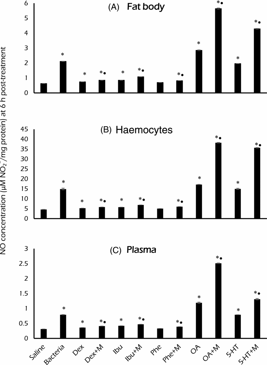
Nitric oxide concentrations, expressed as μM nitrite/mg protein, in (A) fat body, (B) haemocytes and (C) plasma of third instar Sarcophaga argyrostoma post-injection with Micrococcus luteus, either alone or in couple with an EBI (Dex, Ibu or Phe) or with a BMA (OA or 5-HT) at 6 h PI. Values are presented as mean ± SE (n = 3). *significant differences in comparison with the saline-injected larvae at P<0.05; •significant differences in comparison to the corresponding group of the larvae treated with chemical alone.

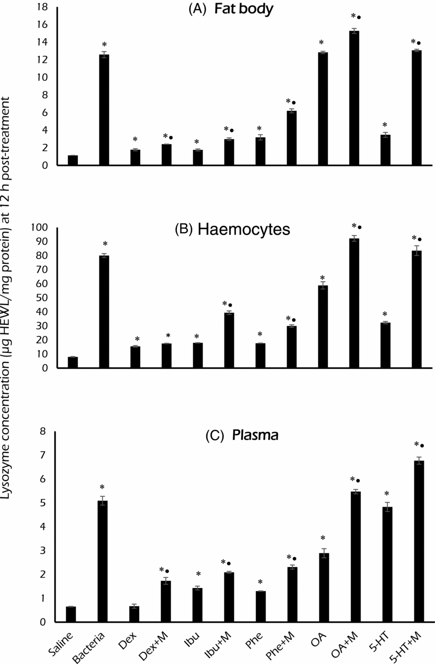
Lysozyme concentrations, expressed as μg HEWL/mg protein, in (A) fat body, (B) haemocytes and (C) plasma of third instar Sarcophaga argyrostoma post-injection with the Micrococcus luteus, either alone or in couple with an EBI (Dex, Ibu or Phe) or with a BMA (OA or 5-HT) at 12 h PI. Values are presented as mean ± SE (n = 3). *significant differences in comparison with the saline-injected larvae at P<0.05; •significant differences in comparison to the corresponding group of the larvae treated with chemical alone.

Nitric oxide and modulating actions of EBIs and BMAs
In all experiments in which third instar S. argyrostoma were treated with the different tested EBIs, Dex, Ibu and Phe, almost yielded similar results; that is, inhibitor treatments led to reductions or depletion in concentrations of NO with time progression (LSD, P<0.001). We chose 6 h PI as an optimum biosynthetic time to establish the main findings (Fig. 1). (The whole dataset is given in Appendix S2A.) However, treatment with BMAs enhanced the biosynthesis of NO, measured in terms of nitrite ion concentration (NO) (Fig. 1 and Appendix S2A).
Constitutive NO levels
A constitutive low level of NO has been observed in tissues of the saline-injected larval S. argyrostoma (negative controls), with no fixed pattern over time, with levels in a descending order haemocytes>fat body>plasma (ca. 4, 0.6 and 0.3 μM NO − 2/mg protein, respectively). These values were elevated by amounts of NO post-challenge with M. luteus (positive control). This increase reached maximal values of ca 25, 2 and 1 μM NO − 2/mg protein in haemocytes, fat body and plasma, respectively, after various time intervals in these tissues, i.e. 24, 6 and 12 h post-injection (PI), respectively (Appendix S2A). However, NO concentration decreased with increase in time PI.
Immune-suppressive actions of EBIs on NO
NO in the fat body decreased slowly in larvae treated with EBIs (Dex, Ibu and Phe) with the increase in the experimental period PI (up to 72 h) with either M. luteus alone or in combination with Dex, Ibu or Phe. However, NO increased up to 24 and 12 h PI with M. luteus combined with Ibu and Phe, respectively, then followed by a continuous decline (Appendix S2A). Maximum concentration of 1.08 μM nitrite/mg protein was obtained after 6 h of treatment with M. luteus combined with Ibu. In all treatments, combined treatment (EBI+M. luteus) induced significant elevation of NO compared to individual treatment. Additionally, combined-treatment larvae showed significant increase in NO levels compared to non-treated (negative and positive control) larvae (Fig. 1).
A similar pattern of concentration criteria occurred in NO of the haemocytes and plasma. There was, generally, a gradual decline in the NO with the progression of time post-treatment. NO significantly increased in treated larvae challenged with M. luteus combined with either Dex, Ibu or Phe compared to treatment only with any of these EBIs. Maximum NO of haemocytes was obtained after 12 h of treatment with M. luteus combined with Phe; while maximum NO concentration of plasma was obtained after 6 h of treatment with M. luteus combined with Ibu. The highest NO was attained in the haemocytes, followed by fat body (Fig. 1). NO of treated larvae was higher (P<0.05) than that of control larvae.
Immune-enhancing actions of BMAs on NO
There was no change in the concentration of the fat body NO after treatment of S. argyrostoma third instars with the biogenic monoamine OA in all the experimental periods. However, NO concentration increased up to 6 h of treatment with both M. luteus combined with OA and individual treatment with 5-HT, and increased up to 12 h PI with M. luteus combined with 5-HT, after which the concentration began to decrease (Appendix S2A). In all treatments, combined treatment with M. luteus and the BMAs (OA and 5-HT) significantly enhanced the NO concentration compared to treatment only with the monoamines. Treated larvae showed a significant increase in the concentration of NO compared to control larvae (Fig. 1). There was no change in the concentration of NO of the haemocytes and plasma after treatment of S. argyrostoma with OA. Combining M. luteus with either OA or 5-HT significantly increased the haemocytes or plasma NO concentration compared to treatment with either one alone. The highest NO concentration was obtained in the haemocytes, followed by plasma and finally fat body in all treatments with either the BMA alone or in combination with M. luteus. Treatment with the BMAs, either alone or in combination with M. luteus, induced a significant enhancement in the NO concentration compared to the non-treated larvae (Fig. 1).
The fitting equations of the relationship and correlation coefficients (r) of the experimental times with the different treatment effects on NO in the different tested tissues are given in Appendix S2A. The levels of NO in the third instar S. argyrostoma after injection of M. luteus, EBIs (Dex, Ibu and Phe), or BMAs (OA and 5-HT) were significantly affected by the experimental periods (F 5, 144 = 2599; P<0.0001, F 5, 144 = 877; P<0.0001, F 5, 144 = 892; P<0.0001, F 5, 144 = 228, P<0.0001 and F 5, 144 = 812; P<0.0001, respectively). In addition, the type of tissue (fat body, haemocytes and plasma) markedly affected the lysozyme activities after treatment with Dex (F 2, 144 = 470256, P<0.0001), Ibu (F 2, 144 = 167067, P<0.0001), Phe (F 2, 144 = 67849, P<0.0001), OA (F 2, 144 = 24005, P<0.0001) and 5-HT (F 2, 144 = 37271, P<0.0001) (Appendix S3A).
Lysozymes and modulating actions of EBIs and BMAs
Comparable to NO data presentation (selection of a specific data point, 6 h), we adopted 12 h PI data as an optimum biosynthetic time to present the main results of EBIs actions in Lys (Fig. 2) and the whole dataset is given in Appendix S2B.
Lysozyme concentration of the saline-treated S. argyrostoma larvae showed a fluctuating pattern with the time PI, while the IrLys concentration in haemocytes, fat body and plasma displayed maximal values after 6, 12, 12 h PI, respectively, then decreased gradually with time in case of treatment with M. luteus. The constitutive levels order of lysozyme concentration of non-treated larvae, in a descending order, was haemocytes>fat body>plasma (Appendix S2B).
Similar to NO concentration, lysozyme concentration in S. argyrostoma larvae decreased with the increase in time PI (up to 72 h) with the three EBIs studied alone (Dex, Ibu and Phe) or in combination with M. luteus in fat body, haemocytes and plasma. Combined treatment significantly enhanced lysozyme concentration compared to the individual treatment (Fig. 2). In all treatments (individual or combined), approximately complete inhibition of the lysozyme concentration was observed up to 48 h PI with Dex and Phe alone, and up to 72 h PI, except with Dex combined with M. luteus. The highest concentration was recorded from haemocytes, followed by the fat body, and finally the plasma (Fig. 2). The same trend was present in larvae treated with the two BMAs (OA and 5-HT) alone or in combination with M. luteus (Fig. 2). However, no complete inhibition of lysozyme concentration was recorded up to the end of the experimental period, i.e. 72 h PI. Maximum lysozyme concentration was observed in the haemocytes while minimum was observed in plasma. Larvae treated with either EBIs, BMAs or M. luteus either alone or in combination showed a significant increase in the lysozyme concentration compared to the control larvae (Fig. 2).
The fitting equations of the relationship and correlation coefficients (r) of the experimental times with the different treatment effects on lysozyme biosynthesis in the different tested tissues are given in Appendix S2B. The lysozyme activity in the third instar S. argyrostoma after injection of M. luteus, EBIs (Dex, Ibu, and Phe), or the BMAs (OA and 5-HT) were significantly affected by the experimental periods (F 5, 144 = 528.8; P<0.0001, F 5, 144 = 308.1; P<0.0001, F 5, 144 = 375; P<0.0001, F 5, 144 = 187.8, P<0.0001 and F 5, 144 = 162.6; P<0.0001, respectively). In addition, the type of tissue (fat body, haemocytes and plasma) markedly affected the lysozyme activities after treatment with Dex (F 2, 144 = 2275.5, P<0.0001), Ibu (F 2, 144 = 1856.2, P<0.0001), Phe (F 2, 144 = 1980.9, P<0.0001), OA (F 2, 144 = 2835.1, P<0.0001) and 5-HT (F 2, 144 = 2019.5, P<0.0001) (Appendix S3B).
Discussion
NO plays various roles in insect immunity; perhaps as a cytotoxic component and also as a nitrinergic signal molecule (Müller, Reference Müller1997; Alderton et al., Reference Alderton, Cooper and Knowles2001). In this study, the de novo production of NO suggests that it is an immune response molecule against bacteria that is mainly biosynthesized in haemocytes compared to fat body, if the rates of NO release from the two tissues into haemolymph are equal, but that remains to be confirmed. For the dipteran A. gambiae, Hillyer and Estévez-Lao (Reference Hillyer and Estévez-Lao2009) reported a fat body–haemocyte comparison that agrees with the data presented in this study. The haemocytes appear to express more iNOS than fat body.
Our data show a constitutive level of lysozyme and IrLys concentrations in larvae, which were increased in immune-challenged larvae. The plasma constitutive lysozymes perform the early immune responses against the invading bacteria by degrading bacterial cells and debris (Park et al., Reference Park, Park, Shin, Park, Kim, Oh and Joo1997) and releasing fragments of PGN, a key-step in bacterial pattern recognition (Imler and Bulet, Reference Imler, Bulet, Kabelitz and Schroder2005). Gillespie et al. (Reference Gillespie, Koshinsky and Khachatourians1993) ascribed the lysozyme activity of crude haemolymph preparations to its ability to lyse cell walls of M. lysodeikticus. This bacterium is highly sensitive to lysozymes that preferentially hydrolyse its mucopeptide cell-wall structure (Kocur et al., Reference Kocur, Pačova and Martinec1972; Jenzano et al., Reference Jenzano, Hogan and Lundblad1986); and hence, it is widely used in routine screening of antibacterial lysozyme activity. IrLys activity was evident in other insects at 6 h PI, and reached a maximum at 48 h PI and then declined slightly (Abraham et al., Reference Abraham, Nagaraju, Salunke, Gupta and Datta1995; Kim and Yoe, Reference Kim and Yoe2003). In larval S. argyrostoma, haemocytes and fat body biosynthesize IrLys de novo. Haemocytes produced and stored IrLy than fat body, probably for both biosynthesis and storage, as reported in other insects, including R. prolixus (Azambuja et al., Reference Azambuja, Feder, Mello, Gomes and Garcia1999) and Manduca sexta (Rao and Yu, Reference Rao and Yu2011). In these cases, use of concentration criterion for comparison is valid only if the rates of release of lysozyme from these two tissues are equal. In third instar Drosophila, a subepidermal population of sessile haemocytes was observed to be released into the circulation in response to parasitization. These sessile haemocytes are a novel haematopoietic tissue that constitutes the main source of certain blood cells (Márkus et al., Reference Márkus, Laurinyecz, Kurucz, Honti, Bajusz, Sipos, Somogyi, Kronhamn, Hultmark and Andó2009; Leitão and Sucena, Reference Leitão and Sucena2015). It is likely that sessile haemocytes were present in the isolated fat body, which would contribute to the activities that were observed in that tissue. Sessile haemocytes are perhaps not a major factor, but a factor nonetheless.
A potential crosstalk may exist between the signalling cascades regulating both IrLys biosynthesis and NO production (and perhaps each with other humoral immune effectors). For instance, lysozymes can intrinsically stimulate production of H2O2, which in turn activates H2O2-dependent pathways leading to upregulation of iNOS and formation of NO (Mink et al., Reference Mink, Jacobs, Cheng, Kasian, Santos-Martinez and Light2009). Likewise, LPS-stimulated macrophages showed enhanced phagocytosis and NO, lysozyme and PGE2 biosynthesis (Qin et al., Reference Qin, Ototake, Noguchi, Soma, Yokomizo and Nakanishi2001).
In the present study, eicosanoids and certain BMAs acted as immune mediators in larval S. argyrostoma. EBI treatments led to suppression of NO and lysozymes post-bacterial challenge, when each EBI was injected with M. luteus. We infer that eicosanoids act in mediating and coordinating biosynthesis of iNOS and lysozyme. A few studies are available on eicosanoid mediation of lysozyme immune response in insects, e.g. in Bombyx mori, D. melanogaster, and Spodoptera exigua (Morishima et al., Reference Morishima, Yamano, Inoue and Matsuo1997; Yajima et al., Reference Yajima, Takada, Takahashi, Kikuchi, Natori, Oshima and Kurata2003; Shrestha and Kim, Reference Shrestha and Kim2009). There are no reports of eicosanoids mediating NO metabolism and its role in insect immunity. However, injection of the larval S. argyrostoma with any of the EBIs—Dex, Ibu or Phe—caused elevation of lysozyme concentration over the level of the saline-injected controls. Similar results were reported before in other insects, e.g. for nodulation and phenoloxidase activity in Pimpla turionellae by the EBI esculetin (Durmus et al., Reference Durmus, Büyükgüzel, Terzi, Tunaz, Stanley and Büyükgüzel2008). Dex failed to inhibit the increase of haemocyte population of bacteria-injected S. exigua (Kim and Kim, Reference Kim and Kim2010).
A noteworthy problem with using pharmaceutical inhibitors to test biochemical pathways is the likelihood of non-specific or non-physiological impacts (Stanley Samuelson et al., Reference Stanley-Samuelson, Jensen, Nickerson, Tiebel, Ogg and Howard1991). This shortcoming has been addressed by generating dose-response curves, showing that EBIs act in a dose-related manner (Miller et al., Reference Miller, Nguyen and Stanley-Samuelson1994; Srikanth et al., Reference Srikanth, Park, Stanley and Kim2011). This indicates the EBIs acted in a physiological, rather than pharmaceutical, mechanism. The linear negative relationships we obtained for EBIs, in particular, support the idea that eicosanoids mediate the tested immune reactions to bacterial infections in flesh flies. Dex is a glucocorticosteroid that inhibits PLA2 specifically. It binds to certain nuclear steroid receptors and interferes with NF-κB activation and apoptotic pathways (Yamamoto and Gaynor, Reference Yamamoto and Gaynor2001). Dex down regulates the expression of several inflammation-regulatory genes and has immunosuppressive properties. For the other two pharmaceutical inhibitors, Phe binds to and inhibits the enzymatic activity of LOXs and reduces the production of leukotrines. Also, it combines with COX, and thereby, prevents its substrate–enzyme combination with AA and the formation of eicosanoids, PGs and thromboxanes. While on the contrary, Ibu is a nonsteroidal anti-inflammatory agent with pharmacologic actions thought to act through inhibition of PGs synthesis by inhibiting COXs, which converts AA to cyclic endoperoxides, precursors of PGs (Stanley and Kim, Reference Stanley and Kim2014). Because these chemicals suppressed the levels of both IrLys and NO, we suggest that their actions may be accomplished by inhibiting the AA cascade in S. argyrostoma.
In Drosophila, PLA2 is involved in the LPS-dependent activation of the immune deficiency pathway (imd), an equivalent to the NF-κB signalling pathway in mammals, via stimulation of AA and other mediators. Dex inhibits this imd-LPS-dependent activation; thus, a certain crosstalk exists between the PLA2-generated AA cascade and the LPS-stimulated imd pathway in insect immunity (Yajima et al., Reference Yajima, Takada, Takahashi, Kikuchi, Natori, Oshima and Kurata2003). In S. exigua, the plasmatocyte-spreading peptide acts via biosynthesis of eicosanoids, where PLA2 inhibitors reversibly block plasmatocyte spreading (Srikanth et al., Reference Srikanth, Park, Stanley and Kim2011). It is also possible that eicosanoid signalling concatenate with other cellular/humoral process mediators (such as BMAs), in insect immunity.
We investigated the actions of BMAs in the present work. These compounds act as neurotransmitters, neuromodulators in response to acute stress and neurohormones in the insect nervous system (Roeder, Reference Roeder2005). Co-injection of OA or 5-HT (a serotonin receptor agonist) with M. luteus elevated NO concentrations in haemocytes and fat body. However, OA, but not 5-HT, elevated IrLys concentrations in haemocytes and fat body. Hence, OA affects the S. argyrostoma IrLys responses and may mediate other stress responses. The immune-enhancing action of OA was reported earlier (Brey, Reference Brey1994). It can also boost resistance to infection when co-incubated with the pathogen (Baines et al., Reference Baines, Desantis and Downer1992; Baines and Downer, Reference Baines and Downer1994). Following LPS activation, Pieris rapae haemocytes synthesize elevated levels of 5-HT in a dose-dependent manner (Qi et al., Reference Qi, Huang, Li, Wu, Xia and Ye2016). In mollusc haemocytes, NE (analogous to OA) enhances the LPS-induced production of NO, a process that is regulated via a α/β-adrenoceptor-cAMP/Ca2+ pathway. NO exerts a reversible feedback homeostatic action on the biosynthesis of NE (Jiang et al., Reference Jiang, Zhou, Wang, Yang, Wang, Wu and Song2014). OA concurrently adapts the insect immune system to maintain maximal functioning under fluctuating physiological conditions (Adamo, Reference Adamo2008). Hence, we suggest that injection of OA alone, and similarly 5-HT, may augment the background tissue levels of both NO and IrLys in response to injection stress in the larvae. The latter observation may be supported by pronounced expression of 5-HT, for example, by naïve haemocytes in some insects (Qi et al., Reference Qi, Huang, Li, Wu, Xia and Ye2016).
BMAs act via cell surface receptors, which leads to increased concentrations of intracellular second messengers, such as cAMP and inositol triphosphate, as seen in Periplaneta americana (Orr et al., Reference Orr, Gole and Downer1985; Baines and Downer, Reference Baines and Downer1994), cAMP in D. melanogaster (Williams et al., Reference Williams, Wiklund, Wikman and Hultmark2006), Galleria mellonella (Marin et al., Reference Marin, Dunphy and Mandato2005), and S. exigua (Kim and Kim, Reference Kim and Kim2010) and Ca2+ in Chilo suppressalis (Huang et al., Reference Huang, Wu, Li, Adamo and Ye2012). Furthermore, OA and 5-HT are pleiotropic and implicated in various physiological processes and behaviours. These biogenic amines could be acting via the modulation of dorsal vessel physiology. Both OA in Apis mellifera and Bactrocera oleae (Papaefthimiou and Theophilidis, Reference Papaefthimiou and Theophilidis2011) and 5-HT in A. gambiae (Hillyer et al., Reference Hillyer, Estévez-Lao and Mirzai2015) are cardio-acceleratory, known to increase the heart rate. This would increase haemolymph flow, perhaps increasing the dissemination and delivery of the tested antimicrobial factors. We suspect similar mechanisms operate in larval S. argyrostoma, the subject of future work.
Conclusion
The observation that EBIs retard the activities of iNO and irLys in flesh fly larvae indicate that their actions are exerted through a common mechanism; specifically, inhibition of COXs. Accordingly, eicosanoids act in biosynthesis of NO and lysozymes. Nonetheless, BMAs enhance flesh fly immunity. We interpret this finding in terms of immune-reactive activities. However, understanding the molecular regulatory mechanisms underlying the modulation of eicosanoid and biogenic amines to the targeted genes should provide strong evidence on the interactions and pleiotropic actions of these molecules in the cell.
Future perspective
As a part of ongoing research, we identified four different mRNA sequences coding for c-type lysozymes from the transcriptome shotgun assembly database for the sister flesh fly Sarcophaga (Liopygia) crassipalpis using TBLASTN on NCBI with Lucilia cuprina c-type lysozyme (KNC28071.1). We aim to study their structural and expressional variation in response to immune challenge and the modulation of their expression dynamics by different EBI treatments.
Acknowledgements
We are grateful to Prof. David Stanley (USDA/Agricultural Research Service, MO, USA) for providing many comments/inputs on the draft of this paper. Thanks are also due to Dr Magdi El-Hawagry for helping in fly identification. We gratefully acknowledge the sincere help of Dr Atef A. Ali in statistical analyses.
Supplementary material
To view supplementary material for this article, please visit https://doi.org/10.1017/S1742758417000315