I. INTRODUCTION
Among the various morphologies and textures of inorganic materials, a sphere is the most attractive shape in relation to its potential applications. A solid sphere can be used as a photonic material, whereas mesoporous spheres are used in catalysts and adsorbents; moreover, hollow spheres can be used as catalysts and matrices for drug delivery systems. Reference Yin and Ge1–Reference Liong, Lu, Kovochich, Xia, Ruehm, Nel, Tamanoi and Zink7 The sol–gel method, reported by Stöber et al., is one of the most commonly used methods for synthesizing spherical particles, which synthesizes monodispersed colloidal silica with a spherical shape. Reference Stöber, Fink and Bohn8 This method has been applied for synthesizing zirconia and titania. Reference Dong, Ye, Zhao, Yu, Wang, Zhang, Wan and Guo9–Reference Eiden-Assmann, Widoniak and Maret12 Spray drying, emulsion using an organic template, and layer-by-layer methods are other methods used to obtain spherical particles. Reference Wang, Angelatos and Caruso6,Reference Iskandar, Nandiyanto, Yun, Hogan, Okuyama and Biswas13–Reference Caruso, Susha and Caruso15 These methods are useful owing to their high mass production and capability to produce particles with controlled size. However, they are complicated processes, and postannealing is often required to remove organic substances and obtain crystalline materials. In contrast, a solvothermal-assisted or -based method without a template is an effective synthesis approach because crystalline spherical particles are directly obtained without further treatment. Reference Zheng, Huang, Qin, Zhang and Dai16–Reference Dong, Ye, Zhao, Yu, Wang, Zhang, Wan and Guo19 Therefore, many research groups have reported the synthesis and application of spherical materials; however, there are few universal methods. Overall, the development of new methodology and approach for synthesizing spheres are valuable for broadening knowledge and facilitating the construction of theory relevant to the formation of spherical materials.
Hydrogen titanates (protonated titanates), whose compositions are written as H2Ti m O2m+1 or H4Ti n O2n+2 (except for hydrous titania), are promising materials in the catalyst and environmental-related fields. Reference Zhu, Wang and Xia20–Reference Song, Xu, Li, Wang and Yan24 Moreover, such materials are good precursors of titania and metal titanate, and have well-controlled shapes such as nanotubes and nanosheets. Reference Kasuga, Hiramatsu, Hoson, Sekino and Niihara25–Reference Fukuda, Ebina, Shibata, Aizawa, Nakai and Sasaki30 Hydrogen titanates are usually synthesized by solvothermal treatment of titania or by ion-exchange of alkaline metal titanates. Due to the nature of these methods and the crystalline habits of the titanates, the obtained samples mainly consist of tube-, whisker- or sheet-like particles. Reference Zhu, Wang and Xia20–Reference Fukuda, Ebina, Shibata, Aizawa, Nakai and Sasaki30 The use and synthesis of H2Ti2O5 have been less frequently reported than those of other hydrogen titanates, such as H2Ti3O7 and H2T4O9, and it is formed as sheet-like particles. Reference Song, Xu, Li, Wang and Yan24,Reference Etacheri, Kuo, Van der Ven and Bartlett31,Reference Sutradhar, Pahari, Jayachandran, Stephan, Nair, Subramanian, Bajaj, Mody and Panda32 Although the structure of H2Ti2O5 has not been well established, it is reported to be a layered compound which is an isostructure to K2Ti2O5. Reference Sugita, Tsuji and Abe33 In K2Ti2O5, potassium ions are intercalated between layers consisting of TiO5 polyhedra, Reference Andersson and Wadsley34 indicating that H2Ti2O5 has a characteristic structure, even though Wu et al. conducted simulations to show that H2Ti2O5 was composed of TiO6, Reference Wu, Lei, Zhu, Yang and Xie35 and is a promising material for use as a solid catalyst because titanium-based oxides are usually composed of TiO6 polyhedra.
To date, we have reported the synthesis of spindle and square bipyramid-shaped anatase-type titania particles by solvothermal treatment of an ethylenediamine (EDA) solution of water-soluble titanium complexes. Reference Kobayashi, Kato and Kakihana36,Reference Kobayashi, Kato and Kakihana37 During this research, we found that spherical agglomerates of EDA-intercalated H2Ti2O5 were formed under certain conditions. Herein, we present the synthesis of spherical particles composed of H2Ti2O5 intercalated with polyethyleneamine using the solvothermal method. We also describe their adsorption characteristics, and from the viewpoint of colloidal chemistry, discuss the self-assembly mechanism of H2Ti2O5 particles that results in the formation of spherical agglomerates. Furthermore, their conversion to titania by thermal treatment is demonstrated.
II. EXPERIMENTAL SECTION
A. Materials
Metallic titanium powder (98.0%) and anhydrous citric acid (98.0%) were purchased from Wako Pure Chemical (Osaka, Japan), and an aqueous solution of H2O2 (30.0 wt%) was purchased from Santoku Chemical Industries Co. (Tokyo, Japan). Moreover, NH3 (28.0 wt%), EDA (NH2(C2H4NH)H, 99.0%; denoted as EDA), diethylenetriamine (NH2(C2H4NH)2H, 97%; denoted as DETA), and triethylenetetramine (NH2(C2H4NH)3H, 98%; denoted as TETA) were purchased from Kanto Chemical. All reagents were used as received.
B. Synthesis
Titanium metal powder (0.239 g, 5 mmol) was dissolved in a mixture of 20 cm3 H2O2 and 5 cm3 aqueous NH3 solution. This was performed in a water bath to maintain approximately 288 K. A yellow transparent solution of peroxotitanate complex was formed after 2 h. An equimolar amount of citric acid (0.961 g, 5 mmol) with respect to titanium was added to the prepared solution. To gently eliminate the excess H2O2 and NH3, the solution was left at room temperature for 12 h and then heated on a hotplate at 353 K until a solid gel-like material was formed. An aqueous solution of the citratoperoxotitanium complex could be obtained by adding distilled water and had a pH of approximately 6. Reference Kakihana, Tada, Shiro, Petrykin, Osada and Nakamura38 The aqueous complex solution was heated in a Teflon vessel on a hotplate at 353 K to evaporate the solvent. Next, 15, 16, 17, 18, or 19 cm3 of each amine was added to the Teflon vessel and the total volume of the solution was adjusted to 20 cm3 by adding distilled water, i.e., [Ti] = 0.25 mol dm−3. The solution was stirred using a magnetic stirrer to obtain a clear solution. The concentrations of amine were calculated to be 11.2, 11.9, 12.7, 13.4, and 14.1 M for the 15–19 cm3 additions of EDA, respectively. Moreover, the pH increased with increasing amine concentration from 13.5 at 11.2 M to 14.0 at 14.1 M. Note that even in such highly basic conditions, no precipitation owing to the hydrolysis of titanium species occurred due to the high stability of the citratoperoxotitanium complex. Reference Kakihana, Kobayashi, Tomita and Petrykin39 The Teflon vessel was sealed in a stainless steel jacket and hydrothermally heated at 473 K for 1.5–48 h. After being cooled to room temperature, the precipitate was separated using a centrifuge. After washing the precipitate three times with distilled water using ultrasonication and centrifugation, the resultant powder was dried at 353 K.
C. Characterization
The powders were analyzed through powder X-ray diffraction (XRD; Rigaku RINT 2200, Rigaku Corp., Tokyo, Japan) using Cu Kα radiation. The morphologies of the samples were characterized by field emission scanning electron microscopy (SEM; Hitachi S-4800, Hitachi Corp., Tokyo, Japan) at an accelerating voltage of 5 kV and transmission electron microscopy (TEM; Hitachi H-7650) at an accelerating voltage of 100 kV. Specimens for TEM observation were prepared by sonication of the powders in ethanol and placing a drop of the solution on a carbon film-coated copper grid, followed by drying in an oven at 353 K. The Fourier transform infrared (FT-IR) spectrum was recorded on a spectrometer (Jasco FT/IR-4200, JASCO Corp., Tokyo, Japan) equipped with a diamond prism using the attenuated total reflection method. Elemental analysis for C, H, and N was conducted using a micro corder (JM10, J-Science Group, Kyoto, Japan). Thermogravimetric-differential thermal analysis (TG-DTA; Shimadzu DTG-60H, Shimadzu Corp., Kyoto, Japan) was also conducted. The sample was heated at a rate of 10 K min−1 under a 50 mL min−1 air flow up to a maximum of 1273 K, with Al2O3 powder used as a reference. Specific surface areas and pore size distributions of the samples were determined using Brunauer–Emmett–Teller (BET) and Barret–Joyner–Halenda (BJH) analysis, respectively, from N2 adsorption isotherms using a physisorption analyzer (Micromeritics ASAP 2010, Chiba, Japan). Prior to measurement, the samples were degassed at 363 K for 1 h and 473 K for 2 h.
III. RESULTS AND DISCUSSION
A. Effect of amines
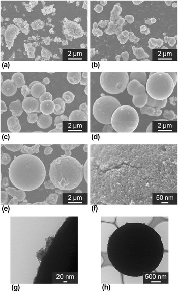
Figure 1 shows SEM and TEM images of the samples obtained by solvothermal treatment of citratoperoxotitanium complex solutions with various EDA concentrations at 473 K for 3 h. The sample obtained from the 11.2 M EDA solution was composed of irregular-shaped particles [Fig. 1(a)]. In the powder formed in the 11.9 M EDA solution, poorly organized spherical particles with diameters of approximately 600 nm were observed [Fig. 1(b)]. Formation of polydispersed spheres were confirmed in the samples obtained from the 12.7, 13.4, and 14.1 M EDA solutions [Figs. 1(c)–1(e)]. The diameters of the formed spheres increased with increasing EDA concentration from 2.0 μm at 12.7 M to 5.5 μm at 14.1 M. According to SEM and TEM observations of the surface of a spherical particle [Figs. 1(f) and 1(g)], the particles were composed of minute particles of 10 nm in diameter. Furthermore, TEM analysis revealed that the spheres were not hollow [Fig. 1(h)].
(a–f) SEM and (g, h) TEM images of powders synthesized at 473 K for 3 h. [EDA] = (a) 11.2, (b) 11.9, (c, f–h) 12.7, (d) 13.4, and (e) 14.1 M

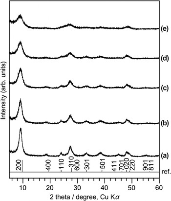
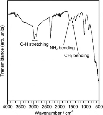
Figure 2 shows XRD patterns of the samples, which correspond to the SEM images shown in Fig. 1. Irrespective of EDA concentration, peaks appeared at the same manner, though the intensities weakened with increasing EDA concentration. Although a peak shift was confirmed, as described below, the patterns could be assigned as H2Ti2O5·H2O. Reference Sugita, Tsuji and Abe33 Peaks of (200) and (020) existed at 9.8° and 48.0°, respectively, in the reference pattern. However, in the samples, the peak for (200) appeared at 9.1°, which was lower than that of the reference, and that for (020) was observed at 48.3°, which was slightly higher than that of the reference. Lattice parameters of the sample obtained from the 12.7 M EDA solution were calculated to be a = 19.476 Å, b = 3.768 Å, and c = 2.922 Å as an orthorhombic system [Fig. 2(c)]. The value of b was almost consistent with the reported value (3.784(3) Å). In contrast, it was revealed that the a axis of the obtained sample was significantly longer and the c axis was significantly shorter than that of the reference (a = 18.08(3), c = 2.998(2) Å). According to previous research, layers of titanium polyhedra are stacked in the a axis direction and protons exist between the layers in H2Ti2O5·H2O. Reference Sugita, Tsuji and Abe33,Reference Wu, Lei, Zhu, Yang and Xie35 Therefore, protonated EDA ions, rather than H+, were intercalated between the layers in the samples; this intercalation caused the lattice constant change. Reference Sasaki, Izumi and Watanabe21,Reference Wu, Lei, Zhu, Yang and Xie35 The intercalation of EDA was also supported by the FT-IR spectrum, showing peaks at 1500, 1600, and 2800–3000 cm−1, corresponding to the CH2 bending, NH2 bending, and C–H stretching modes in EDA, respectively (Fig. 3). Reference Krishnan and Plane40 Elemental analysis and TG-DTA (Fig. S1) revealed that the contents of C, H, N, and Ti in the sample obtained from the 12.7 M EDA solution were 4.06, 4.30, 2.43, and 45.66 wt%, respectively. This indicates that 0.18 mole of EDA per 1 mol of Ti were intercalated in, or adsorbed on, the surface (based on the assumption that all of nitrogen was derived from the EDA). This result may be supported by the relatively minor lattice constant change calculated from the XRD patterns. The contents of EDA in the samples are almost the same irrespective of EDA concentration, implying that a certain equilibrium composition existed in the present system.
XRD patterns of powders obtained at 473 K for 3 h. [EDA] = (a) 11.2, (b) 11.9, (c) 12.7, (d) 13.4, and (e) 14.1 M. ref.: H2Ti2O5·H2O (PDF #47-0124). Reference Sugita, Tsuji and Abe33

FT-IR spectrum of spherical agglomerates synthesized from a 12.7 M EDA solution at 473 K for 3 h

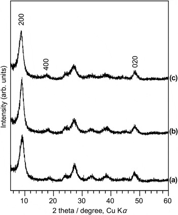
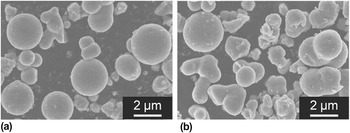
Figure 4 shows the XRD patterns of the samples synthesized by solvothermal treatment of 7.9 M DETA and 5.7 M TETA solutions (prepared using 17 cm3 of each amine) of the titanium complex at 473 K for 3 h. The pH values of the solutions were 13.4 for DETA and 13.1 for TETA. Both patterns could be assigned to H2Ti2O5·H2O, though the peak positions were shifted. Lattice parameters of each sample were calculated to be a = 19.696 Å, b = 3.759 Å, and c = 2.900 Å for the sample obtained using DETA [Fig. 4(b)] and a = 20.243 Å, b = 3.762 Å, and c = 2.880 Å for the sample obtained using TETA [Fig. 4(c)], as an orthorhombic system. This indicates that crystals along the a axis expanded, whereas those along the c axis shrank, with an increase in the molecular size of the polyethyleneamine used. This result is in good agreement with discussions on amine, rather than proton, intercalation. Because K2Ti2O5 is classified as a monoclinic system, the lattice can be distorted when a large cation is intercalated into H2Ti2O5·H2O. Therefore, a decrease in length along the c axis was possibly due to distortion of the crystal. Figure 5 shows SEM images of the corresponding samples shown in Fig. 4. Spherical agglomerates with a diameter of 2–2.5 μm for DETA [Fig. 5(a)] and 1–2 μm for TETA [Fig. 5(b)] were obtained; therefore, concentrated amine solution was apparently necessary to obtain spherical particles.
XRD patterns of samples obtained from (a) 12.7 M EDA [same as Fig. 2(c)], (b) 7.9 M DETA and (c) 5.7 M TETA aqueous solutions at 473 K for 3 h.

SEM images of samples obtained from (a) 7.9 M DETA and (b) 5.7 M TETA aqueous solutions at 473 K for 3 h.

B. Effect of treatment time
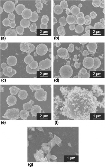
Figure 6 shows SEM images of the powders synthesized by solvothermal treatment of the citratoperoxotitanium complex in 12.7 M EDA solutions at 473 K for various times. Spherical particles were formed even with a short treatment time [1.5 h, Fig. 6(a)]. The diameter was approximately 2 μm, which is almost the same as that of the samples obtained at 3 and 6 h [Figs. 6(b) and 6(c)]. Spherical agglomerates with a rough surface were observed in the sample obtained at 12 h [Fig. 6(d)]. In the sample obtained at 24 h, the formation of spherical agglomerates and small particles on the spheres was confirmed [Fig. 6(e)]. The sample obtained at 48 h was composed of equiaxed nanoparticles and spheres [Figs. 6(f) and 6(g)]. Note that extended treatment led to the formation of irregular-shaped agglomerates of bipyramid-shaped anatase particles. Reference Kobayashi, Kato and Kakihana36,Reference Kobayashi, Kato and Kakihana37 XRD patterns revealed that the samples obtained with less than 24 h of treatment consisted of EDA-intercalated H2Ti2O5 (Fig. S2). Above 12 h, the formation of anatase was also detected. Considering previous research and the equilibrium shape of anatase, equiaxed nanoparticles could be assigned as anatase. Reference Kobayashi, Kato and Kakihana37,Reference Lazzeri, Vittadini and Selloni41,Reference Oliver, Watso, Kelsey and Parker42 The peaks for EDA-intercalated H2Ti2O5 were not shifted irrespective of treatment time. Therefore, spherical particles of EDA-intercalated H2Ti2O5 with the same composition are obtained at 473 K over 1.5–12 h.
SEM images of particles synthesized using 12.7 M EDA solutions at 473 K for (a) 1.5, (b) 3 [same as Fig. 1(c)], (c) 6, (d) 12, (e) 24, and (g) 48 h. (g) is a magnified image of (f).

C. Formation mechanism
Considering only surface energy, the shape of agglomerates is inevitably spherical because it involves the minimum amount of energy. However, irregular or other shaped agglomerates are often observed due to the effect of various factors, such as an electrostatic barrier caused by electrical double layers and other van der Waals forces among primary particles. For example, Look et al. investigated the origin of the formation of spherical aggregations and reported that when the particle surface potential was low, the repulsive interactions among particles were also low, resulting in the formation of a nonuniform agglomerate by Brownian aggregation. Reference Look, Bogush and Zukoski43 Herein, irregular-shaped agglomerates were formed using low concentration amine solutions and a 14.8 M (28.0 wt%) NH3 aqueous solution, as previously reported. Reference Kobayashi, Petrykin, Tomita and Kakihana44 Spherical agglomerates were synthesized using 12.7 M EDA solutions with [Ti] = 0.125 and 0.5 M (Fig. S3). This result indicates that the ratio of EDA to titanium is not crucial for obtaining spheres, i.e., the formation of a spherical shape in the present method is not due to micelle formed by polyethyleneamine and titanium species. During synthesis using solution methods, the reaction pathways are often dependent on pH value. However, there is no clear relationship between pH and the formation of the sphere. Herein, the ionic strength of the solutions wherein the spheres were formed was high, because high valence and ion concentration produces high ionic strength. When the ionic strength of a solution is high, the electric double layer is compressed and the ionic atmosphere becomes dense, resulting in the flocculation of precipitates (Derjaguin-Landau-Verwey-Overbeek [DLVO] theory). Spherical agglomerates could not be obtained when the solution was gently stirred by a magnetic stirrer during solvothermal treatment (Fig. S4). Therefore, herein, it is expected that the coagulation of primary particles (caused by van der Waals forces) was not dominant under conditions where spheres were formed because agglomeration by the van der Waals force is rigid and flocculation is easily reversed. It is speculated that the relatively weak force, i.e., the flocculation caused by high ionic strength, in solutions leads to the formation of spherical agglomerates, Reference Chen, Cao, Huang, Imperia, Cheng and Caruso45,Reference Kanie, Seino, Matsubara, Nakaya and Muramatsu46 though DLVO theory is usually applied to nanoscopic phenomena.
D. Adsorption properties of spherical agglomerates
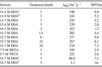
Physical properties of the samples are summarized in Table I. The BET specific surface areas, which were calculated from nitrogen isotherms (Figs. S5 and S6), of the samples composed of amine-intercalated H2Ti2O5 spheres were approximately 200 m2 g−1. Pore diameters determined by the BJH method (Figs. S5 and S6) indicated that the obtained samples had mesoporous structures of diameters between 2.4 and 8.0 nm. The pore size decreased with increasing EDA concentration or molecular weight of the amine used, or with a decrease in treatment time. Longer treatment times promoted the growth of primary particles, resulting in the formation of agglomerates with large voids. As mentioned above, ionic strength is determined not only by ion concentration but also by ion charge, and the effect of the charge on ionic strength is greater than that of the concentration. Therefore, with a greater number of amines, polyethyleneamine yielded a higher ionic strength, i.e., rigid flocculation was induced using polyethyleneamine. According to these findings, a dense agglomerate was formed and the pore size decreased when using a higher amine concentration or an amine product with a greater number of amines. These results are consistent with discussions on the formation mechanism of spheres in this study.
Physical properties of the samples.

a BET surface areas.
b Pore diameters calculated by BJH method.
c The samples were composed of irregular-shaped agglomerates.
d The sample was composed of irregular-shaped and spherical agglomerates.
e Calcined at 773 K.
f Calcined at 1273 K. “na” means the pore diameter could not be determined from the N2 isotherm measurement.
E. Conversion to titania by subsequent thermal treatment
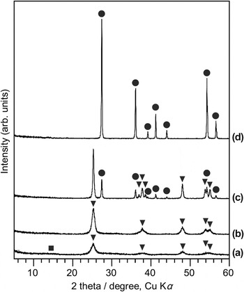
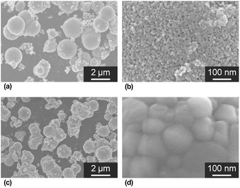
The amine-intercalated H2Ti2O5 particles obtained with the present method are good candidates for application in an ion-exchange matrix or an electrode. However, if titania with the same shape is obtained, the particles could be utilized in a support or photocatalyst. Figure 7 shows XRD patterns of the samples obtained by 1 h calcination of EDA-intercalated H2Ti2O5 powders synthesized by the solvothermal treatment of 12.7 M EDA solutions of the titanium complex at 473 K for 3 h. Crystalline anatase-type titania with a trace amount of TiO2(B) was formed at 673 K [Fig. 7(a)]. At 773 K, an almost single phase of anatase was obtained [Fig. 7(b)]. Formation of rutile was confirmed at 973 K as a secondary phase [Fig. 7(c)]. Single phase rutile could be obtained by calcination at 1273 K [Fig. 7(d)]. Figure 8 shows representative SEM images of anatase and rutile samples obtained at 773 and 1273 K, respectively, at 1 h using EDA-intercalated H2Ti2O5 samples. Both samples were composed of spherical agglomerates. Note that irregular-shaped agglomerates of anatase particles were formed by hydrothermal treatment of an aqueous suspension of polyethyleneamine-intercalated H2Ti2O5 spheres at 473 K. The diameter of anatase spheres obtained at 773 K was almost the same as the agglomerates before calcination, and pores were observed on their surface [Fig. 8(a)]. However, for rutile, spheres of a smaller size and with a sintered surface were obtained [Fig. 8(b)]. Nitrogen adsorption measurements revealed that the surface areas were drastically decreased by calcination, as shown in Table I and Fig. S7, resulting from the growth of primary particles and the shrinking of spheres. However, the sample obtained at 773 K maintained a relatively high surface area and mesopores, giving it the potential for application as a photocatalyst or adsorbent. Mesopores were not observed on the rutile sample, which is consistent with the SEM observations. These physical properties were dependent on the properties of the samples obtained by solvothermal treatment, i.e., the physical properties of the titania spheres can be controlled by tuning the solvothermal conditions.
XRD patterns of samples obtained by calcination of spherical agglomerates corresponding to sample (c) in Figs. 1 and 2 at (a) 673, (b) 773, (c) 973, and (d) 1273 K for 1 h. Triangle, circle, and square symbols indicate the reflections from anatase-type titania, rutile-type titania and TiO2(B), respectively.

Representative SEM images of calcined samples corresponding to samples in Fig. 7. (a, b) Anatase agglomerations obtained at 773 K for 1 h [corresponding to samples (b) in Figs. 7(c) and 7(d)] rutile agglomerations obtained at 1273 K for 1 h (corresponding to samples (d) in Fig. 7). (b, d) Magnified images of a surface of spherical agglomeration.

IV. CONCLUSIONS
Micrometer-sized mesoporous spheres composed of polyethyleneamine-intercalated H2Ti2O5 were obtained by solvothermal treatment of concentrated polyethyleneamine solution containing a titanium complex at 473 K. A prolonged solvothermal treatment resulted in the formation of anatase. The titanates formed were intercalated with polyethyleneamine. The BET surface area of the samples composed of spheres was approximately 200 m2 g−1. The diameter and pore size of the spheres were controlled by amine concentration, molecular size of the amine used, and treatment time. On the basis of colloidal chemistry and DLVO theory, the formation mechanism was considered to be induced by the high ionic strength produced by concentrated amine ions, causing flocculation and resulting in the self-assembly of primary particles. Polydispersed spheres were thereby formed. It was revealed that the obtained agglomerates could be used as a template for the synthesis of titania with a spherical shape, and that this may be utilized as a precursor for metal titanates such as K2Ti2O5. Thus, the present study has demonstrated a new approach for the synthesis of spherical agglomerates by a solvothermal-based method, and open new prospectives for the synthesis of inorganic spheres.
Supplementary Material
To view supplementary material for this article, please visit http://dx.doi.org/10.1557/jmr.2016.67.
ACKNOWLEDGMENTS
This work was supported in part by a Grant-in-Aid for Scientific Research (No. 22107002) on Innovative Areas of “Fusion Materials: Creative Development of Materials and Exploration of Their Function through Molecular Control” (No. 2206) from the Ministry of Education, Culture, Sports, Science and Technology, Japan (MEXT). M. Kobayashi acknowledges the support of a Research Fellowship for Young Scientists from the Japan Society for the Promotion of Science (JSPS).