A14-year-old boy presented to a community hospital emergency room with a complaint of right flank pain. A CT scan of his abdomen was performed, which demonstrated a renal infarct. He was subsequently admitted to the paediatric service for further evaluation. During his workup for a possible thromboembolic event, a transthoracic echocardiogram with a microcavitation study was notable for a possible atrial shunt and rapid filling of the left atrium. Evaluation for hypercoagulable disorders was negative.
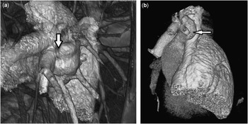
The patient was subsequently discharged from the community hospital with an appointment for an outpatient transoesophageal echocardiogram and cardiac catheterisation. The transoesophageal study showed an intact atrial septum; however, based on the findings of the transthoracic study, selective branch pulmonary artery agitated saline and blood contrast injections were performed in the catheterisation laboratory. There was a large right-to-left shunt with complete opacification of the left atrium and ventricle with right pulmonary artery angiography (Fig 1a). Interestingly, this was also accompanied by transiently decreased left ventricular systolic function, possibly due to sudden volume load. Further evaluation with angiography revealed a moderate-sized fistulous connection from the middle right pulmonary artery to the right upper pulmonary vein (Fig 1b). A CT scan of the chest was performed to further define the anatomy to allow for surgical planning (Fig 2). Owing to the size of the defect, surgical ligation was performed. He was seen during follow-up at 2, 6, and 12 months postoperatively. He reported that he was unaware of his exercise intolerance before the procedure, but he has since noticed a significant improvement in his endurance with athletics, particularly in comparison with his brothers at home and teammates on the football team.
( a ) Angiogram of the right pulmonary artery in the right anterior oblique plane demonstrating a fistulous connection from the mid right pulmonary artery to the right upper pulmonary vein. ( b ) Angiogram of the fistula in a right anterior oblique plane with cranial and caudal projections, with a 0.25-Rosen wire positioned in the fistula to the right upper pulmonary vein.

( a and b ) CT scan with three-dimensional re-formatting, demonstrating the pulmonary arteriovenous fistula.

A pulmonary arteriovenous fistula is defined as a direct communication between branches of the pulmonary artery and pulmonary vein, without an intervening pulmonary bed.Reference Khurshid and Downie 1 The incidence is 2–3/100,000 and is more common in females. More than 80% of pulmonary arteriovenous fistulas are congenital, and 47–80% of these are associated with Osler–Weber–Rendu disease/hereditary haemorrhagic telangiectasia.Reference Wozniak, Mielczarek and Sabiniewicz 2 Interestingly, when discussing direct fistulous connections with the left atrium, there is a strong male predominance estimated at 3:1.Reference Diaz, Marquez and Gentile 3 The presentation can vary depending on the size of the shunt and can include exertional dyspnoea, cyanosis, cerebrovascular accidents, and systemic embolisation.Reference Wozniak, Mielczarek and Sabiniewicz 2 , Reference Diaz, Marquez and Gentile 3 Studies including children and adults report an incidence of neurological events related to pulmonary arteriovenous fistula as 37% for transient ischaemic attacks and 18% for stroke.Reference White, Lynch-Nyhan and Terry 4 Systemic, non-cerebral embolisms account for 5–10% of all paradoxical events; however, recent studies demonstrate that this is a rare occurrence, even in patients with hypercoagulopathy.Reference Bartz, Cetta and Cabalka 5 , Reference Dao and Tobis 6
There are limited studies discussing cases of pulmonary arteriovenous fistulas in children. The presentations range in age, shunt size, and symptomatology. The case report by Diaz et alReference Diaz, Marquez and Gentile 3 describes the case of a 4-year-old boy and 16-year-old boy. Both of them had decreased oxygen saturations. Husain et al described a 29-year-old woman who presented with a cerebrovascular accident. She was subsequently found to have an isolated fistula from the right pulmonary artery to a right pulmonary vein. Of note, her oxygen saturations, similar to our case, were normal.Reference Husain, Vuppali and Hussain 7 Previous case reports have demonstrated that lesions smaller than 2 cm are typically asymptomatic.Reference Pejhan, Rahmanijoo, Farzanegan and Rahimi 8 On the other hand, based on our review of the literature, shunts as small as 7 mm can cause paradoxical brain embolism.Reference Wozniak, Mielczarek and Sabiniewicz 2 , Reference Dao and Tobis 6 , Reference Kumar and Sivasankaran 9 – Reference Ahn, Choi and Park 11 The present case is the first documentation of an adolescent presenting with a renal thromboembolic event due to a pulmonary arteriovenous fistula.
Acknowledgements
The authors thank the staff of Novant Health Pediatric Cardiology and Levine Children’s Hospital at Carolinas Medical Center for their assistance.
Financial Support
This research received no specific grant from any funding agency or from commercial or not-for-profit sectors.
Conflicts of Interest
None.
Ethical Standards
The authors assert that all procedures contributing to this work comply with the ethical standards of the relevant national guidelines on human experimentation and with the Helsinki Declaration of 1975, as revised in 2008, and has been approved by the institutional committees – Novant Health and Carolinas Health System.