INTRODUCTION
Pycnogonids (the sea spiders) are a small group of marine invertebrates that display significant diversity in larval development (Bain, Reference Bain2003; Bogomolova, Reference Bogomolova2007). However, complete larval development (from hatching through formation of adult appendages) has not been described for the vast majority (>98%) of pycnogonids, making their developmental modes unknown as well (Bain, Reference Bain2003).
Bain (Reference Bain2003) proposed four modes of development. The most common form, the ‘typical protonymphon’ development, involves a free-moving six-legged larva that acquires its adult limbs sequentially (Okuda Reference Okuda1940; King, Reference King1973; Behrens, Reference Behrens1984; Bain, Reference Bain1991, Reference Bain2003; Vilpoux & Waloszek, Reference Vilpoux and Waloszek2003). Larvae following the ‘atypical protonymphon’ pathway develop all eight adult legs simultaneously while inside the mantle cavities of molluscs or on sedentary polychaetes (Ohshima, Reference Ohshima1933; Arnaud, Reference Arnaud1978; Ogawa & Matsuzaki, Reference Ogawa and Matsuzaki1985; Salazar-Vallejo & Stock, Reference Salazar-Vallejo and Stock1987; Bain, Reference Bain2003). The ‘encysted’ postembryonic mode of development is characterized by a larva that develops in the gastrocoel of cnidarians and becomes free-moving with the first juvenile stage (Hilton, Reference Hilton1916; Lebour, Reference Lebour1945; Russel, Reference Russel and Hedgpeth1990; Bain, Reference Bain2003; Lovely, Reference Lovely2005). The ‘attaching’ developmental mode involves a larva that has only one pair of appendages (as opposed to three) and remains attached to the male throughout most of its development (Meinert, Reference Meinert1899; Hooper, Reference Hooper1980; Nakamura, Reference Nakamura1981; Bain, Reference Bain2003). Recently, an additional developmental mode was described, the ‘lecithotrophic protonymphon,’ in which the larva remains on the male for a longer period than the ‘attaching’ larva and has reduced larval appendages (Bogomolova & Malakhov, Reference Bogomolova and Malakhov2006; Bogomolova, Reference Bogomolova2007; Cano & López-González, Reference Cano and López-González2009).
Although Bain's terminology for pycnogonid postembryonic development is commonly used in the literature, it confuses larval type with mode of development and should be modified (Cano & López-González, Reference Cano and López-González2009). The terms ‘typical protonymphon,’ ‘atypical protonymphon,’ and ‘encysted larva’ suggest that morphological differences exist between the larvae following these developmental patterns. However, Bain insists that there are only two types of larvae (the attaching larva and the protonymphon), and that these three development modes all share the same protonymphon. This implies that a sea spider's developmental mode cannot be determined based on characteristics of the first larval stage. However, several studies have shown that a species' developmental mode can often be inferred from the larva's morphology (Bogomolova & Malakhov, Reference Bogomolova and Malakhov2003, Reference Bogomolova and Malakhov2004; Bogomolova, Reference Bogomolova2007; Cano & López-González, Reference Cano and López-González2009). Unfortunately, larval morphology has not been described for most species of sea spiders (Bain, Reference Bain2003).
This paper illustrates the variety of larval morphologies within and between three families (Phoxichilidiidae, Ammotheidae and Pycnogonidae) and five genera of intertidal sea spiders. An alternative terminology is proposed for the developmental modes of pycnogonids based on morphological characteristics of larvae with known developmental patterns. The larval morphologies of eight pycnogonid species are described for the first time and used to infer postembryonic developmental mode. Insight into pycnogonid postembryonic developmental modes can lead to a better understanding of larval dispersal, host preference(s), adult biology and distribution, and the phylogenic position of the sea spiders.
MATERIALS AND METHODS
Males with egg masses of the species Pycnogonum rickettsi Schmitt, 1934 were collected during January 2009 from North Cove, Cape Arago, Oregon. Specimens of Pycnogonum stearnsi Ives, 1892 were collected in March 2009 from Asilomar, Monterey, California. Egg-carrying males of both species were found on the columns of the sea anemone Anthopleura xanthogrammica Brandt, 1835. Specimens of Achelia gracilipes Cole, 1904 were collected from bryozoans (Crisia sp.) at Sunset Beach, Cape Arago, Oregon during April 2009. Males of Achelia simplissima Hilton, 1939 and Eurycyde spinosa Hilton, Reference Hilton1916 were found on rocks with large aggregations of the spirorbid worm Spirorbis bifurcatus Knight-Jones, 1978 at North Cove, Cape Arago, Oregon during January 2010. All other species were collected at Lighthouse, Cape Arago, Oregon in June 2009. Specimens of Achelia chelata Hilton, 1939, Nymphopsis spinosissima Hall, 1911, and Anoplodactylus viridintestinalis Cole, 1904 were found on the sandy tops of large boulders. The studied material can be found at the Oregon Institute of Marine Biology, Oregon. Males carrying egg masses were kept in Petri dishes and checked daily for hatched larvae.
Scanning electron microscopy (SEM) was used to examine the first postembryonic stages of all species. Upon hatching, larvae were relaxed in 7.5% MgCl for twenty minutes and fixed in osmium tetroxide and seawater according to protocol. Larvae were then dehydrated in an ascending alcohol series (10, 30, 50, 70, 85, 95 and 100%), immediately critical point dried, and coated with gold. Larvae were examined with a Tescan Vega SBU scanning electron microscope. Larval characteristics, including the shape and size of the body, proboscis, mouth, spines, appendages, and cheliphores, as well as the presence of pores and sensilla, were described and measured from SEM images. For each species, body size was measured in ten larvae prior to dehydration. There was no difference in average measurements of body size before and after dehydration; therefore, measurements for all other characteristics were made from SEM photographs. For the species Anoplodactylus viridintestinalis, the quality of the preparations did not allow for descriptions of all characteristics. However, this species is included because it exhibits a distinctive larval morphology.
RESULTS
The first larval stage in all studied species is characterized by having three pairs of appendages, the cheliphores and II and III appendages (Figure 1A). The appendages are typically tripartite. The most anterior and largest pair of larval appendages are the cheliphores. They point forward over the proboscis and terminate with a set of chelae, or claws, which are directed downwards. Unless specified, the proboscis is held horizontal to the body, directed forward, and cannot be seen from the dorsal side as it is hidden by the cheliphores. The edges of the larval body hang over the bases of the II and III appendages. These two appendages are always similar in length and ornamentation; therefore, characteristics of the II and III appendages will only be described once but will refer to both appendages. The cuticle of the larval body is often wrinkled, a possible artefact of the dehydration process.
Family Ammotheidae
GENUS ACHELIA
Larval morphology of Achelia gracilipes (N = 15)
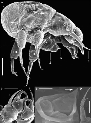
The body is rounded and 110–130 µm in length (measured as the distance between the base of the proboscis and the abdomen) (Figure 1A). The proboscis is conical in shape, 65 µm long and 60 µm wide at the base. The mouth is unopened, tripartite, and 13 µm in diameter (Figure 1B). There is a thin, smooth, raised lip around the mouth that ends in a single pointed denticle (Figure 1A, B).
Larva of Achelia gracilipes. (A) Side view of larva (scale bar: 50 µm); (B) tripartite mouth with raised lip (arrow) ending in a single denticle (5 µm); (C) cheliphores and spinning spines (50 µm); (D) tip and tooth of terminal article of larval appendages (20 µm); (ch) cheliphore, (II) second larval appendage, (III) third larval appendage; (1) spinning spine, (2) proboscis, (3) fixed chela finger, (4) moveable chela finger, (5) trifurcate smooth sensillum, (6) quadfurcate sensillum, (9) raised lip.
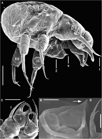
The basal article of the cheliphores is 60 µm in length and is slightly wider than long (Figure 1A, C). There is a spinning spine located on the distal external edge of the article. It is the same length (85 µm) as the chelae (Figure 1A, C). The hooked fingers of the chelae overlap when closed. The inner edges of the moveable fingers have a number of small sharp denticles (Figure 1C), while the outside edges bear a single small denticle. The fixed finger bears a single large denticle located on the inner edge. On the dorsal and ventral sides, there are multiple slit-like pores located at the base of the cheliphores, as well as at the base of the chelae. The pores are about 1.5–2 µm long and located in a circular 3 µm long depression of the cuticle.
The basal articles of the II and III appendages are much shorter (20 µm in length) than the second articles (50 µm) (Figure 1A). The terminal claw is 80 µm long with one or two large teeth located on the inside edge halfway up the article (Figure 1D). There is a short (20 µm) spine located at the base of the appendages. There are pores located on the ventral side of the articles.
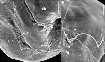
Nine sensilla were found in the larva, between 25 and 30 µm long each (Figure 2). On the anterior dorsal side of the body there is an unpaired median sensillum (Figure 2A). At the ventral posterior end of the body there is a pair of sensilla. These three sensilla are quadfurcate. Two pairs of trifurcate posterior dorsolateral sensilla and slit-like pores are located behind the second and the third pair of appendages on the dorsal side of the body (Figure 2A, B). In between this last pair of sensilla, is a pair of simple sensilla (Figure 2A). Located above each of these simple sensilla is a slit-like pore (Figure 2B). The pores and sensilla are distributed bilaterally symmetrical. In the centre of the dorsal side of the body is a pair of 6 diamond-shaped pores. These pores are not slit-like, but are depressions that are 3–4 µm long (Figure 2A).
Dorsal view of Achelia gracilipes. (A) Dorsal pores and sensilla (scale bar: 20 µm); (B) trifurcate sensillum and slit-like pore (arrow) (5 µm); (ch) cheliphore, (5) trifurcate sensillum, (6) median dorsolateral quadfurcate sensillum, (7) cluster of 6 dorsal pores, (8) bifurcate sensillum.

Larval morphology of Achelia chelata (N = 15)
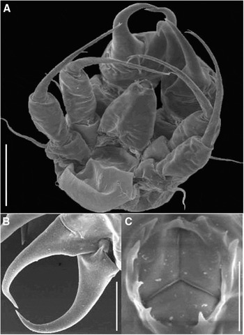
The body is 105–115 µm in length and is more circular than ovoid (Figure 3A). The conical proboscis is 45 µm in length and 30 µm at the widest part. The mouth is circular and on average 14 µm in diameter (Figure 3C). It is closed in a distinct Y-shape. The surrounding raised lip bears up to ten denticles.
Achelia chelata larva. (A) Ventral view (scale bar: 50 µm); (B) fixed and moveable finger of chela (30 µm); (C) tripartite closed mouth and denticles on lip (10 µm).

The cheliphores are smooth with the moveable finger rounded, and the fixed finger ending in a sharp hook (Figure 3B). The basal article is 60 µm in length and the chelae are 100 µm long. There is no spinning spine present.
There are short (50–60 µm) spines at the base of the II and III appendages. The basal article of the appendages is very short (16 µm) and has one slit-like pore (2 µm) located distally on the ventral side. The second article, which has two slit-like pores on its ventral side, is 50 µm long. The terminal claw is almost twice as long as the second article (90–100 µm). The surface of the claw is sparsely decorated with thin denticles. At about 1/3 of the length from the tip there is one large tooth on the ventral side of the claw. The tip of the claw is bifurcate.
Five sensilla were found in the larva, between 10 and 15 µm long each. On the anterior dorsal side of the body there is an unpaired median sensillum. There is a pair of posterior dorsolateral sensilla located behind the third pair of appendages on the dorsal side of the body. At the ventral posterior end of the body there are paired sensilla. The dorsally located sensilla are smooth and bifurcate, while the ventrally located sensilla are trifurcate. Located above each of these sensilla is a slit-like pore.
Larval morphology of Achelia simplissima (N = 10)
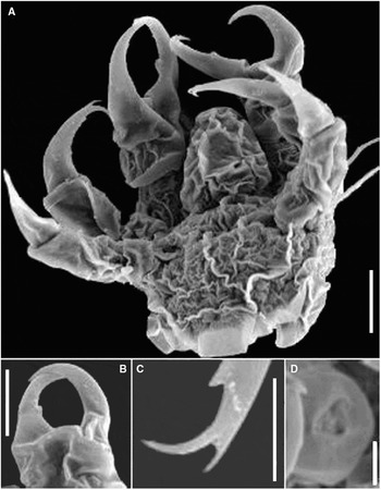
The body is much smaller than the previous two species, only 33 µm long (Figure 4A). The proboscis is 17 µm long, and 15µm wide at its base (Figure 4A). It is cylindrical in shape, tapering slightly to the end with a diameter of 11 µm. The mouth (5 µm in diameter) is open and tripartite. The three surrounding lips are smooth, without denticles (Figure 4D).
Achelia simplissima larva. (A) Ventral view of larva (scale bar: 10 µm); (B) chela (10 µm); (C) tip and teeth on terminal article of II and III appendages (5 µm); (D) tripartite mouth (5 µm).

The basal article of the cheliphore is wider than it is long (15 µm long and 21 µm wide) (Figure 4B). The chelae are almost twice the length of the basal article (26 µm in length). The chelae are rounded, with overlapping fingers when closed. The fingers are smooth, except for one small tooth on the inner edge of the fixed finger. There are no spinning spines present.
The II and III appendages have a basal article 6 µm long, and a second article and terminal claw 16 µm long each (Figure 4A). The appendages are smooth. About one quarter of the way from the tip of the claw is a small denticle (Figure 4C). The tip of each claw is bifurcate. There is a pore on the inside of the distal edge of the second article of the II and II appendages. There is a short (6–10 µm) spine at the base of each leg (Figure 4A).
The larva has one large median dorsolateral pore that is not slit-like. There was no evidence of sensilla anywhere on the larva.
GENUS NYMPHOPSIS
Larval morphology of Nymphopsis spinosissima (N = 15)
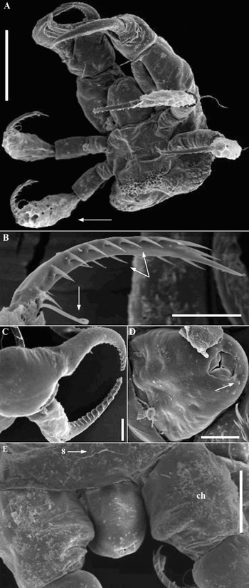
The larval body is rectangular; 140–150 µm long (Figure 5A). The proboscis is very short (55–65 µm) and rounded; it does not even reach the chelae (Figure 5A, D & E). The tripartite mouth is 15 µm in diameter and is open (Figure 5D). The surfaces of the lips are smooth and at their crevices there are three pores of proboscis glands.
Nymphopsis spinosissima larva. (A) Ventral view showing II and III appendages with extra article (arrow) (scale bar: 100 µm); (B) terminal article of the II and III appendages showing basal spine (arrow) and two rows of denticles (double arrows) (20 µm); (C) chela showing ornamentation (20 µm); (D) proboscis and tripartite mouth with glands (arrow) at each corner (20 µm); (E) view of proboscis from dorsal side with cheliphores (ch) offset and a median dorsolateral unpaired bifurcate sensillum (8) (50 µm).

The cheliphores are almost twice the length of the body, 290–300 µm long (Figure 5A). Cheliphores are set slightly apart, so that the proboscis is visible from the dorsal side (Figure 5E). The basal article is 85–90 µm long, while the chelae are just over 200 µm long (Figure 5C). At the base of the first article is a much reduced spinning spine, 22–26 µm long (Figure 5A). The chelae are ovoid and the fingers do not overlap when closed. The inside blade of each finger bears 11–13 teeth.
The appendages II and III are not tripartite; instead they are quadripartite (Figure 5A). The basal article of the II and III appendages is 22 µm long, and bears a short spine at the base (65–80 µm). The second article is between 40 and 45 µm long. The third and extra article is very long (100 µm). It is covered by a different sort of cuticle compared to the rest of the body. The surface of the cuticle has many small entwined threads (Figure 5A). The fourth article, the claw, is curved inwards and is 95 µm in length. The claw has two rows of pointy denticles, one on either side of the blade (Figure 5B). They are between 7 and 10 µm long and there are seven in each row. At the base of the inner edge of the claw is a small (35 µm) spine.
There are seven sensilla; each is bifurcate (Figure 5E). The sensilla are 15–25 µm long. There is an unpaired sensillum located just behind the proboscis on the dorsal side of the larva (Figure 5E). There is a pair of dorsolateral sensilla and slit-like pores located just behind the II appendages. Located 1/3 of the way from the posterior end of the body and in line with the middle of the cheliphores is another pair of sensilla and slit-like pores on the dorsal side of the larva. The final pair of sensilla is located ventrally at the posterior end of the larva.
GENUS EURYCYDE
Larval morphology of Eurycyde spinosa (N = 10)
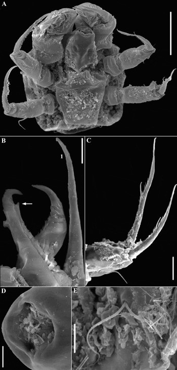
The larva is slightly wider than long (115 µm and 110 µm, respectively), and rectangular in shape (Figure 6A). The proboscis is conical, 45 µm long and 50 µm wide at its base. The mouth is 13 µm in diameter. There are at least four indents just below the rim of the mouth that are 3–4 µm across. The mouth is tripartite, with pores at the corners of the three lips (Figure 6D). The lips lack denticles.
Eurycyde spinosa larva. (A) Ventral view of larva (scale bar: 50 µm); (B) chela with tooth (arrow) and spinning spine (1) (10 µm); (C) II and III appendages (20 µm); (D) mouth (5 µm); (E) bifurcate sensillum on dorsal posterior (5 µm).

The cheliphores are 85–95 µm long. The chelae are half of their size and have a large (65–70 µm) spinning spine (Figure 6B). The inner side of the fixed finger has a tooth 1/3 of the way from the tip. The moveable finger has many small denticles on the bottom half of the inner edge (Figure 6B).
The II and III appendages are 150–180 µm in length. The first article is half the length of the second article (45 µm long). At the base of this article is a thin but long spine (30–50 µm). The terminal article is twice the length of the second article (90–115 µm) (Figure 6C). The inner blade has many short denticles, and a long (10–13 µm) tooth halfway from the base.
There were seven bifurcate sensilla (19–24 µm long) found on the dorsal side of the larva (Figure 6E). They are in the same position as in the larvae of Nymphopsis spinosissima. There were no pores found.
Family Pycnogonidae
GENUS PYCNOGONUM
Larval morphology of Pycnogonum stearnsi (N = 20)
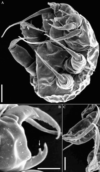
The larval body is dorsally rounded, ventrally flat, and slightly wider than long. The length of the body is 70–80 µm and the width is 70–100 µm (Figure 7A). The cuticle is wrinkled dorsally but smooth ventrally. There is a centrally located pore on the ventral side of the body and also one on the dorsal side. The proboscis is cone shaped and ends in a sharp point (Figure 7A). It is 33–40 µm long and 23 µm in diameter at the widest point. Lips are not visible.
Pycnogonum stearnsi larva. (A) Ventral view of larva and pointed proboscis (arrow) (scale bar: 20 µm); (B) chela with spinning spine and short fixed finger with a single tooth (arrow) (10 µm); (C) terminal articles of II and III appendages showing fine denticles (10 µm).

The cheliphores are almost as long as the body (60–65 µm). The basal article is cylindrical and wider than long. At the distal edge of each basal article is a spinning spine (70 µm long) that extends out past the chelae (Figure 7A). On the ventral and dorsal sides of the basal article is a pore 3–5 µm long. The chelae are almost half the width of the basal article. The moveable finger is hook-shaped, tapering to a point, and almost twice as long as the fixed finger (Figure 7B). It has a series of thin spines along the inner and outer edges (Figure 7B). The distal inside edge of the fixed finger has one blunt tooth.
The II and III appendages are 90–100 µm in length. The basal article is the shortest (15 µm long) but widest (22 µm) article of each appendage. At the distal end of this article is a smooth spine (30 µm in length). On the ventral side of each basal article is a large pore. The second article is narrower than the first but is about 30 µm long. The terminal article is 50–55 µm long. It tapers to a point and has a slight curve towards the ventral side of the larva. The inside edge of the claw is ornamented with a dense row of thin denticles (Figure 7C). The outside edge is smooth.
There were five bifurcate sensilla on the dorsal side of the larva. At the anterior end of the body is an unpaired median sensillum. There is a pair of smooth sensilla located behind the cheliphores. At the posterior end of the body behind the III appendages is another pair of smooth sensilla, with a large pore located between them. There are two large pores, not slit-like, located at the posterior of the larva, below the final pair of sensilla.
Larval morphology of Pycnogonum rickettsi (N = 20)
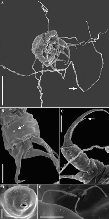
The larval morphology of Pycnogonum rickettsi is very similar to that of Pycnogonum stearnsi. The larval body is rectangular in shape and slightly larger (90–105 µm long and 100 µm wide) than that of P. stearnsi (Figure 8A). There is no ventrally located pore present. However, the cuticle is also wrinkled on the ventral side. The proboscis is cylindrical in shape and almost half the length of the body (50 µm long and 32 µm wide) (Figure 8A, D). The tip of the proboscis tapers slightly to a blunt point 20 µm in diameter. The mouth (4 µm in diameter) is open and surrounded by a single raised lip (Figure 8E).
Pycnogonum rickettsi larva. (A) Ventral view showing long cheliphore spinning spines (arrow) and II and III appendage spines (scale bar: 100 µm); (B) chela with sharply hooked fixed finger and large pore on the basal article of the cheliphore (arrow) (20 µm); (C) terminal article of II and III appendages showing denticle (arrow) (20 µm); (D) open mouth showing single raised lip (10 µm); (E) trifurcate sensillum (5 µm).

The cheliphores are also larger (80–90 µm long) than those of P. stearnsi (Figure 8A, B). The basal articles have two pores, one ventral and the other dorsal. The spinning spine on the distal end of the basal article is the longest of any of the species described. It is more than two times the length of the body (200–250 µm long). The chelae are large, and both fingers have thin spines on the inner blades (Figure 8B). The fixed finger has three teeth, two on the inner edge and one near the tip of the outer edge. The fixed finger is shorter than the moveable finger and ends in a strong hook (Figure 8B).
The II and III appendages are 120–140 µm in length (Figure 8C). The basal article is 15–20 µm long, the second article is 30–40 µm long, and the terminal article is 80–95 µm long. There is a long coiled spine (150–280 µm long) at the base of each basal article. There is a single denticle on the inner edge of the terminal article about 1/3 from the tip (Figure 8C).
There were three sensilla on the dorsal side of the larval body. There is one trifurcate sensillum located at the anterior end of the larva. There are paired bifurcate sensilla located behind the III appendages. There is one large pore between this pair of sensilla and two more large pores just posterior to them. There are two large pores located behind each of the II appendages.
Family Phoxichilidiidae
GENUS ANOPLODACTYLUS
Larval morphology of Anoplodactylus viridintestinalis (N = 10)
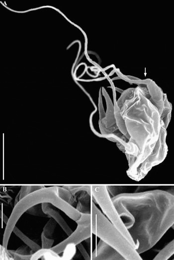
This larva is very small, with a body length of 26–36 µm (Figure 9A). The mouth is circular with no hint of a tripartite form. The mouth appears to be open and is 1.5 µm in diameter (Figure 9C). The lip surrounding the mouth was smooth with straight edges without any denticles.
Anoplodactylus viridintestinalis larva. (A) Side view showing filamentous strands (arrow) as the terminal articles of the II and III appendages (scale bar: 20 µm); (B) chela (5 µm); (C) open mouth (5 µm).

The chelae (20 µm) are smooth and without ornamentation (Figure 9B). The moveable finger has a slight hook at the end (Figure 9C). There are no spines at the base of any of the appendages. Instead, the terminal articles of the appendages II and III are long (100 µm), smooth threads instead of claws (Figure 9A). With these threads, the legs can be four times the length of the larval body.
There was no indication of pores or sensilla on the larval body.
DISCUSSION
Dogel (Reference Dogel1913) suggested three forms of pycnogonid postembryonic development based on the ‘way of life of the larva’ (free-moving ectoparasites, endoparasites, and feeding lecithotrophically while on the adult male). Bain (Reference Bain2003) proposed four modes of development, three of which (‘typical protonymphon,’ ‘encysted larva’ and ‘attaching’) correspond to those suggested by Dogel. Bain defined the fourth mode, the ‘atypical protonymphon,’ based on three criteria: the number of larval appendages (three pairs or one pair), the larva's way of life (free-moving, parasitizing, encysting, or attaching) and the formation of the adult walking legs (sequential, simultaneous or partially simultaneous). While Bain's criteria are more inclusive than Dogel's, the terminology confuses larval morphology with developmental mode. Bain's terms imply that the larval mode of development cannot be determined from protonymphon morphology. However, it has been shown that pycnogonid species having the same mode of development also have larvae with similar morphologies (Bogomolova & Malakhov, Reference Bogomolova and Malakhov2003, Reference Bogomolova and Malakhov2004, Reference Bogomolova and Malakhov2006; Bogomolova, Reference Bogomolova2007; Cano & López-González, Reference Cano and López-González2009). Therefore, I propose new terms for the larval types and postembryonic developmental modes of pycnogonids. The term ‘ectoparasitic’ will replace ‘typical protonymphon’ since larvae undergo development as ectoparasites on cnidarians (King, Reference King1973; Behrens, Reference Behrens1984; Bain, Reference Bain1991; Okuda, Reference Okuda1940; Vilpoux & Waloszek, Reference Vilpoux and Waloszek2003; Bain, Reference Bain2003). ‘Endoparasitic’ will be used in place of ‘atypical protonymphon’ because larvae develop while inside bivalves and sedentary polychaetes (Ohshima, Reference Ohshima1933; Arnaud, Reference Arnaud1978; Ogawa & Matsuzaki, Reference Ogawa and Matsuzaki1985; Salazar-Vallejo & Stock, Reference Salazar-Vallejo and Stock1987; Bain, Reference Bain2003). The term ‘encysting’ replaces ‘encysted larva’ and ‘attaching’ will be used instead of ‘attaching larva.’ Finally, ‘prolonged attaching’ will be used in place of ‘lecithotrophic protonymphon’ because these larvae remain on the male for much longer than those following the ‘attaching’ mode of development (Bogomolova & Malakhov, Reference Bogomolova and Malakhov2006; Bogomolova, Reference Bogomolova2007; Cano & Lopez-Gonzalez, 2009).
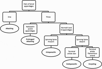
Drawings and descriptions of larvae for which developmental modes are known reveal four morphological characteristics indicative of a species' pattern of development. The first and most obvious characteristic is larval body size. Large larvae (>300 µm) are typical of the ‘attaching’ and ‘prolonged attaching’ developmental modes. Because these larvae are non-feeding while they develop, their large sizes may be due to their substantial yolk reserves (Nakamura, Reference Nakamura1981; Bain, Reference Bain2003; Bogomolova & Malakhov, Reference Bogomolova and Malakhov2006; Bogomolova, Reference Bogomolova2007; Cano & López-González, Reference Cano and López-González2009). Similarly, very small feeding larvae (<80 µm) are typical of the ‘encysting’ mode of development (Meinert, Reference Meinert1899; Bain, Reference Bain2003; Lovely, Reference Lovely2005). Second, the number and development of larval appendages may also suggest the mode of development. For example, all species shown to have the ‘attaching’ mode of development have a larva with the II and III appendages absent (Bain, Reference Bain2003). Although Hooper (Reference Hooper1980) described an attaching larva with all appendages present, it is evident that this ‘larva’ is actually at a very advanced stage of development. Similarly, all larvae with reduced II and III appendages, as in the species Ammothea glacialis Hodgson, 1907 and Nymphon grossipes Fabricius, 1794, have been shown to follow the ‘prolonged attaching’ mode of development (Bogomolova & Malakhov Reference Bogomolova and Malakhov2003; Bogomolova, Reference Bogomolova2007; Cano & López-González, Reference Cano and López-González2009). In larvae following these developmental modes, the function of attachment to the male is by the cheliphores or spinning spines and larvae are not free-moving until the end of development. This may explain why the II and III larval appendages, which are used in other larvae for attachment and movement to their host, are absent or reduced in these larvae (Cano & López-González, Reference Cano and López-González2009). Third, the presence or absence of a spinning spine is also indicative of a species' postembryonic mode of development. Species shown to have the ‘ectoparasitic’ mode of development always have larvae with spinning spines that are used in attachment (Okuda, Reference Okuda1940; Behrens, Reference Behrens1984; Vilpoux & Waloszek, Reference Vilpoux and Waloszek2003; Gillespie & Bain, Reference Gillespie and Bain2006). For example, the species Pycnogonum litorale Ström, 1762 (family Pycnogonidae) and Tanystylum orbiculare Wilson, 1880 (family Ammotheidae) both follow the ‘ectoparasitic’ development and have long spinning spines that they use as ‘safety lines’ to return to their food source if they become dislodged (Russel, Reference Russel and Hedgpeth1990; Bain, Reference Bain2003; Vilpoux & Waloszek, Reference Vilpoux and Waloszek2003). Both the larvae of Nymphonella tapetis Ohshima, 1927 and Ammothella spinifera Cole, 1904 lack spinning spines, presumably because attachment organs are not needed for an endoparasitic lifestyle (Ohshima, Reference Ohshima1933; King, Reference King1973; Salazar-Vallejo & Stock, Reference Salazar-Vallejo and Stock1987). Finally, the morphology of the terminal articles on the II and III appendages is suggestive of a species' postembryonic mode of development. For instance, only species shown to have an ‘encysting’ development have larvae with modified terminal articles: in the place of claws they have long filamentous strands up to five times the length of their bodies (Hilton, Reference Hilton1916; Lebour, Reference Lebour1945; Bain, Reference Bain2003; Lovely Reference Lovely2005). A larva with characteristics of the encysting mode of development has been found in the plankton, making it possible that these long strands are an adaptation to the pelagic environment by decreasing the rate of larval sinking (Malakhov & Bogomolova, Reference Malakhov and Bogomolova2001). They may also be provisional structures for movement to the larva's hydroid host (Russel, Reference Russel and Hedgpeth1990; Lovely, Reference Lovely2005). Therefore, when considered together, these four morphological characteristics (size, number and development of appendages, presence/absence of spinning spines, modifications of the terminal appendage articles) appear to correlate with the mode of postembryonic development in pycnogonids (Figure 10).
Key to postembryonic developmental mode based on larval characteristics (number of larval appendages, degree of development of larval appendages, presence/absence of spinning spine, and morphology of the terminal article of the II and III appendages). Larval characteristics are in boxes and developmental modes are in ovals.

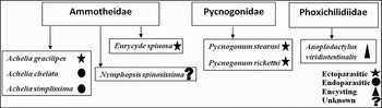
The larvae of Achelia gracilipes, Eurycyde spinosa, Pycnogonum rickettsi and Pycnogonum stearnsi have morphologies characteristic of the most common developmental pathway, the ‘ectoparasitic’ mode. These larvae are of a medium size (70–130 µm long), the three pairs of larval appendages are not reduced, the II and III appendages end in claws (not filamentous strands), and spinning spines are present. Morphologically, these larvae are very similar to the larvae of Nymphon brevirostre Hodge, 1863, Nymphon micronyx Sars, 1888, Nymphon longitarse Krøyer, 1844, Tanystylum orbiculare, Tanystylum duospinum Hilton, 1939, Ammothea alaskensis Cole, 1904 and Pycnogonum litorale, which have been shown to have the ‘ectoparasitic’ mode of development (Morgan, Reference Morgan1891; Okuda, Reference Okuda1940; Russel, Reference Russel and Hedgpeth1990; Tomaschko et al., Reference Tomaschko, Wilhelm and Bückmann1997; Wilhelm et al., Reference Wilhelm, Bückmann and Tomaschko1997; Bain, Reference Bain2003; Vilpoux & Waloszek, Reference Vilpoux and Waloszek2003; Gillespie & Bain, Reference Gillespie and Bain2006; Bogomolova, Reference Bogomolova2007). Therefore, based on body size, number of larval appendages, presence of a terminal claw and of spinning spines, it is likely that the larvae of P. rickettsi, P. stearnsi, A. gracilipes and E. spinosa undergo an ‘ectoparasitic’ developmental mode (Figures 10 & 11).
Predicted mode of development for eight species of pycnogonids based on larval morphology showing within and between family and genera differences. The five species examined in the family Ammotheidae exhibit larval morphologies indicative of two types of postembryonic development (‘ectoparasitic’ and ‘endoparasitic’). The two species of larvae from the family Pycnogonidae both display characteristics suggestive of an ‘ectoparasitic’ postembryonic development. Anoplodactylus viridintestinalis from the family Phoxichilidiidae has a larva indicative of the ‘encysting’ developmental mode.

The family Ammotheidae exhibits the most diversity in postembryonic development modes of all eight pycnogonid families; both the ‘ectoparasitic’ and ‘endoparasitic’ modes of development are typical of species in this family (Bain, Reference Bain2003). It is unsurprising that larvae of Achelia chelata and Achelia simplissima display significant differences in morphology from the larvae of Achelia gracilipes. The generally small body sizes (less than 100 µm long) and absence of spinning spines suggest that these larvae follow an ‘endoparasitic’ mode of development instead of an ‘ectoparasitic’ mode (Figures 10 & 11). The life history of the juveniles and adults of A. chelata supports an ‘endoparasitic’ mode of development. Juveniles and adults of A. chelata have been found parasitizing the mantle cavities of molluscs (Benson & Chivers, Reference Benson and Chivers1960; personal observation of the author). Although larvae have not been found inside the hosts, reproductive adults were present. Larvae could have been overlooked because of their size. Small size coupled with a lack of spinning spines suggests that A. simplissima and A. chelata undergo an ‘endoparasitic’ postembryonic mode of development (Figures 10 & 11).
Larvae of the species Anoplodactylus viridintestinalis from the family Phoxichilidiidae show characteristics suggestive of an ‘encysting’ mode of development (Figures 10 & 11). The size of the larval body is extremely small (30 µm) compared to most other pycnogonid larvae (King, Reference King1973). In addition, larvae of A. viridintestinalis have terminal articles modified into strands that are up to four times the length of the body. All other species of the genus Anoplodactylus that have been described follow the ‘encysting’ developmental pathway and have a small larva with modified terminal articles and no appendage spines (Hilton, Reference Hilton1916; Lebour, Reference Lebour1945; Bain, Reference Bain2003; Lovely, Reference Lovely2005).
Larvae of the species Nymphopsis spinosissima have some unusual morphological characteristics, but most notably, an extra article on both the II and III larval appendages. The number and development of larval appendages seems to be correlated with developmental mode (Bogomolova & Malakhov, Reference Bogomolova and Malakhov2006; Bogomolova, Reference Bogomolova2007; Cano & López-González, Reference Cano and López-González2009). Therefore, the modified larval appendages of N. spinosissima may be an important characteristic indicative of a new form of larval development (Figure 11). Additionally, the larval cheliphores of N. spinosissima are unusually positioned: offset on either side of the body, they allow the short proboscis to be seen from the larva's dorsal side. Although present, the spinning spine is much reduced. All terminal articles show an unusual amount of ornamentation. There are no larval descriptions for any species of the genus Nymphopsis; however, no other larvae in the literature have offset cheliphores and quadriarticulated, heavily ornamented appendages (Bain, Reference Bain2003). Unfortunately, the larvae of N. spinosissima did not develop past the first larval stage, and so further research will need to be conducted to determine if this species does indeed follow a new mode of development.
Larvae of most species of sea spiders have not yet been described (Bain, Reference Bain2003). Future research describing larval morphologies and developmental modes will provide a better understanding of which larval characteristics are most helpful in determining postembryonic developmental modes. Larval morphology suggests that two types of postembryonic development (ectoparasitic and endoparasitic) are evident in members of the genus Achelia. Results suggest that the ‘ectoparasitic’ mode of development is present in both the families Ammotheidae and Pycnogonidae. The ‘encysting’ development typical of the family Phoxichilidiidae is suggested for the species Anoplodactylus viridintestinalis. This paper is the first description of larvae from these eight species of Oregon pycnogonids.
ACKNOWLEDGEMENTS
The author would like to thank Joshua Lord, Svetlana Maslakova, Craig Young, and Alan Shanks for advice and editing suggestions. This study was supported by the National Science Foundation (GK-12 grant number DGE-0638731). SEM materials were provided by Craig Young.