Introduction
In silks, proteins are the structural components, and water is the solvent. Protein and water combine and separate—under ambient pressures and temperatures—to make a surprisingly tough thread (Figure 1). Spider silk is a case in point for the superb mechanical performance that can be achieved with this process. This, and the ability to collect the material under highly controlled conditions, makes spider drag-line silk an excellent starting material in the quest to unravel animal silks and their many trade secrets. Good silk research requires comprehensive biological understanding as well as the application of the fullest possible range of tools and techniques developed in the polymer sciences. This feature, in addition to its enormous diversity, makes silk a powerful model for inspiration to guide the design of novel polymeric materials and matter.
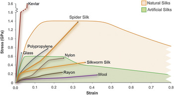
Silk tensile properties in comparison to other industrial and natural fibers and artificial silks. Colored backgrounds indicate the impressive toughness of silk and vast range of properties achievable by varying spinning conditions (see legend) (adapted from References Reference Vollrath, Porter and Holland3 and Reference Holland, Terry, Porter and Vollrath57).

Importantly, silk already has a venerable history as well as a strong following in our present society, which happily spends a hundred billion dollars annually on silk products. In particular, the silk threads of the Bombyx silkworm, a mulberry leaf-eating moth larva, have for millennia formed the basis of a hugely lucrative textile industry and attracted extensive industrial research and development (R&D).Reference Altman, Diaz, Jakuba, Calabro, Horan, Chen, Lu, Richmond and Kaplan1, Reference Omenetto and Kaplan2 The silk threads produced by spiders, on the other hand, have (so far) not led to any important commercial or technological advances, in spite of being intrinsically stronger and tougher than their silkworm counterparts.3 A few basic generalizations may be useful to highlight some key differences and similarities of the two types of fiber. Both silk types share many important features (such as fundamental molecular composition and flow characteristics of the spinning feedstock) despite a totally independent origin and evolution.Reference Holland, Vollrath, Ellison and Abbott4–Reference Eisoldt, Smith and Scheibel6 However, millions of years of selection/natural R&D have resulted in spiders yielding silks specialized for application in lightweight net structures (webs), while moths use silks optimized for solid multilayer non-woven mesh composites (cocoons).Reference Vollrath7, Reference Chen, Porter and Vollrath8 Fundamentally, moth silks are all of one basic design-type, while spider silks cover a huge range of types. Moreover, moths have only one set of glands, each producing one set of silk, while each individual spider is able to produce several different silk types, each originating in a dedicated gland.
This overview will avoid speculation and hype about the wonders of spider silk and consider rationally what we have learned from studying the different silks as a biomaterial and what further study and technological application might have in store. In particular, we start from the position that research on spider silks has already resulted in a wide range of fundamental insights into biopolymer production, processing, properties, and functions that would have been impossible to derive solely from the study of moth silks.Reference Holland, Terry, Porter and Vollrath9–Reference Dicko, Kenney, Vollrath, Kajava, Squire and Parry12 This is due to the massive diversity and apparently more attractive mechanical properties of spider silks because of the wider range of properties required in webs.Reference Vollrath and Porter13
Comparative studies are beginning to shed light on silk formationReference Holland, Terry, Porter and Vollrath9 as well as the functional structure-property relationships.Reference Vollrath and Knight10 This includes the importance of structural hierarchy for the performance characteristics of a silk as well as the effects of processes during silk formation on fiber performance (Figures1 and 2). These processes can be studied not only by examining the spider’s and the worm’s natural in vivo silk production systems, but also by extracting the silks from their native glands in order to examine their flow and transition characteristics in vitro under controlled conditions.Reference Holland, Terry, Porter and Vollrath9 In this context, the rheological behavior of spun silks that have been dissolved and purified for re-spinning are of considerable interest, as these artificial “silks” have material properties that differ significantly from their native silk precursors (Figure 1). A series of simple questions shall give a rapid overview of the field followed by selected aspects of special interest.
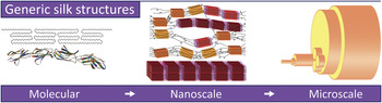
The hierarchical nature of silks. Molecular components are long chain proteins/protein complexes, typically of several 100 KDa. These consist of highly repetitive peptide sequences that facilitate variable secondary structure formation and the potential for high hydrogen bonding density. The nanoscale is dependent on processing, which determines the degree of disordered and ordered domains the proteins adopt (grading from yellow to red boxes). The microscale builds upon the nanoscale, combining nanofibrils into large and microfibrillar structures that run parallel to the long axis of the fiber. Finally, depending on the animal that spins the silk, there are skin-core features and additional coatings that may be applied ranging from neurotoxins to adhesive binders.

Why does silk research matter?
It might seem surprising that the independent origin and parallel development of biomaterials with different natural end-user requirements has led to (1) fundamental molecular designs that are very similar, (2) extrusion spinning processes that have overlapping features, and (3) flow characteristics of the spinning feedstocks that are virtually identical.Reference Holland, Terry, Porter and Vollrath9, Reference Vollrath and Knight10, Reference Lucas and Rudall14–Reference Holland, Porter and Vollrath19 However, if we consider that Nature tends to economize (energy being her principal “currency”) and typically uses a limited range of raw materials to assemble her molecules, then this convergence becomes less surprising. At the same time, the independent origin of the materials adds value to any insights it might provide for bioinspired material designs. After all, a good model system should be not only relevant but also generic in the lessons it provides.
Sustainability of materials and production processes is becoming ever more important for human industry and commerce. Nature’s silks are well placed to provide relevant and generic insights.Reference Holland, Vollrath, Ryan and Mykhaylyk20–Reference Vollrath and Porter22 Silks have the potential to add important insights to fundamental questions concerning protein-folding and function because silk feedstocks are composed of highly structured protein complexes. Additionally, feedstock is produced quickly (hours), can be stored readily mid- to long-term (weeks to months), and can be converted rapidly from liquid to solid (milliseconds), while the naturally functional lifespan of a silk can be long (months), and the commercial deployment as fiber in manmade textiles ranges from years to millennia. Thus within silks, we find a huge range of highly tuned fibrous proteins that undergo a number of on-demand conversions. From a research perspective, this is important, as these materials can be uniquely probed and investigated because all of these conversions occur in environments that are experimentally accessible with a minimum of trauma.
What has silk research taught us so far?
Natural processing of native silk feedstocks
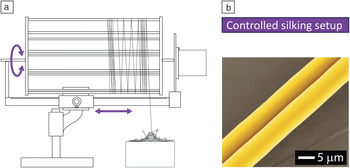
Much like the important engineering polymers, such as ultrahigh molecular weight polyethylene and Kevlar, the properties of silks rely heavily on processing. So much so that it is possible to spin silks to have almost identical material properties despite having significantly different amino acid (chemical) compositions.Reference Liu, Sponner, Porter and Vollrath23 An elegant tool to investigate processing is forced silking of immobilized spiders by experimentally drawing silk from the animal onto a bobbin (Figure 3). Reeling speed as well as body temperature have both been shown to affect fiber diameter as well as all conceivable mechanical properties.Reference Vollrath, Madsen and Shao24, Reference Shao and Vollrath25 This was supported by structural changes in the silk observed using real-time micro x-ray diffractionReference Riekel, Madsen, Knight and Vollrath26, Reference Riekel and Vollrath27 and Raman spectroscopy of fibers in the process of being spun.Reference Young, Sirichaisit, Shao and Vollrath28
Diagram of the reeling apparatus (a) and false color scanning electron microscopy micrograph of a spider dragline silk (b). The reeling apparatus is under controlled translation and speed (purple arrows). This enables the processing conditions within the spider to be altered as a function of reeling speed. The animal undergoing silking is safely restrained by mesh pinned to an expanded polystyrene block that can be heated or cooled (center bottom).

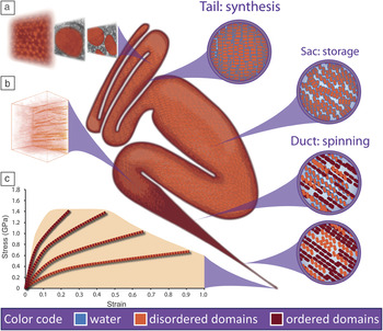
In order to understand exactly how a liquid protein gel with modulus in the kPa range transforms into a final solid fiber with modulus on the order of several GPa within a matter of seconds, we must look inside the animalReference Vollrath and Knight10, Reference Knight, Knight and Vollrath29 (Figure 4). Microscopic analysis has revealed that silk is produced and processed by specialist glands in the spider and silkworm in a remarkably similar manner in spite of their divorced developmental origins.Reference Craig30 Silk proteins are manufactured and excreted by epithelial cells into the sac of the gland where they are stored at high concentrations in solution (typically ≥20% dry weight).Reference Knight and Vollrath31, Reference Akai32 Upon spinning, this protein solution (feedstock) travels down an elongated tapering tubular duct and undergoes a stress-induced phase transition before exiting the spinneret as a fiber.Reference Vollrath and Knight10, Reference Asakura, Umemura, Nakazawa, Hirose, Higham and Knight17 On a molecular level, this transition involves the exchange of a silk protein’s closely bound water for more stable amide–amide hydrogen bonds, resulting in protein aggregation and precipitation.Reference Porter and Vollrath33
A typical silk gland, be it from a spider or silkworm, consists of a tail, sac, and duct. The tail secretes most of the feedstock, which is an aqueous liquid of hydrated silk molecules. The cells lining the tail’s wall assemble the silk proteins, which then tightly pack into small vesicles (a) that transport and excrete the molecules into the lumen (tail circular inset). The sac stores the silk molecules in a hydrated metastable state prior to spinning (as an aquamelt, sac circular inset). From the sac, the feedstock flows into a duct, typically tapered and of substantial length, allowing silk proteins to flow in patterns that resemble liquid crystalline flow and resulting in dehydration, aggregation, and precipitation into nanofibrillar structures ((b) and duct circular insets). Changes in these processing conditions (such as speed of spinning) greatly affect the range of fiber properties possible with silk (c). This can be directly related to the relative amounts of ordered and disordered hydrogen bonding generated during processing—see in (c) how strength increases and toughness decreases with increasing relative amounts of ordered domains.Reference Vollrath and Knight10, Reference Asakura, Umemura, Nakazawa, Hirose, Higham and Knight17, Reference Holland, Vollrath, Ryan and Mykhaylyk20, Reference Shao and Vollrath25

Morphological and birefringence patterns revealing the shape changes in the packing of the silk proteins from spherical to ellipsoid provided initial evidence that mechanical deformation (i.e., rheological processing) primarily fuels the stress-induced phase transition.Reference Knight, Knight and Vollrath29, Reference Akai32, Reference Magoshi, Magoshi and Nakamura34, Reference Akai, Imai and Tsubouchi35 The origin for the introduction of mechanical energy has been attributed to both wall shear along the ductReference Viney36 and extensional flow in the draw-down taper.Reference Asakura, Umemura, Nakazawa, Hirose, Higham and Knight17, Reference Knight, Knight and Vollrath29, Reference Knight and Vollrath37, Reference Asakura, Yao, Yang, Zhu and Hirose38 Not surprisingly, this natural extrusion “spinning” process is not dissimilar to industrial processes where hyperbolic dies are used in synthetic polymer processing to reduce disorientation by maintaining a constant elongational flow field.Reference Chen, Cuculo and Tucker39 It has been suggested that this may be utilized by these animals to maintain orientation of the silk proteins while preventing premature phase transition until they encounter the higher shear rates found in the draw-down taperReference Kerkam, Viney, Kaplan and Lombardi40 and allow for the gradual changes in the chemical environment (pH, ionic balance). This induces a pre-gelation in the material, thereby activating it for processing and enabling it to develop its hierarchical structures (Figure 2).Reference Zhong, Zhou, Shao, Chen, Chen, Hu, Deng and Yao41–Reference Meyer and Jeannerat50
Thus it appears that mechanical deformation is the catalyst for silk spinning and accordingly rheology has increasingly become the tool of choice not only to characterize the mechanical properties of silk feedstocks, but to probe the phase transition itself.Reference Holland, Terry, Porter and Vollrath9, Reference Holland, Vollrath, Ryan and Mykhaylyk20, Reference Ochi, Nemoto, Magoshi, Ohyama and Hossain51–Reference Holland, Urbach and Blair56
Initial studies revealed great similarities in the flow properties of silk feedstocks despite significant differences in chemical composition; a result of either deep-rooted phylogenetic origin (e.g., spider versus moth) or more recent adaptation to selection (e.g., domesticated versus wild silkworm).Reference Holland, Terry, Porter and Vollrath9, Reference Kojic, Bico, Clasen and McKinley53 Importantly, this apparently generic similarity extends to synthetic polymer melts. Extrapolating, we must hypothesize that natural selection has converged, from different starting points, on a singularly efficient way of rapidly processing proteins into high performance fibers. This allows the use of tools and techniques developed by the polymer sciences to be adapted to study silk, enabling silk to serve as an inspiration for novel developments in polymer processing. Furthermore, by using the flow characteristics of natural silks as the key design criteria, it is now possible to predict the quality of artificial silks as they are being developed, and it has highlighted the need for higher fidelity artificial feedstocks.Reference Holland, Terry, Porter and Vollrath57
But how does such a complex hierarchical structure as observed in the spun fiber develop as a result of shear? Here lies the path to the current state of the art for silk rheology, integrating novel tools and techniques to characterize the molecular mechanisms behind spinning. Spectroscopic (infrared, nuclear magnetic resonance) and scattering studies (small angle x-ray scattering) reveal molecular alignment of silk proteins as a result of shear prior to a stress-induced phase transition,Reference Boulet-Audet, Vollrath and Holland55, Reference Ohgo, Bagusat, Asakura and Scheler58, Reference Rössle, Panine, Urban and Riekel59 thus confirming previous birefringence measurements inside the duct.Reference Asakura, Umemura, Nakazawa, Hirose, Higham and Knight17, Reference Knight, Knight and Vollrath29 Moving to larger scales in the silk hierarchy, recent observations of native silk feedstocks under flow using confocal microscopyReference Arevalo, Urbach and Blair60, Reference Schmoller, Fernandez, Arevalo, Blair and Bausch61 have revealed that shear stress alone is a sufficient mechanism for the formation of the large-scale fibrillar features present in the silk fiber.Reference Holland, Urbach and Blair56, Reference Putthanarat, Stribeck, Fossey, Eby and Adams62–Reference Hakimi, Knight, Knight, Grahn and Vadgama64 This lends fuel to the hypothesis that silk fibers can assemble in the total absence of any external chemical stimuli, such as changes to pH or ions,Reference Foo, Bini, Hensman, Knight, Lewis and Kaplan46 and that flow elongation and shear are the main evolutionary drivers for silk fibrillogenesis.
Thus apparent complexity in silk spinning is actually a result of a controlled exposure of hidden functionality within the silk proteins. However, natural selection acts at all levels within biological systems, striving for both elegance in reducing complexity but also efficiency of the individual components.
While there are several direct comparisons between the physical properties of silk fibers and industrial analogs,Reference Guan, Porter and Vollrath65, Reference Drodge, Mortimer, Holland and Siviour66 there are only a handful of studies of direct comparisons between silk and synthetic feedstocks.Reference Tsukada, Freddi and Crighton67 The recent development of shear-induced polarized light imagery enables analysis of the kinetics of polymer crystallization/fibrillation and the calculation of a specific work parameter such as shear energy density.Reference Mykhaylyk68 As a result, upon comparison with a typical polymer (high density polyethylene), silk feedstock was shown to require 1000 times less total energy in order to generate fibrils.Reference Holland, Vollrath, Ryan and Mykhaylyk20
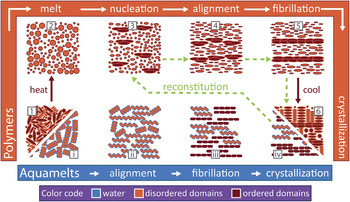
We proposed that Nature achieves this massive energy saving by employing a specific type of material, an “aquamelt.” An aquamelt is a naturally hydrated polymeric material that is able to solidify at environmental temperatures as a result of a controlled stress input perhaps enhanced by a subtle chemical contribution (Figure 5).Reference Holland, Vollrath, Ryan and Mykhaylyk20 This highly energy efficient solidification is brought about by the unique interaction of these materials with their shell of hydration, which is lost during processing, thus causing a phase separation into solvent water and solid polymer.Reference Porter and Vollrath69 This process is a feature that we hypothesize to be inherent to all de novo proteins due to their peptide-based chemistry.Reference Porter and Vollrath70
Schema for fiber formation in polymers (orange box, numbers) and their proposed subclass aquamelts (blue box, roman numerals). Horizontal arrows depict stages that require sample shearing, with aquamelts requiring one-tenth the work required in order to bring about fibrillation (as represented by arrow length). Colored objects in the drawings represent components responsible for the formation of fibers in the systems: long chain molecules (their radius of gyration or shape) in polymers and proteins (secondary and tertiary structures) in aquamelts. These are color-coded to represent ordered, disordered, and hydrated domains. Green dotted arrows represent the path reconstituted (artificial) silk currently follows in order to be reprocessed into a fiber. Adapted from Reference Reference Holland, Vollrath, Ryan and Mykhaylyk20.

Thus, for silk, the removal of associated water and the resultant change in solubility serves as the mechanism to “lock in” any fibrillation that occurs during spinning, a mechanism that is currently both practically (i.e., reconstitution) and potentially theoretically irreversible.Reference Porter and Vollrath69 It is our opinion that only by fully understanding aquamelts will it be possible to resolve some of the issues surrounding reconstitution and spin truly biomimetic silks.Reference Vollrath, Porter and Holland71 Furthermore, using aquamelts as a source of bioinspiration may also help uncover new ways to manipulate hydrogen bonding during processing in both biological (e.g., collagens or celluloses) and synthetic polymers and access Nature’s low energy processing routes.
Mechanical properties of natural silk fibers
Interestingly, it turns out to be rather difficult to emulate natural silk feedstocks and processing with synthetic or reconstituted silk products and to spin fibers with properties approaching those of natural silks (Figure 1).Reference Vollrath, Porter and Holland3 Yet, success in this field would be a prerequisite for any commercial de novo manufacture of silk-based or silk-inspired polymer fibers because of the close link between processing conditions and material properties.
Why does it matter? The attractive combination of strength and toughness in the mechanical properties of silk is often called the “Holy Grail” vision for synthetic fibers, and attempts are being made to copy these properties in semi-natural materials, for example, by reprocessing spun silks through a dissolving and purification phase followed by a controlled re-spinning by extrusion into a dehydrating solidification bath. Natural silkworm silks unraveled from cocoons are available in large quantities at attractive pricing, although they are less interesting mechanically than spider silks. Yet spider silks are difficult to harvest commercially in quantity. To match the quality of a spider silk with the quantity and availability of silkworm silk, three main routes have been tried: (1) genetically engineer spider silk genes into silkworms; (2) insert the relevant genes into microorganisms; or (3) reconstitute silkworm silk feedstock from naturally spun fibers and try to improve its mechanical properties through physical or chemical processing.Reference Fu, Shao and Fritz72, Reference Chung, Kim and Lee73 So far, none of these approaches has been successful, most probably because of the inability to appropriately control the material’s macromolecular structure before and during spinning. Importantly, rheological investigations continue to show that typical reconstituted silks are orders of magnitude away from the typical flow characteristics of both spider and silkworm silks.Reference Holland, Terry, Porter and Vollrath57
Spider dragline silk stands out in comparisons of engineering stress-strain curves (Figure 1). Kevlar is stiffer (modulus of 112 GPa) and stronger (failure stress of 3 GPa) than dragline silk (modulus of about 10 GPa and strength of about 1.5 GPa), but golden orb-spider dragline silk is tougher in terms of energy to breakage (Kevlar 40 MJm–3 and dragline silk up to about 250 MJm–3). While specific spider silks can be considered outliers, silkworm silks also have excellent polymeric properties (such as a good weight–strength ratio), although these are not all that different from those of typical synthetic polymeric fibers such as medium or high tenacity nylon or polyethylene terephthalate. Nevertheless, even here, there are key differences. For example, high-rate (bullet) impact testing that compared Bombyx mori cocoon silk with medium tenacity nylon demonstrated that the two materials differ in their ability to share strain energy between the ordered and disordered domains, which suggests differences in their micro- and nanostructures.Reference Drodge, Mortimer, Holland and Siviour66 Interestingly, the silk had superior energy absorption compared to the nylon, and the energy imparted by the passage of a longitudinal wave varied little with pre-stress in the silk (unlike the nylon).
Thus silks, which are also sustainable and biocompatible, look attractive as natural replacements for synthetic fibers such as nylon. However, the materials engineer will legitimately point out that silk must struggle in engineering applications (such as the often quoted bulletproof-vest applicationReference Hudspeth, Nie, Chen and Lewis74) that rely on different relationships of stiffness and strength than those provided by a silk. Moreover, most polymers have bulk engineering properties well below their fiber properties; for example, in bulk nylon, strength reduces to below 100 MPa. Consequently, it might be reasonable to expect that silk might have comparable fracture mechanics problems, as indeed is the case.Reference Porter, Guan and Vollrath75 Importantly, in analogy to man’s glass-fiber/resin matting, Nature uses the combination of its silk threads as fully integrated fiber components in sericin-glue composite structures, which maximizes the benefit of the inherent material properties of the ultrathin silk fibers.Reference Chen, Porter and Vollrath8, Reference Porter, Guan and Vollrath75, Reference Chen, Porter and Vollrath76
More generally, silk fibers can have almost any possible set of mechanical properties available to polymers: from soft hydrated gels, through rubber-like hydrated states, to strong tough draglinesReference Vollrath, Porter and Holland3 (Figure 1). Control of properties through structure looks at first sight to be highly complex because of the involvement of many different combinations of peptide sequences and secondary structure combinations at the nanometer scale. Early empirical models of secondary structures of semi-crystalline morphology that are made up of ordered, nanometer scale, hard polymer domains in a softer matrix of more disordered polymer chainsReference Termonia77 and more recent, more complex nanoscale molecular dynamics simulationsReference Keten, Xu, Ihle and Buehler78 can reproduce the mechanical properties of silks quite well, without actually predicting bulk properties. Mean-field models may give better insights, especially if they are based on the evolutionary principle of “energy management” and reduce the apparent complexity to the key structural elements of order for stiffness, disorder for toughness, and hydration of the disorder for rubberlike fractionsReference Porter and Vollrath69, Reference Porter and Vollrath79–Reference Porter, Vollrath and Shao81 (Figure 4c). This first-principles approach radically simplifies the subject of silk protein modeling and indeed allows accurate predictions of bulk viscoelastic properties.Reference Porter and Vollrath33 However, the key question of strength remains a mystery and, until the fracture mechanics of polymers in general and silk in particular are well understood, silk properties will remain unresolved. However, a recent study has demonstrated that the outstanding properties of silk threads can be traced to their extreme thinness (Figure 3).Reference Porter, Guan and Vollrath75
Attention tends to focus on the mechanical properties of silk, and much progress is being made to understand the fundamental principles. However, in addition to a compelling combination of stress and strain mechanics, some spider silks (such as a typical dragline) have the intriguing property of exceptional contraction in a mild solvent. This so-called “supercontraction” manifests itself when a silk fiber contracts on exposure to water by up to 50% of its original length and is a property that we neither fully understand nor have learned to harness.Reference Liu, Sponner, Porter and Vollrath23 At the molecular level, supercontraction is usually attributed to recoiling of elongated amino acid proline-containing sequences in the silk protein that are sensitive to water, and supercontraction increases with increasing proline volume fraction.Reference Liu, Sponner, Porter and Vollrath23 Recent studies have also shown that other protein constituents can participate in supercontraction as well, since only a fraction of contraction can be induced specifically by taking the silk through its glass transition conditions, and then the remaining fraction by exposure to liquid water.Reference Guan, Porter and Vollrath65 Clearly such an interesting feature as supercontraction holds potential lessons for bioinspired materials, and efforts are being made to harness the process (e.g., for potential actuator functions via controlled cycles of ambient humidity).Reference Blackledge, Boutry, Wong, Baji, Dhinojwala, Sahni and Agnarsson82
Potential for further important discoveries
It can be claimed that much progress has been made in the last decade toward unraveling the secrets of silk. Yet many crucial secrets remain to be discovered. While seemingly contrary to regular reports in the media, so far no-one has managed to produce a material that matches a typical spider silk in performance; indeed, nothing has been published that suggests even a half- or quarter-way success (Figure 1).Reference Vollrath, Porter and Holland3 Toward this goal of silk-like performance in a bioinspired analogue, new and substantial key insights are required, and they will have to come from (1) principally the feedstock production processes, followed by (2) the storage conditions and, finally, (3) specific tuning details of the spinning processes.
For the production of potential silk feedstocks, the use of micro-organisms (such as yeasts or E.coli) is widespread, but such systems are not without their problems. First, the molecules thus produced are usually derived short peptide sequences rather than full length proteins because of constraints in the technologyReference Chung, Kim and Lee73 (with the exceptions of the short-chain helix-silks of beesReference Sutherland, Campbell, Weisman, Trueman, Sriskantha, Wanjura and Haritos83). Second, the extraction of silk-analogue molecules from the cell solution (if micro-organisms are used)Reference Xia, Qian, Ki, Park, Kaplan and Lee84 or milk (if goats are used)Reference Lazaris, Arcidiacono, Huang, Zhou, Duguay, Chretien, Welsh, Soares and Karatzas85 requires treatments that in turn would degrade the molecules, if indeed they were true silk-feedstock proteins. Any exposure to inappropriate chemicals will quickly denature such live proteins, and thus render them unspinnable using the natural, biological spinning techniques.Reference Holland, Terry, Porter and Vollrath57
Any analysis of natural silk storage conditions will have to consider the new insights into aquamelt-processing combined with the concept of pre-charged protein functionality; until this is done, our understanding of silk feedstock structure-function relationships will remain in a state of great flux. A valuable test would be the ability to create the molecule in such a way that it keeps its electronic structure in the storage state until such time that a switch into a spinnable state is required. Presently, there is no way of doing this in bulk chemistry, which would be required for the “bucket” processing necessary for industrial production.
Finally, silk spinning is another area that still requires further substantial R&D. While a simple spinning rig can produce nice fibers,Reference Yan, Zhou, Knight, Shao and Chen86 no rig so far has been able to deploy a bioinspired process in order to produce fibers matching spider thread properties. However, in order to honestly claim success for true silk spinning, the interaction of feedstock and extrusion processes should at least match the principal criteria of the natural process (i.e., sustainable production of the feedstock, extended storage under aqueous conditions at ambient temperatures, and a rapid phase transition/conversion without the use of unnatural chemicals, temperatures, and pressures). This is the essence of bioinspired silk production.
Where might any discoveries lead us?
Materials are a cornerstone of our society but are also becoming a major challenge facing the global economy in the 21st century. Materials manufacture and processing result in over 20% of the world’s carbon emissions, with many structural materials sourced from nonsustainable supplies.Reference Pauli87 A prime example is polymers, accounting for 5% of crude oil usage; and peak oil is predicted to have a dire impact on the price of this cheap, ubiquitous commodity. This situation is compounded as consumer demand for high-performance materials is forever increasing, as is the demand for sustainable and environmentally friendly products. Many people believe that Nature can contribute much to the discussion, since natural materials tend to be supremely energy efficient as well as recyclable. Today’s biopolymers, specifically silks, offer both inspiration and solutions to the current challenges facing the synthetic polymer industry, provided we understand how to process them correctly.
Silk is proving to be a superb model system that gives access to the most powerful of R&D: millions of years of trial and error development in millions of interconnected but partially independent laboratories all undergoing the most rigorous quality control by natural selection. But, will we ever be able to truly copy (mimic) silk production processes, or will these remain on the level of inspiration? In the case of bioinspiration, the engineer “simply” takes specific aspects of a biological model and re-engineers them together into a functional unit that can be mass-produced. In the case of bio-mimicry, the engineer in collaboration with the biologist aims to copy a biological system. In the case of silk spinning, bio-mimicry should be a realistic goal (one would have thought) given that the extrusion spinning of a fiber from a feedstock should be easier to achieve than growing a filament (such a hair) from a cellular organ.
Silks, unlike any other biological material, are not defined by their structure nor by their properties, but rather by their processing: silks by definition are spun.Reference Vollrath and Porter22 This distinguishes silks both conceptually and practically from other natural biopolymers, which are grown. Thus silks, because of their production processes, have more in common with synthetic polymers than any other biological material. Simply put, silks are nanostructured materials shaped by the specific conditions of the extrusion process. This has implications well outside the field of polymer-based textile or engineering materials, and studies are providing fundamental insights into amyloids (responsible for Alzheimer’s disease) as well as adding to a deeper understanding of the roles of hydration and H-bonds in protein performance.Reference Dicko, Kenney, Vollrath, Kajava, Squire and Parry12, Reference Porter and Vollrath69, Reference Porter and Vollrath79, Reference Kenney, Knight, Wise and Vollrath88
Summary of the case for silk research
There are many applications for silks ranging from garment textilesReference Wynne89 to high-technology medical implants such as conduits for nerve or ligament tissue repair patches.Reference Huang, Begum, Barber, Ibba, Tee, Hussain, Arastoo, Yang, Robson, Lesage, Gheysens, Skaer, Knight and Priestley90, Reference Chaudhury, Holland, Thompson, Vollrath and Carr91 Much natural value-adding R&D on the raw material has been ongoing for millennia, and today continues to see significant trade as well as venture capital investment. Some newspaper stories are clearly unsubstantiated, such as using spider silks for bulletproof vests. Other ideas will be hard-pushed to provide a return for the investor, such as inserting spider silk genes into the silkworm genome, since both kinds of silks derive most of their properties from the spinning process, and silkworms with spider genes will still spin like silkworms and most likely produce inferior fibers.Reference Shao and Vollrath25
While spider silks and their use in lightweight web structures have attracted much interest for developing high-performance fibers,Reference Vollrath, Porter and Holland3 silkworm cocoons provide ideal practical examples for natural non-woven reinforced composites.Reference Chen, Porter and Vollrath8 Here, structure-property relations are another fine example of Nature’s ability to manage energy efficiently and effectively, with mechanical properties being controlled by the gradual fragmentation of the bonded-fiber nonwoven structure under load.Reference Chen, Porter and Vollrath21 Initial properties such as stiffness are determined by the packing density of the fibers and binder, and as the bonding connectivity between the fibers is reduced, the composite stiffness reduces up to a percolation threshold, where there is no continuous bonded pathway to sustain the load, and the cocoon fails as the fiber simply disentangles. While cocoon properties per se may not be of great specific interest to a materials engineer, the insights derived from silk and cocoon properties can be used very effectively in a remarkably wide range of different composite materials, from concrete to nanofiber mats and polymer bonded explosives.Reference Chen, Porter and Vollrath21
Conclusion
There are many similarities between natural and synthetic fiber-spinning: both start with liquid feedstocks, which share comparable flow properties,Reference Holland, Terry, Porter and Vollrath9 both are melts (albeit with significant differences) that are extruded through convergent dye designs,Reference Knight and Vollrath37 and both utilize spinning to fabricate highly ordered semicrystalline fibrous structures.Reference Vollrath and Porter22 There are also many similarities in the mechanical performance of these nanostructured biopolymers compared with synthetic fibers of comparable dimensions. The difference in both cases is the amount of time expended for R&D, which in biological materials extends to hundreds of millions of years and in manmade materials amounts to a few millennia at best. Hence, for the polymer engineer, silk can be a powerful model for future material design, having an attractive combination of strength and toughness coupled with environmentally benign and ultralow energy aqueous processing over a 40°C range; these are all qualities synthetic polymers are yet to match.Reference Holland, Vollrath, Ryan and Mykhaylyk20
Acknowledgments
We are grateful to Ms. Juan Guan for SEM imagery, AFSOR (Grant No. F49620–03–1-0111) and ERC (SP2-GA-2008–233409) for generous funding of all aspects of the work, and to the UK research councils (EPSRC, BBSRC) and Magdalen College Oxford for funding specific studies.

Fritz Vollrath is a senior researcher in the Department of Zoology at the University of Oxford. His professional development ranged from studies of zoology and physics at the Universities of Göttingen and Freiburg to postdoctoral research and fellowships with the Smithsonian Institution and Oxford University. He was an assistant professor at Basle University, held the Chair of Zoology at Aarhus University, and ultimately returned to Oxford on a senior research fellowship. His fieldwork covers Central and South America, Australasian, and Africa, while his business interests include spin-out companies furthering the commercialization of academic silk research. Vollrath can be reached at the Department of Zoology, University of Oxford, South Parks Road, Oxford OX1 3PS, UK; tel. 44-1865-358220; and email fritz.vollrath@zoo.ox.ac.uk.

David Porter is a senior researcher at the University of Oxford in the Department of Zoology. He obtained his PhD degree in the Physics Department at Loughborough University and then worked for Dow Chemical on the modeling of polymer properties. He also worked as a fellow at QinetiQ in Farnborough, where he focused on materials modeling of a wide range of materials and is now in the group of Fritz Vollrath at Oxford—studying structure-property relations in silk and other interesting biomaterials. Porter can be reached at the Department of Zoology, University of Oxford, South Parks Road, Oxford OX1 3PS, UK; tel. 44-1865-271216; and email david.porter@zoo.ox.ac.uk.

Chris Holland is an EPSRC Early Career Fellow in the Department of Materials Science and Engineering at the University of Sheffield. Studying at Oxford, he earned his BA in biological sciences, an MSc in integrative biosciences, and a DPhil degree in silk rheology in the Oxford Silk Group. Afterward, Holland was awarded a fellowship from Magdalen College. He is now continuing his research on the thermomechanical characterization and evolution of silks alongside other natural materials at Sheffield. Holland can be reached at the Department of Materials Science and Engineering, University of Sheffield, Sir Robert Hadfield Building, Mappin Street, Sheffield S1 3JD, UK; tel. 44-7712-051921; and email christopher.holland@sheffield.ac.uk.