INTRODUCTION
Myxozoa are highly modified endoparasitic cnidarians (Jimenéz-Guri et al. Reference Jimenéz-Guri, Okamura and Holland2007a ; Collins, Reference Collins2009; Holland et al. Reference Holland, Okamura, Hartikainen and Secombes2011) gaining attention due to their impact on fish health, aquaculture production and economy. Myxozoa are divided into two subgroups: Myxosporea and Malacosporea, based on morphological, biological and molecular differences. Myxosporeans are highly diverse, with around 2400 species currently described (Zhang, Reference Zhang2011). In contrast, malacosporeans consist of two genera with only three nominal species Buddenbrockia plumatellae, Buddenbrockia allmani and Tetracapsuloides bryosalmonae (Schröder, Reference Schröder1910; Canning et al. Reference Canning, Tops, Curry, Wood and Okamura2002, Reference Canning, Curry and Okamura2008). Recent molecular studies conducted in bryozoan and fish hosts (Bartošová-Sojková et al. Reference Bartošová-Sojková, Hrabcová, Pecková, Patra, Kodádková, Jurajda, Tyml and Holzer2014; Hartikainen et al. Reference Hartikainen, Gruhl and Okamura2014) unveiled high diversity, with a putative total of 16 species identified from relatively limited geographic regions of Europe, North America and Borneo, indicating that malacosporean biodiversity is far greater than previously suggested.
Malacosporeans alternate between two hosts, fish and freshwater bryozoans, however, T. bryosalmonae is the only malacosporean species for which the full life cycle has been experimentally completed, independently verified and confirmed by DNA sequencing (Anderson et al. Reference Anderson, Canning and Okamura1999; Feist et al. Reference Feist, Longshaw, Canning and Okamura2001; Morris and Adams, Reference Morris and Adams2006a ; Grabner and El-Matbouli, Reference Grabner and El-Matbouli2008). Two further species, B. plumatellae and an unidentified Buddenbrockia sp. have been successfully transmitted from bryozoan to cyprinid hosts (Grabner and El-Matbouli, Reference Grabner and El-Matbouli2010a ). In the bryozoan host, malacosporeans develop either sac-like or vermiform stages in which spores are produced for transmission. However, B. plumatellae, as presently recognized, contrasts with all other malacosporeans in exhibiting both these morphotypes. Tetracapsula bryozoides (junior synonym of B. plumatellae) having sac-like plasmodia was combined with species descriptions of vermiform stages and renamed B. plumatellae as 18S rDNA sequence divergence was considered insufficient to separate them into different species (Canning et al. Reference Canning, Tops, Curry, Wood and Okamura2002, Reference Canning, Okamura and Curry1996). Sac and vermiform stages are suspected to have evolved multiple times in the evolution of the Malacosporea (Hartikainen et al. Reference Hartikainen, Gruhl and Okamura2014).
In this study, we describe a new malacosporean species from Fredericella sultana and provide evidence for its transmission from bryozoan to common carp Cyprinus carpio. Different life cycle stages are described and confirmed by 18S rDNA sequencing. This malacosporean represents a member of Tetracapsuloides based on sequence similarity and phylogenetic clustering, however, the ultrastructure of its vermiform stage in the bryozoan is highly similar to B. plumatellae. Many traits used for species identification and diagnosis are uninformative within malacosporeans. In an effort to simplify classification, defining features that could be used to discriminate between malacosporeans species are reviewed. A summary of the biology, ecology, morphology and molecular phylogeny is used to identify robust traits for use in malacosporean taxonomy and to make suggestions for future taxonomic revisions.
MATERIALS AND METHODS
Origin of bryozoans
Hard substrates including stones, decaying wood and twigs were collected from Motovidlo pond, South Bohemia, Czech Republic (N 48°99′94·48″, E 14°38′05·30″), a site of semi-intensive culture of common carp C. carpio. The substrates were collected in July 2013, when the water temperature was ~22 °C. They were covered with bryozoans, dominated by Plumatella repens and Fredericella sultana. These species were confirmed by morphology and DNA analysis (see the section ‘DNA analysis’ below). Bryozoan colonies were not examined immediately after collection for malacosporean infection.
Origin of specific pathogen free (SPF) carps
The carp used in the experimental infections were obtained as fertilized eggs from the Faculty of Fisheries and Protection of Waters (University of South Bohemia, Czech Republic). Carp eggs were hatched and reared under SPF conditions using dechlorinated tap water with continuous ozone and UV treatment at the aquaculture facility of the Institute of Parasitology (Biology Centre of the Czech Academy of Sciences) on a diet of Artemia reared from cysts (for 14 days) and later, commercially available pellets of different grain size. The SPF fish held in our aquaculture facility are periodically screened for ecto- and endo-parasites by light microscopy and for myxozoan infections using polymerase chain reaction (PCR) with general myxozoan primer combinations (Holzer et al. Reference Holzer, Bartošová, Pecková, Tyml, Atkinson, Bartholomew, Sipos, Eszterbauer and Dyková2013). Carp were approximately 1 year old at the beginning of the experiment.
Experimental design
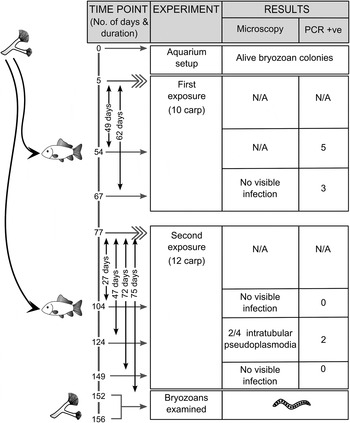
The substrates together with the attached bryozoan colonies were carefully rinsed using de-chlorinated water, and placed into a 40 L aquarium containing dechlorinated, aerated tap water. For cohabitation, SPF fish were introduced into the tank and maintained in a sieve basket (mesh size: 2·5 mm). This allowed the fish to swim, but prohibited direct interaction of the fish with the bryozoans. The basket permitted water flow and therefore malacosporean spores could circulate throughout the whole aquarium in the water current caused by aeration. The water temperature was maintained at 18–19 °C. Fish were fed 3–4 times a week on Skretting coarse fish food, a commercial pellet diet. Bryozoans were not fed but presumably were maintained by fish feces, circulating bacteria and algae. Fish and bryozoans were cohabited for a total of 156 days. During this period, two sets of SPF fish were exposed in succession and examined by light microscopy and/or PCR. The first exposure of ten SPF fish was from day 5 to day 67 of the experiment (62 days total) (Fig. 1). Six fish were examined at day 54 (49 days into experiment) and the last four on day 67 (65 days into experiment). The second exposure involved 12 fish and lasted from day 77–149 (72 days total). Fish from the second exposure were examined more frequently, four fish each at 27 days, 47 days and 72 days post exposure (day 104, 124 and 149 into the experiment respectively). Bryozoan colonies were thoroughly checked for malacosporean infection under the microscope at the end of the experiment, on days 152–156.
Experimental setup of Tetracapsuloides vermiformis n. sp. with wild bryozoan colonies and specific pathogen free common carp. Microscopic and PCR results are recorded in the table. Solid curved arrows indicate transmission of the parasite from bryozoan to fish host. Fish exposures (twice) are indicated by fishtail arrows. Lines with numbers and horizontal arrows indicate each time point. +ve = PCR positive result, N/A = data not available.

Microscopic analysis
Bryozoan colonies were checked for infection with an Olympus SZX7 stereomicroscope. Malacosporean vermiform stages dissected from the hosts were collected for light microscopy (n = 3), ultrastructural (n = 1) and molecular analyses (n = 3). Video documentation of the motility of vermiform malacosporean stages within their hosts was recorded with an Olympus U-TVO.5XC-3 camera using Quick PHOTO CAMERA 2·3 software. Light microscopy images of vermiform stages and spores released from bryozoans were obtained using an Olympus DP70 digital camera on an Olympus BX51 microscope. Squash preparations of fish kidneys were screened at 400–1000x magnification, and digital images of pseudoplasmodia identified in the renal tubules. Measurements of different parasite stages were taken from digital images using ImageJ 1·48q (Wayne Rasband, http://imagej.nih.gov/ij, Java 1·7·0_45; 64 bit) with a graticule for size reference.
One vermiform stage obtained from a bryozoan and three infected common carp kidneys were processed for transmission electron microscopy (TEM) according to the protocol of Dyková et al. (Reference Dyková, Kodádková, Buron, Fiala and Roumillat2013). Ultrathin sections were examined with a JEOL JEM 1010 electron microscope operating at 80 kV and images were obtained with the Megaview II imaging system by AnalySIS software.
DNA analysis
Small pieces of kidneys as well as 4 µL of blood from each SPF experimental carp (n = 22) were used for molecular analyses. Furthermore, three vermiform malacosporean stages and eight uninfected bryozoan zooids were used for molecular analyses. The material was collected in 400 µL TNES urea buffer (10 mm Tris–HCl pH = 8, 125 mm NaCl, 10 mm ethylenediaminetetraacetic acid, 0·5% sodium dodecyl sulfate and 4 m urea). After an overnight Proteinase K digestion (20 µg mL−1), a standard phenol-chloroform method was used for DNA extraction. Extracted DNA was re-suspended in 50–100 µL RNAse/DNAse-free water.
To amplify and sequence the malacosporean 18S rDNA, general malacosporean primer pairs mala-f with mala-r and budd-f with budd-r (Grabner and El-Matbouli, Reference Grabner and El-Matbouli2010a ) were used. Primers for mitochondrial 16S rDNA 16S-L (Cunningham and Buss, Reference Cunningham and Buss1993) with 16S-H (Schroth et al. Reference Schroth, Jarms, Streit and Schierwater2002) were used to confirm the identity of the bryozoan host. Before use in the experiment, fish were randomly sampled from the SPF fish stock at different times and kidneys, gills and blood were examined by PCR using general myxozoan primer combinations (Holzer et al. Reference Holzer, Bartošová, Pecková, Tyml, Atkinson, Bartholomew, Sipos, Eszterbauer and Dyková2013) to verify the absence of myxozoan infection.
PCRs were performed in 10 µL reactions with 0·4 µL Red Taq-DNA polymerase (Top-Bio, Czech Republic) with 1× buffer containing 1·5 mm MgCl2, 0·2 mm dNTP, 0·5 µ m of each primer, 1 µL of DNA and PCR grade water. PCR cycle parameters were: denaturation of DNA at 95 °C for 3 min followed by 30 cycles of amplification at 95 °C for 50 s, annealing temperature (65 °C for mala-f/mala-r, 58 °C for budd-f/budd-r and 50 °C for 16S-L/16S-H) for 50 s, elongation at 72 °C for 1·5 min and a final extension at 72 °C for 8 min. PCR products were purified by using the Gel/PCR DNA Fragments Extraction Kit (Geneaid Biotech Ltd., USA) and eluted in 20 µL of RNAse/DNAse-free water. Sequencing was performed commercially (SeqMe, Czech Republic).
The obtained 18S rDNA sequences were aligned with all other malacosporean 18S rDNA sequences available on GenBank (December 2014), using MAFFT v6·864b (Katoh et al. Reference Katoh, Misawa, Kuma and Miyata2002) and the L-INS-i method, with a default gap opening (–op = 1·53) and gap extension (–ep = 0·0) penalty, within Geneious v8·0 (Kearse et al. Reference Kearse, Moir, Wilson, Stones-Havas, Cheung, Sturrock, Buxton, Cooper, Markowitz, Duran, Thierer, Ashton, Meintjes and Drummond2012). Sequences were carefully checked for their quality and a subset of predominantly full-length 18S rDNA sequences were used to produce a similarity matrix in PAUP* v4.b10 (Swofford, Reference Swofford1993).
Phylogenetic analysis
Both maximum parsimony (MP) and maximum likelihood (ML) phylogenetic tree estimation methods were used to investigate malacosporean phylogenetic relationships. MP analysis was performed in PAUP* v4.b10, using a heuristic search with random taxa addition, the ACCTRAN option, TBR swapping algorithm, all characters treated as unordered, a Ts/Tv ratio of 1:2 and gaps treated as missing data. ML analysis was calculated in RAxML v7·0·3 (Stamatakis, Reference Stamatakis2006) using the GTR+Γ model of evolution determined as optimal by ModelTest 3·7 (Posada and Crandall, Reference Posada and Crandall1998). Bootstraps were based on 1000 replicates for both analyses.
Three subsets of the molecular data were used for intraspecific, intrageneric and intergeneric analyses. The first one included 36 malacosporean species with almost complete 18S rDNA sequences (maximum sequence length 1616 bp; Supplementary alignment file 1) to produce a general overview of all malacosporean 18S rDNA relationships. The second dataset was composed of 18 B. plumatellae 18S rDNA sequences from vermiform and sac-like bryozoan-related stages and from fish hosts (maximum sequence length 1632 bp; Supplementary alignment file 2) to better understand sequence heterogeneity within this species. For the same reason, a third comparison was performed with 19 T. bryosalmonae 18S rDNA sequences (maximum sequence length 1414 bp; Supplementary alignment file 3) with gaps treated as missing data. All sequences were selected from GenBank with regard to the sequence length, and an assumption of minimal sequencing error.
Evaluation of informative characters for malacosporean taxonomy
The malacosporean literature was studied and information regarding the morphology, development, biology and ecology of malacosporean species was summarized. The information is compiled in supplementary tables (Tables S1, S2 and S3) and was analysed in relation to 18S rDNA divergence (Table 1) and malacosporean phylogeny to determine the usefulness of characters for malacosporean taxonomy/species identification.
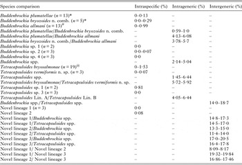
The 18S rDNA sequence divergence of among all malacosporean species including newly described species Buddenbrockia bryozoides n. comb. and Tetracapsuloides vermiformis n. sp. The range of sequence divergences are based on pair wise comparisons. For intraspecific divergence, only malacosporean species with two or more 18S rDNA sequence isolates were included (compare Table S4)

For individual divergences provided for B. plumatellae, B. bryozoides n. comb. and Tetracapsuloides bryosalmonae are provided from Tables S6 and S5, respectively. Undescribed species are similar to Fig. 4. Symbols used in this table: * = compare Table S5; # = compare Table S7 and @ = compare Table S6.
RESULTS
Experimental transmission
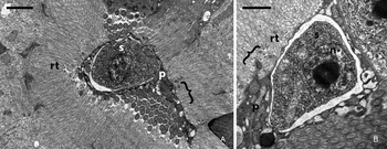
Two lots of common carp were successfully infected with a malacosporean parasite from wild bryozoans by cohabitation. Infection in eight out of ten fish (80% prevalence) was detected by PCR between 49 and 62 days post exposure (p.e.) (first exposure). During the second exposure, all fish (n = 4) were PCR negative for malacosporean infection at day 27, suggesting that the parasite did not produce infective spores in the bryozoans during the first 27 days p.e. At day 47 p.e., intratubular pseudoplasmodia were detected in the kidneys (Fig. 2A and B) in two out of four fish (50% prevalence) and the infection was confirmed by PCR, reflecting the invasion of parasite in the fish host. At day 72 p.e. all fish (n = 4) were PCR negative, suggesting that the infection was transitory or that a smaller fraction of fish had become infected in the second exposure compared with the first. The exact duration of development in the fish host is unknown as mature malacospores were not observed.
Transmission electron microscopy of intratubular pseudoplasmodia of Tetracapsuloides vermiformis n. sp. found in the kidney of common carp. (2A-B) Pseudoplasmodia composed of primary cell (p) containing 1-2 secondary cells (s) and their nuclei (n). Brackets indicate the connecting regions between renal tubule (rt) microvilli and the primary cell of pseudoplasmodia. Scale bar for 2A-B = 1 μm.

First thorough examination for infection within all bryozoan colonies was performed on 75 days after the second exposure of fish by using a stereomicroscope. Live bryozoan zooids were sufficiently transparent to allow detection of infection without dissection. Only one species of bryozoan (F. sultana) was present, and the identity of infected zooids was confirmed morphologically and by DNA sequencing (GenBank accession no. AB365637). Seven vermiform malacosporean stages were detected inside five zooids from two small colonies (Movie S1, Fig. 3A). These were identified and collected over a period of five days for morphological description and 18S rDNA sequencing. All bryozoans had died by the end of the exposure studies.
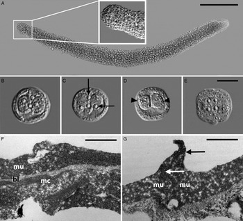
Light microscopy (3A-E) and transmission electron microscopy (3F-G) of vermiform stages of Tetracapsuloides vermiformis n. sp. found within the coelom of Fredericella sultana. (3A) Mature vermiform stages with scalloped proximal part (inset) and mature malacospores visible inside it. (3B-E) Spherical to subspherical malacospores with (3B-D) two distinct sporoplasms (black arrowheads) and (3B-C, E) four polar capsules (black arrows). (3F) Body wall of vermiform stage with three distinct layers: outermost mural cells (mu), middle basal lamina (bl) and innermost muscle cells (mc). (3G) Body wall showing prominent gap junction (arrow) between mural cells with irregular projection (black arrow). Scale bar for 3A = 200 μm, 3B-E = 10 μm, 3F-G = 1 μm.

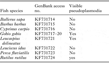
The 18S rDNA sequences of three vermiform stages from bryozoans (acc. no. KX013242) were 100% identical as were the sequences obtained from ten of the cohabited carp. Intratubular pseudoplasmodia were observed in two fish and the remaining eight fish were PCR positive (acc. no. KX013243). The malacosporean species from the present experiment shared 99·3–100% sequence identity with Tetracapsuloides sp. 3, which was detected in eight different fish species in Bartošová-Sojková et al. (Reference Bartošová-Sojková, Hrabcová, Pecková, Patra, Kodádková, Jurajda, Tyml and Holzer2014) (total 11 sequences, GenBank acc. no. KF731714-KF731724) (Table 2).
Visible infection (pseusoplasmodia in kidney tubule) record of Tetracapsuloides vermiformis n. sp. found by Bartošová-Sojková et al. (Reference Bartošová-Sojková, Hrabcová, Pecková, Patra, Kodádková, Jurajda, Tyml and Holzer2014) from different fish species

Species description
Phylum: Cnidaria Hatschek, 1888
Unranked subphylum: Myxozoa Grassé, 1970
Class: Malacosporea Canning et al. Reference Canning, Curry, Feist, Longshaw and Okamura2000
Order: Malacovalvulida Canning et al. Reference Canning, Curry, Feist, Longshaw and Okamura2000
Family: Saccosporidae Canning, Okamura and Curry, 1996
Genus: Tetracapsuloides Canning et al. Reference Canning, Tops, Curry, Wood and Okamura2002
Species: Tetracapsuloides vermiformis n. sp.
Tetracapsuloides vermiformis n. sp.
Type invertebrate host: Fredericella sultana (Blumenbach, 1779)
Potential fish hosts: Cyprinus carpio L. (present study, GenBank acc. no. KX013243), and seven other fish species reported in Bartošová-Sojková et al. (Reference Bartošová-Sojková, Hrabcová, Pecková, Patra, Kodádková, Jurajda, Tyml and Holzer2014) (Table 2). Type fish host status is not stipulated as detection of spore development was not observed.
Type locality: Motovidlo pond, South Bohemia, Czech Republic (Latitude: 48°99′94·48″ N, Longitude: 14°38′05·30″ E).
Other localities: Different parts of Thaya and Morava rivers, Jindřiš, České Budějovice (all Czech Republic); Danube River, Štúrovo (Slovakia); Hortobágy (Hungary); Iseo lake (Italy) (Bartošová-Sojková et al. Reference Bartošová-Sojková, Hrabcová, Pecková, Patra, Kodádková, Jurajda, Tyml and Holzer2014).
Etymology: Species name is based on the vermiform shape of the parasite in the bryozoan host.
Description of parasite stages from the bryozoan host (F. sultana): Vermiform stages are located inside the host metacoel and measure 0·99 × 0·08 mm (n = 1, immature stage) and 1·21 × 0·09 mm (n = 1, mature stage) (length × width). Smaller vermiform stages tapering at both ends and lacking spores, larger stage has a scalloped protrusion at one end and tapers at the other (Fig. 3A, 3A inset) and is filled with different developmental stages including mature spores. Mature spores are spherical to subspherical (Fig. 3B–E), and appear to have four (possibly more) flexible valve cells and four spherical polar capsules (indicating four capsulogenic cells). Each spore possesses two sporoplasms each containing a single secondary cell. Measurements (average ± standard deviation, range in parenthesis) based on light microscopy: diameter of spore 18·3 µm ±0·48 (17·62–18·88 µm) (n = 10), length of polar capsules 2·04 µm ±0·21 (1·69–2·58 µm) (n = 15), width 1·95 µm ±0·12 (1·87–2·01 µm) (n = 15), sporoplasm length 9·33 µm ±1·24 (8·0–11·29 µm) (n = 10) and width 6·91 µm ±0·497 (6·37–7·87 µm) (n = 10).
All vermiform stages are unattached and freely moving within the bryozoan metacoel. Stages show erratic movement, such as slow bending, straightening and rapid contracting motions along the entire length of the body to form a spiral, independent from the peristaltic host gut movement (Movie S1).
Ultrastructural examination of a longitudinally sectioned, immature vermiform stage revealed a body wall consisting of mural cells, basal lamina and myocytes (exterior to interior) (Fig. 3F). Numerous myofibrils are present within myocytes, which are oriented either along the longitudinal axis of the vermiform stages or are slightly inclined with respect to the longitudinal axis of the vermiform stage (Fig. 3F). Polar capsules were not observed in immature vermiform stages. The outer surface of the mural cell layer frequently possesses irregular projections at cell junctions. Sometimes these junctions are very prominent (Fig. 3G).
Description of parasite stages from C. carpio: Early sporogonic pseudoplasmodia containing refractile granules are located in the lumina of the renal tubules (Fig. 2A and B). In TEM sections, pseudoplasmodia are composed of electron-light primary cells harbouring electron-dense secondary cells with a prominent nucleus (Fig. 2A and B). Abundant mitochondria occur in all cells, and numerous ribosomes are present in secondary cells. Irregular projections from primary cells provide attachment to the microvilli border of the epithelial cells of the renal tubule (Fig. 2A and B). Sporoplasmosomes were not observed.
Prevalence in fish hosts: C. carpio: 45·5% (10/22) (experimental prevalence reported here); Ballerus sapa: 11% (1/9); Ballerus barbus: 14·3% (1/7); C. carpio: 9% (1/11); Gobio gobio: 9% (1/11) (Dyje River, Czech Republic), 25% (1/4) (Morava River, Czech Republic), 67% (4/6) (Jindřiš Farm, Czech Republic); Leucaspius delineatus: 100% (2/2); Leucaspius idus: 75% (3/4); Rutilus rutilus: 14% (1/7); Perca fluviatilis: 25% (2/4) (Bartošová-Sojková et al. Reference Bartošová-Sojková, Hrabcová, Pecková, Patra, Kodádková, Jurajda, Tyml and Holzer2014).
Pathology: No significant macroscopic or ultrastructural changes observed in the fish kidneys (few parasites noted). Pathology in bryozoan host unknown.
Deposition of type-materials: Five grids of vermiform stages from F. sultana for TEM; one DNA sample of infected bryozoan host, one DNA sample of infected carp kidney deposited in the collection of the Institute of Parasitology, Biology Centre of the Czech Academy of Sciences (Collection ID: IPCAS Prot no. 35) and two 18S rDNA sequences, deposited in GenBank (acc. no. KX013242-KX013243).
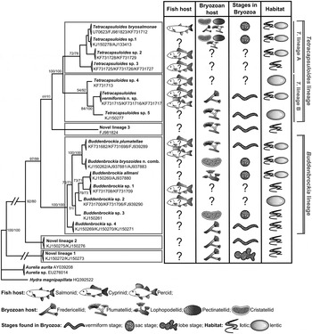
Phylogenetic position of T. vermiformis n. sp.: In the phylogenetic tree based on malacosporean 18S rDNA sequences (Fig. 4), T. vermiformis n. sp. clusters in one of two Tetracapsuloides subclades (see below), with its closest relative being another vermiform malacosporean (Tetracapsuloides sp. 5; Fig. 4) from Plumatella sp. (GenBank acc. number KJ150277) (Hartikainen et al. Reference Hartikainen, Gruhl and Okamura2014) with 2·86–2·94% sequence divergence with T. vermiformis n. sp (Table S4).
Maximum likelihood phylogenetic tree based on a total of 37 18S rDNA sequences from all available malacosporean species (n = 17) with free-living cnidarians (Aurelia aurita, Aurelia sp. and Hydra magnipapillata) as outgroup. Bootstrap support values at each node indicate maximum parsimony/maximum likelihood support based on 1000 replicates. The original length of long branches was shortened by 80% (indicated as -//-). Biological and morphological characteristics mapped to the tree include type of fish and bryozoan hosts, morphology of bryozoan-related stages and habitat types. Host and habitat type were included only for species records with molecular confirmation. Undescribed species from (Bartošová-Sojková et al. Reference Hartikainen, Gruhl and Okamura2014) (B) and (Hartikainen et al. 2014) (H) are listed as Tetracapsuloides sp. 1 = Tetracapsuloides sp. 1 by (B) and Tetracapsuloides sp. by (H); Tetracapsuloides sp. 2 = Tetracapsuloides sp. 5 by (B); Tetracapsuloides sp. 3 = Tetracapsuloides sp. 4 by (B); Tetracapsuloides sp. 4 = Tetracapsuloides sp. 2 by (B); Tetracapsuloides sp. 5 = Novel lineage 3 by (H); Buddenbrockia sp. 1 = Buddenbrockia sp. 3 by (B); Buddenbrockia sp. 2 = Buddenbrockia sp. 2 by (B) and Buddenbrockia sp. 3 by (H); Buddenbrockia sp. 3 = Buddenbrockia 1 by (H); Buddenbrockia sp. 4 = Buddenbrockia sp. 1 by (B) and Buddenbrockia sp. 2 by (H); Novel lineage 1 and 2 = Novel lineage 1 and 2 by (H) and Novel lineage 3 = Novel lineage by (B) and Buddenbrockia sp. 4 by (H). Symbols used in the figure: ? = information unavailable; X = uncertain existence; ? (within the fish) = fish reported without mature spore.

Remarks: T. vermiformis n. sp. vermiform stages show a striking similarity with B. plumatellae regarding three layered organisation (basal lamina, muscle cells and mural cells) and body movement, but not with T. bryosalmonae sac-like stages, which are devoid of any muscle cells and movement (Canning et al. Reference Canning, Curry, Feist, Longshaw and Okamura2000, Reference Canning, Tops, Curry, Wood and Okamura2002). Vermiform stages have been reported from all lineages (see Fig. 4) and this is the second report from Tetracapsuloides, however, ultrastructural details are provided for the first time here.
Intratubular pseudoplasmodia were detected in the kidney tubules (Fig. 2A and B) of experimental carp Cyprinus carpio L. (present study) (GenBank acc. no. KX013243) and three other fish species (Bartošová-Sojková et al. Reference Bartošová-Sojková, Hrabcová, Pecková, Patra, Kodádková, Jurajda, Tyml and Holzer2014) (Table 2). Although various fish hosts were reported with pseudoplamodia within the renal tubules, no mature spores have yet been observed. This leads to uncertainties regarding the vertebrate host status and indeed the possibility of accidental infections in various cases.
The 18S rDNA phylogeny and sequence divergence
The molecular phylogenetic tree (Fig. 4) provides evidence for 17 species. The two nominal genera of Malacosporea, Tetracapsuloides and Buddenbrockia are clearly separated by high bootstrap support (MP/ML 97/99). The species allocated to Tetracapsuloides cluster in two subclades (Tetracapsuloides lineage A and B) and those allocated to Buddenbrockia cluster in a single clade, sister to Tetracapsuloides. Based on a 1616 bp 18S rDNA sequence alignment distance matrix (Table 1 and Table S4), the sequence divergence between Tetracapsuloides and Buddenbrockia is 14–18·7%. Sequence divergences were observed in variable regions (45–117 bp, 356–385 bp, 464–481 bp, 540–665 bp, 734–743 bp and 1304–1426 bp) and were highest between novel lineage 3 and Buddenbrockia spp. (17·0–20·5%) (Table 1, Table S4 and Supplementary alignment file 1). Intrageneric divergence within Buddenbrockia spp. and Tetracapsuloides spp. (both described and undescribed species) are 2·14–5·04% and 1·45–6·44%, respectively. There are at present, three well-supported novel lineages in the phylogenetic tree: novel lineage 1 (100/100), which lies basal to all other malacosporeans; novel lineage 2 (92/80), a sister group of all Tetracapsuloides and Buddenbrockia lineages and the less supported (69/93) novel lineage 3, previously ascribed to Buddenbrockia (Hartikainen et al. Reference Hartikainen, Gruhl and Okamura2014), and grouping here as a separate, weakly supported lineage sister to Tetracapsuloides (Fig. 4).
Tetracapsuloides lineages A and B form distinct clades with strong support (100/100). The 18S rDNA sequence divergence between these two lineages (up to 6·44%) does not exceed other intrageneric (compare Table 1 and Table S4) divergences in malacosporeans. In view of this, we regard the new malacosporean described here and those compromising lineage B as species of Tetracapsuloides rather than denoting a new genus. Descriptions of additional members of this clade will likely clarify the interrelationships of this group. Clarification of the forms (sac-like or vermiform) that develop in bryozoan hosts may determine whether the two clades can be distinguished on this basis.
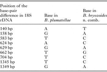
Buddenbrockia plumatellae from various fish hosts were found to have up to 0·13% sequence divergence to the vermiform stages (Table S5), which indicates conspecificity of these two stages. Within the sac-stages of B. plumatellae sequence divergence was up to 0·29%, and between the latter and vermiform/fish stages it ranged from 0·59 to 1%. In the phylogenetic analysis, B. plumatellae from fish and vermiform stages cluster together and are distinct from the sac-forming stages (100/100), which were initially described as Tetracapsula bryozoides (Canning et al. Reference Canning, Okamura and Curry1996), with strong bootstrap support (Figs 4 and 5). Additionally, 18S rDNA sequences demonstrate variation in nine base pairs between vermiform/fish stages and sac-like stages (Table 3).
Maximum likelihood phylogenetic tree based on a total of fifty two 18S rDNA sequences from all Buddenbrockia spp. showing the distinct clustering of B. plumatellae (vermiform and fish stages) and B. bryozoides n. comb. (= previous B. plumatellae sac-like stages). Bootstraps support values at each node indicate maximum parsimony /maximum likelihood support based on 1000 replicates. The original length of long branches was shortened by 50% (indicated as -//-). For undescribed species names are identical to Figure 4. Symbol used in the figure: ? = fish reported without mature spore.

Position and nature of consistent nucleotide changes in a 1632 bp alignment of 18S rDNA sequences (five Buddenbrockia plumatellae vermiform stages, eight B. plumatellae fish stages and five Buddenbrockia bryozoides n. comb. sac stages) (compare Supplementary alignment file 2)

DISCUSSION
Experimental transmission of T. vermiformis n. sp.
Tetracapsuloides vermiformis n. sp. was successfully transmitted from naturally infected F. sultana to common carp twice (Fig. 1). Pseudoplasmodia of T. vermiformis n. sp. were formed in fish renal tubules in SPF fish exposed to infected bryozoans. Mobile vermiform stages were produced in the bryozoan F. sultana. Three important aspects remain unclear in this experiment: (i) the duration of parasite development in fish is imprecisely known as the release of the infective stages from wild bryozoans was not observed, (ii) we were unable to find mature malacospores in carp, which leaves the fish type host unconfirmed and (iii) it is uncertain whether the vermiform stages found at the end of the experiment are a result of an infection sustained in the bryozoans since their collection or that of a reinfection from experimentally infected fish.
Although not precisely known, we can nevertheless surmise that the duration of the intrapiscine development of T. vermiformis n. sp. is comparable with other malacosporeans. Fish were parasite-free (PCR confirmed) at 27 days post exposure, after which intratubular psuedoplasmodia developed (confirmed visually and by PCR on day 47). In T. bryosalmonae, early presporogonic stages were observed in the kidney of brown trout (Salmo trutta) and rainbow trout (Oncorhynchus mykiss) after 21–28 days (Feist et al. Reference Feist, Longshaw, Canning and Okamura2001; Morris and Adams, Reference Morris and Adams2008) and spores were produced around 63 days p.e. (Morris and Adams, Reference Morris and Adams2008). Buddenbrockia plumatellae sporogonic stages in carp and minnow were detected 35–56 days p.e. (Grabner and El-Matbouli, Reference Grabner and El-Matbouli2010b ). Vermiform stages were present for five days in the bryozoan coelom. Previous results suggest variation in development within bryozoan hosts. Buddenbrockia plumatellae developed mature vermiform stages 17 days after collection from the wild and only six days later developed mature spore-forming vermiform stages (Canning et al. Reference Canning, Tops, Curry, Wood and Okamura2002). Single cell stages of T. bryosalmonae were observed after 42 days of continuous exposure to infected fish and developed into sac-like stages within two days (Morris and Adams, Reference Morris and Adams2006a ). These collective results indicate that malacosporeans may generally undergo a potentially long period of covert infection and rapid final maturation to spore-forming stages in the bryozoan host.
Malacosporean characters and species discrimination
Below we assess the most common traits included in malacosporean species descriptions and evaluate their use for species discrimination.
Morphology of plasmodia in bryozoan host: Sizes of the plasmodia overlap between species and can also vary according to the parasite's development. This feature is thus unreliable for differentiating species (see Table S3). Furthermore, vermiform and sac-like are found scattered in lineages of both Buddenbrockia and Tetracapsuloides (Fig. 4) suggesting these morphotypes have been lost and gained repeatedly during malacosporean evolution (Fig. 4 and Hartikainen et al. Reference Hartikainen, Gruhl and Okamura2014) and are not helpful for discriminating closely-related species.
Spore morphology in bryozoan host: The sizes of mature spores and polar capsules clearly overlap between malacosporeans (Table S3). External ornamentation with small posterior protrusions (Morris et al. Reference Morris, Morris and Adams2002), as well as reports of rough spore surfaces (Grabner and El-Matbouli, Reference Grabner and El-Matbouli2010b ; Okamura et al. Reference Okamura, Gruhl, Reft, Okamura, Gruhl and Bartholomew2015), may represent processing artefacts. In contrast to morphological variation among actinospores from different invertebrate hosts of myxosporeans (Lom and Dyková, Reference Lom and Dyková2006), malacospores from bryozoan hosts are found to be virtually identical in size and shape in different species and are hence not useful for species discrimination.
Pseudoplasmodia and spores from fish host: Monosporic pseudoplasmodia within fish renal tubules are a characteristic for all malacosporeans described so far. However, data are extremely scarce. Comprehensive information is only available for T. bryosalmonae. Spore morphology in fish thus has an uncertain value for species diagnosis.
Life cycle and development: The life cycle and development of T. bryosalmonae have been relatively intensively investigated (Ferguson and Needham, Reference Ferguson and Needham1978; Kent and Hedrick, Reference Kent and Hedrick1986; Morris et al. Reference Morris, Adams, Feist, McGeorge and Richards2000; Longshaw et al. Reference Longshaw, Le Deuff, Harris and Feist2002; Morris and Adams, Reference Morris and Adams2006a , Reference Morris and Adams b , Reference Morris and Adams2007a , Reference Morris and Adams2008), but some insights are also available for species of Buddenbrockia (Canning et al. Reference Canning, Okamura and Curry1996, Reference Canning, Curry, Feist, Longshaw and Okamura2000, Reference Canning, Tops, Curry, Wood and Okamura2002, Reference Canning, Curry, Hill and Okamura2007, Reference Canning, Curry and Okamura2008; Okamura, Reference Okamura1996; Morris et al. Reference Morris, Morris and Adams2002; McGurk et al. Reference McGurk, Morris and Adams2006; Morris and Adams, Reference Morris and Adams2007b ; Grabner and El-Matbouli, Reference Grabner and El-Matbouli2010a ). For example, T. bryosalmonae has been shown to switch between uni- or pluri-cellular stages in covert and overt infections (respectively) according to environmental conditions (Morris and Adams, Reference Morris and Adams2006a ; Tops et al. Reference Tops, Lockwood and Okamura2006) however, it is unknown if developmental stages and triggers are taxonomically useful. In general, there is insufficient information on comparative life cycles and developmental traits for species discrimination.
Specificity for bryozoan host: Buddenbrockia spp. have so far been associated with a single host species (Tables S1 and S2). Tetracapsuloides sp. 1 uses three host families and T. bryosalmonae infects four different bryozoan families. Despite our present limited knowledge, with more data this trait could be relevant for discrimination of some malacosporean species.
‘Specificity’ for vertebrate (fish) hosts: Malacosporean species appear to be able to infect a wide range of fish hosts. Invasion of fish can be relatively unspecific (Kallert et al. Reference Kallert, Eszterbauer, Grabner and El-Matbouli2009, Reference Kallert, Bauer, Haas and El-Matbouli2011), which could lead to positive PCR results in unsuitable hosts (Holzer et al. Reference Holzer, Bartošová, Pecková, Tyml, Atkinson, Bartholomew, Sipos, Eszterbauer and Dyková2013). Partial development may occur in any fish species but in unsuitable hosts infection could be a dead end, without the production of infective transmission stages. For example, Grabner and El-Matbouli (Reference Grabner and El-Matbouli2008) found various types of salmonids were infected by T. bryosalmonae, however only brook trout and brown trout were able to transmit the parasite to bryozoan hosts. In addition, spore formation was observed only in rainbow trout from North America but not in the same host in Europe though both T. bryosalmonae isolates cause proliferative kidney disease in the fish (Kent and Hedrick, Reference Kent and Hedrick1986; Feist et al. Reference Feist, Longshaw, Canning and Okamura2001; Grabner and El-Matbouli, Reference Grabner and El-Matbouli2008). Similarly, according to Bartošová-Sojková et al. (Reference Bartošová-Sojková, Hrabcová, Pecková, Patra, Kodádková, Jurajda, Tyml and Holzer2014), a total of eight different fish species were PCR positive for T. vermiformis n. sp. but intraluminal stages were observed only in G. gobio, L. delineatus and C. carpio. At present, infection of fish hosts appears to be limited to fish family level (see Table S2), although individual studies indicate that it may be far more restricted. Identification of the vertebrate host range of malacosporeans requires further investigation into life cycles and experimental trials and cross-infections using SPF bryozoans.
Habitat and geographic distribution: Malacosporean species are described from lentic and lotic habitats and at least some are recorded in both. The majority of malacosporean species have been described from Europe and North America. Sampling other areas like Asia (two records; Braem, Reference Braem1911 and Hartikainen et al. Reference Hartikainen, Gruhl and Okamura2014), South America (one record; Marcus, Reference Marcus1941), Africa and Australia (no records) is needed before species discrimination can be evaluated in a geographical context.
Molecular markers for species discrimination: 18S rDNA – We generally define malacosporean intrageneric divergence as 1–7% and intergeneric divergence as 8–20% (at least, Table 1 and Table S4). The yardstick of ⩽ 1% intraspecific 18S rDNA sequence divergence for myxosporeans (Whipps and Kent, Reference Whipps and Kent2006; Bartošová and Fiala, Reference Bartošová and Fiala2011) mostly concurs with the divergences between malacosporean species with one exception: T. bryosalmonae has up to 1·53% intraspecific variation (Table S6). 28S rDNA – the relatively few 28S rDNA sequences available for malacosporeans demonstrate similar molecular phylogenies as those generated by 18S rDNA (Hartikainen et al. Reference Hartikainen, Gruhl and Okamura2014). ITS – internal transcribed spacer sequences are only available from T. bryosalmonae and were unable to resolve the T. bryosalmonae ‘species complex’ entirely (see the section ‘Phylogenetic ambiguity of Tetracapsuloides bryosalmonae’ below for summary) but provided some evidence for divergence between isolates that may reflect speciation (Henderson and Okamura, Reference Henderson and Okamura2004). Protein coding genes – partial sequences of ribosomal protein rpl27e, elongation factor 2 and DNA repair protein RAD51 are known from B. plumatellae (Jimenéz-Guri et al. Reference Jimenéz-Guri, Philippe, Okamura and Holland2007b ) and were used as evidence of heterospecificity between vermiform and sac-like stages of B. plumatellae. These and other genetic markers have potential for species delimitation and stabilization of 18S rDNA phylogeny in future malacosporean taxonomic revisions and additional sequences are urgently needed from other species. Currently however, the most comprehensively sampled molecular marker for malacosporean classification is 18S rDNA. Caution is required with this marker as the levels of divergence vary within and between putative species and genera, hence 18S rDNA is most informative when combined with other characters such as morphology or host.
Phylogenetic ambiguity of Tetracapsuloides bryosalmonae
ITS-1 rDNA sequence data show that T. bryosalmonae isolates from North America and Canada distinctly cluster together apart from the majority of European isolates with the exception of some French and Italian isolates. This outcome was suggested to be indicative of an invasion of North American strains to Southern Europe (Henderson and Okamura, Reference Henderson and Okamura2004) but ML bootstrap support was relatively low (75). Similarly, 18S rDNA sequences of T. bryosalmonae (n = 19, Supplementary alignment file 3) show inconsistent nucleotide changes with regard to the type of change and the position in the alignment (similar to Okamura et al. Reference Okamura, Anderson, Longshaw, Feist and Canning2001). As a result and despite a sequence divergence of up to 1·53%, our phylogenetic analyses based on 18S rDNA were also unable to resolve relationships, producing subclades of varying compositions with low bootstrap support (trees not shown). Tetracapsuloides bryosalmonae has been shown to infect multiple fish hosts whilst only producing transmissible spores in a few species (Morris and Adams, Reference Morris and Adams2006a ; Grabner and El-Matbouli, Reference Grabner and El-Matbouli2008) (see the section ‘Specificity’ for vertebrate (fish) hosts’ above). Furthermore, the lack of spore formation observed in the same host (rainbow trout) in Europe but not in Northern implies that these isolates may represent multiple species. It is possible that Tetracapsula renicola (Kent et al. Reference Kent, Khattra, Hedrick and Devlin2000; junior synonym of T. bryosalmonae) originally described from Canada represents in fact a different species than European T. bryosalmonae (Morris and Adams, Reference Morris and Adams2006a ). Cross-infection studies could resolve the ultimate species limits of European and Northern American isolates from different hosts. Until this is done, T. bryosalmonae remains a ‘species complex’ as mentioned in Atkinson et al. (Reference Atkinson, Bartošová-Sojková, Whipps, Bartholomew, Okamura, Gruhl and Bartholomew2015).
Recognition of distinct species status of B. bryozoides n. comb.
As a result of the foregoing evaluation of informative characters for species discrimination in malacosporeans we propose a revision of B. plumatellae and its present junior synonym Tetracapsula bryozoides. Based on 18S rDNA sequences, vermiform/fish stages of B. plumatellae differ up to 0·13% and sac stages (Tetracapsula bryozoides) up to 0·29% (see the section ‘18S rDNA phylogeny and sequence divergence’ above) (Table 1 and Table S5). The divergence between vermiform/fish and sac and stages however, ranges from 0·59 to 1·0% (Table 1 and Table S5). A similar value (0·7% divergence in 18S rDNA sequences) was reported by Tops and Okamura (Reference Tops and Okamura2005) between sac (n = 3) and vermiform (n = 3) stages. The strong bootstrap support for distinct lineages led Tops and Okamura (Reference Tops and Okamura2005) to suggest that further study was required to clarify the systematics of sac-like and vermiform malacosporeans. Bartošová-Sojková et al. (Reference Bartošová-Sojková, Hrabcová, Pecková, Patra, Kodádková, Jurajda, Tyml and Holzer2014) reported 18S rDNA divergences within vermiform and sac-like stages as 0·43% and 0·55%, respectively, and a 1·29% divergence between the two morphotypes, all of which were higher than our calculations. Careful trimming of sequence ends and exclusion of B. plumatellae sequences containing large numbers of unidentified nucleotides (N) in the present study likely accounts for the differences between analyses. In our analysis as well as in previous studies based exclusively on 18S rDNA sequence analysis (Tops et al. Reference Tops, Curry and Okamura2005; Bartošová-Sojková et al. Reference Bartošová-Sojková, Hrabcová, Pecková, Patra, Kodádková, Jurajda, Tyml and Holzer2014; Hartikainen et al. Reference Hartikainen, Gruhl and Okamura2014; Fiala et al. Reference Fiala, Bartošová-Sojková, Okamura, Hartikainen, Okamura, Gruhl and Bartholomew2015a ) the two morphotypes appear as separate phylogenetic lineages with high bootstrap support (100/100) (Figs 4 and 5). Additionally, we detected consistent nucleotide changes in nine positions of the alignment (Table 3, Supplementary alignment file 2) between vermiform/fish stages and sac-like stages. Finally, the examination of four protein coding genes also led Jimenéz-Guri et al. (Reference Jimenéz-Guri, Philippe, Okamura and Holland2007b ) to conclude that B. plumatellae sacs and vermiform stages are not conspecific. We therefore reconsider and summarize distinguishing characters between B. plumatellae vermiform/fish stages and sac-like stages below:
-
(1) Morphology and host: sac-like and vermiform stages in the bryozoan do not characterize the same host species (Canning and Okamura, Reference Canning and Okamura2004). Sac-like stages were reported exclusively from Cristatella mucedo (Cristatellidae), while vermiform stages develop in members of the bryozoan family Plumatellidae.
-
(2) Molecular data: some vermiform stages ascribed to B. plumatellae from Fredericellidae (Canning et al. Reference Canning, Tops, Curry, Wood and Okamura2002) and Lophopodidae (Oda, Reference Oda1972) without molecular confirmation are difficult to consider as conspecific. The 18S rDNA sequences from fish with B. plumatellae infection cluster exclusively with vermiform stages and are separated from the sac-forming stages initially described as T. bryozoides in our molecular phylogenetic analyses, with strong bootstrap support (Figs 4 and 5). The 18S rDNA sequences show nine consistent base pair changes between vermiform/fish stages and sac-like stages (Table 3). This suggests that sac-like stages may not require a fish host, or more likely, that the fish host for sac-like stages is still unknown. Furthermore, protein-coding data (rpl23a) confirmed the heterospecificity of the two morphotypes (Jimenéz-Guri et al. Reference Jimenéz-Guri, Philippe, Okamura and Holland2007b ).
Based on these differences, we hereby reject the synonymy of T. bryozoides with B. plumatellae proposed by Canning et al. (Reference Canning, Tops, Curry, Wood and Okamura2002), and due to its phylogenetic positioning, ascribe T. bryozoides to B. bryozoides n. comb., reserving B. plumatellae for the vermiform stages and various isolates obtained from the excretory system of cyprinids.
Species description
Phylum: Cnidaria Hatschek, 1888
Unranked subphylum: Myxozoa Grassé, 1970
Class: Malacosporea Canning et al. Reference Canning, Curry, Feist, Longshaw and Okamura2000
Order: Malacovalvulida Canning et al. Reference Canning, Curry, Feist, Longshaw and Okamura2000
Family: Saccosporidae Canning, Okamura and Curry, 1996
Genus: Buddenbrockia Schröder, Reference Schröder1910
Species: Buddenbrockia bryozoides n. comb.
Buddenbrockia bryozoides n. comb.
Type invertebrate host: Cristatella mucedo (Cuvier 1798)
Vertebrate host: Unknown.
Synonyms: Buddenbrockia plumatellae sensu Canning et al. Reference Canning, Tops, Curry, Wood and Okamura2002, p. 294, not Schröder, Reference Schröder1910; T. bryozoides Canning et al. Reference Canning, Okamura and Curry1996
Type locality: not specified by Canning et al. Reference Canning, Okamura and Curry1996; specimens of Okamura (Reference Okamura1996) originated from different sites in the UK.
Pathology: Unknown.
Description of B. bryozoides n. comb. in the invertebrate host (Bryozoa): Description based on the sac-like stages from Okamura (Reference Okamura1996) and Canning et al. (Reference Canning, Okamura and Curry1996) from C. mucedo, where B. bryozoides n. comb. produces free-floating ellipsoid sac-like stages (size 50–700 µm) within the coelomic cavity of the host. Body wall of sac-like stages composed of a single layer of flat cells. According to light microscopy, mature spore with four valve cells, four polar capsules and two sporoplasms. Size of the mature spores, polar capsules and sporoplasm 19·21 µm, 1·3 × 1·4 µm, 4·5 × 5·0 µm. Polar filament in five turns being S-shaped in cross-section.
Phylogenetic position: Based on 18S rDNA sequence analysis of all available malacosporean sequences to date B. bryozoides n. comb. clusters within the Buddenbrockia lineage with maximum proximity to the vermiform B. plumatellae (Fig. 4) and a clear separation from the latter by 100% bootstrap support.
Morphological similarities of Tetracapsuloides and Buddenbrockia
Until recently, the most important distinguishing character between two nominal malacosporean genera was development of only sac-like stages for Tetracapsuloides and both sac-like and vermiform stages for Buddenbrockia in the bryozoan hosts (Fiala et al. Reference Fiala, Bartošová-Sojková, Whipps, Okamura, Gruhl and Bartholomew2015b ). This distinction is no longer valid after finding vermiform stages in a Tetracapsuloides species (Hartikainen et al. Reference Hartikainen, Gruhl and Okamura2014) with the same ultrastructural organization as Buddenbrockia (three wall layers, present study). Other morphological and biological traits fail to distinguish these two genera (see the section ‘Malacosporean characters and species discrimination’ above and Table S3). On the other hand, considerable 18S rDNA divergence (14·0–18·7% and Table 1) results in distinct clustering in phylogenetic trees (Fig. 4) and confirms distinct and independent lineages of Tetracapsuloides and Buddenbrockia.
CONCLUDING REMARKS
A review of presently available evidence suggests that bryozoan host species, plasmodial morphotypes in bryozoan hosts and molecular data variously provide the most informative data for malacosporean species discrimination. For example, although, B. plumatellae and B. bryozoides n. comb. show minimal (e.g. 0·59–1·0%) 18S rDNA divergence, they are distinguished by distinct biological and morphological characters. Tetracapsuloides bryosalmonae, however, demonstrates more substantial 18S rDNA divergence (1·53%) in morphologically similar isolates suggesting that molecular divergence is not always linked with variation in morphology. For 18S rDNA, sequence divergences of <1%, consistent changes in 18S rDNA sequences and phylogenetic clustering with high bootstrap support may therefore be indicative but insufficient for distinguishing species, requiring additional support from biological or morphological traits. Nevertheless, DNA sequences are absolutely required for the description of new species. Malacosporean taxa can generally be characterized by intraspecific, interspecific/intrageneric and intergeneric 18S rDNA sequence divergences of <1%, 1–7% and 8–20%, respectively. Future malacosporean species descriptions and taxonomic revisions will be benefited from further details of the development and morphology of parasite stages in both bryozoan and fish hosts. In addition, proof of spore formation and cross-infection studies are required to distinguish between true and accidental malacosporean hosts. Data on geographic ranges, life histories and development in fish hosts may eventually also prove useful for classification.
SUPPLEMENTARY MATERIAL
The supplementary material for this article can be found at https://doi.org/10.1017/S0031182016001931
ACKNOWLEDGEMENTS
We are thankful to Dr Ivan Fiala (PARU, BC ASCR, České Budějovice, Czech Republic) for assistance with phylogenetic analyses, Dr Pavla Bartošová-Sojková (PARU, BC ASCR, České Budějovice, Czech Republic) for valuable comments on the manuscript, the Laboratory of Electron Microscopy (BC ASCR, České Budějovice, Czech Republic) for technical assistance in transmission electron microscopy and Tomáš Douda (Animal facility, BC ASCR, České Budějovice, Czech Republic) for rearing fish and handling the experimental setup. We would also like to thank the three anonymous reviewers for their assistance in improving the manuscript.
FINANCIAL SUPPORT
We acknowledge financial support by the Czech Science Foundation (505/12/G112, Center of Excellence).