Tumours originating from cardiac tissues are rare during childhood, with an incidence of 0.08%.Reference Gleason and Hornick 1 Fortunately, a majority of these tumours are benign in nature, such as rhabdomyomas, fibromas, myxomas, and teratomas. Inflammatory myofibroblastic tumour, which has unique clinical, pathological, and molecular characteristics, is a relatively new entity.Reference Gleason and Hornick 1 Most of the cardiac inflammatory myofibroblastic tumour patients are in the age group of 4 months to 17 years.Reference Stark, Sandbank, Rudnicki and Zahavi 2 – Reference Vaideeswar, Patwardhan and Sathe 7 This unique tumoural process has not been subject of any specific research, and the prognosis is not well known.Reference Attili, Chandra, Hemant, Bapsy and Anupama 8 Here we present the case of a 3-month-old child who was admitted to our outpatient clinic with massive pericardial effusion and who had excellent progress after surgical resection of over 1 year.
Case presentation
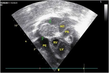
A 3-month-old boy was referred to our outpatient clinic with dyspnoea and severe cardiomegaly in his chest X-ray. He was a term baby with an uneventful pregnancy. His postnatal follow-up was normal. His initial cardiovascular examination including electrocardiography was normal except for cardiomegaly on his chest X-ray. Echocardiographic examination revealed a mass of solid structure of about 17.48 to 30.44-mm dimension with an indefinite shape and blurred boundaries. The mass was showing right atrial lateral wall involvement including patchy echo-intense and hypoechoic areas. It did not cause any obstruction to any vascular or valvular structure. The patient had a massive pericardial effusion without cardiac compression (Fig 1). His laboratory findings were as follows: haemoglobin 7.7 g/dl; complete blood count with a platelet count of 562,000/mm3. Erythrocyte sedimentation rate: 32 mm/hour and C-reactive protein 2.3 mg/dl (n: 0.33 mg/dl). Electrolytes and other biochemical studies were normal. Fluid drainage stopped at 30 ml during diagnostic pericardiocentesis because of haemorrhagic nature. Pericardial effusion biochemistry revealed a pH of 7.4, density of 1010, lactic dehydrogenase of 346 μ/dl, protein of 4 g/dl, and glucose of 85 mg/dl. Microscopic cytopathologic examination of fluid smear showed reactive leucocytes and haemolysed erythrocytes. Thoracic magnetic resonance imaging showed massive pericardial effusion and isointense mass of cardiac tissue, which was primarily located in the pericardial space, at the anterior aspect of the right atrium. Rhabdomyoma or a mass originating from the pericardium was the first-line diagnosis. Cranial and abdominal ultrasonographic evaluations were normal.
Apical four chamber view displaying the mass. PA = pulmonary artery; Ao = aorta; LV = left ventricle; RV = right ventricle; PE = pulmonary embolism.

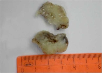
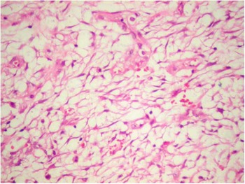
Surgical resection of the mass was planned. After surgical drainage of the effusion, clear visualisation of the mass involving the right atrial appendage occupying over two-thirds of the right atrial wall with definite boundaries was observed. The mass was not obstructing the inferior and superior caval veins. Surgical removal of the mass was successful without any complication of dysrhythmia or bleeding (Fig 2). Early follow-up in the intensive care unit was uneventful, and the patient was extubated on the 1st day with a sinus rhythm and normal echocardiographic findings. Pathological specimen examination revealed inflammatory myofibroblastic tumour with spindle myofibroblastic cell proliferation in a myxoid stroma. There were few inflammatory cells among these structures. İmmunohistochemically, dyes showed that tumoural cells were positive for vimentin and smooth muscle actin but negative for CD31, CD34, demsin HMB45, and anaplastic lymphoma kinase (Fig 3).
Macroscopic view of the mass.

Rarely seen inflammatory cells among spindle myofibroblastic cells (200 × HE).

Discussion
Inflammatory myofibroblastic tumour mostly involves the lungs, liver, stomach, lymph nodes, retroperitoneal tissue, and spleen.Reference Gleason and Hornick 1 World Health Organization classifies inflammatory myofibroblastic tumour as a biologically intermediate tumour with local recurrence and lower metastasis risk.Reference Gleason and Hornick 1 Cardiac involvement is extremely rare.Reference Gleason and Hornick 1 – Reference Vaideeswar, Patwardhan and Sathe 7 Right atrium, right ventricle, tricuspid valve, and interventricular septum are the primary site of involvement.Reference Stark, Sandbank, Rudnicki and Zahavi 2 – Reference Vaideeswar, Patwardhan and Sathe 7 No specific symptom or sign related to inflammatory myofibroblastic tumour has been published; it depends on the location of the tumour. Inflammatory myofibroblastic tumour is thought to release cytokines these cause fever, anorexia, anaemia, hypergammaglobulinaemia, leucocytoclastic vasculitis, and thrombocytosis.Reference Gleason and Hornick 1 , Reference Li, Cerilli and Wick 3 Our patient had anaemia and thrombocytosis.
Macroscopically, inflammatory myofibroblastic tumour may be firm, fleshy, or gelatinous, with a white- or tan-coloured surface. Spindle cell formation in a myxoid stroma is characteristic on histological examination.Reference Gleason and Hornick 1 Pathophysiological survey of inflammatory myofibroblastic tumour is related to immunologic damage or inflammation of the spindle cells and primary myofibroblastic cells.Reference Li, Cerilli and Wick 3 Moreover, it is related to inflammatory reagents such as interleukin 6 and cytokeratin expression or vimentin or muscle actin.Reference Attili, Chandra, Hemant, Bapsy and Anupama 8 Our patient's histological preparations show spindle myofibroblastic cell proliferation in a myxoid stroma. There are few inflammatory cells among these structures. İmmunohistochemically, dyes showed that tumoural cells were positive for vimentin and diabetic ketoacidosis but negative for CD31, CD34, desmin HMB45, and anaplastic lymphoma kinase.
The mainstay of therapy was surgical resection. Previous management protocols including radiotherapy, immunosuppression, and chemotherapy failed.Reference Attili, Chandra, Hemant, Bapsy and Anupama 8 , Reference Andersen, DiBernardo, Linardic, Camitta and Lodge 9 Follow-up of the case is uneventful for more than a year. We plan to follow up for recurrences by echocardiography and testing for thrombosis and C-reactive protein as an inflammatory marker until adulthood.
Conclusion
Cardiac intima-media thicknesss are benign in nature and must be kept in mind in case of cardiac tumours of childhood and adolescence. Surgical removal of tumour is the choice of therapy. So far, no recurrences or metastasis have been reported unless the tumour cannot be resected completely.Reference Andersen, DiBernardo, Linardic, Camitta and Lodge 9 Patients with intima-media thickness after surgery should be informed about the nature of this entity and must be followed up with regular physical examination supported by echocardiography, blood tests for inflammatory markers, platelet counts, C-reactive protein and interleukin 6 levels.