Introduction
Acanthogyrus Thapar, 1927 is a cosmopolitan genus of quadrigyrid acanthocephalans found in freshwater and marine fish (Amin, Reference Amin2005). Acanthogyrus is subdivided in two subgenera: Acanthogyrus Thapar, 1927 with two species and Acanthosentis Verma & Datta, 1929 with 45 known species (Amin, Reference Amin2005, Reference Amin2013; Amin et al., Reference Amin, Heckmann and Zargar2017). These subgenera are distinguished by the number of hooks on the proboscis: 24 in Acanthogyrus (three circles of eight hooks each) and 18 in Acanthosentis (three circles of six hooks each) (Amin & Hendrix, Reference Amin and Hendrix1999).
Acanthogyrus (Acanthosentis) maroccanus (Dollfus, 1951) was described from Luciobarbus setivimensis (Valenciennes) (syn. Barbus setivimensis Valenciennes) from Azrou, Morocco by Dollfus (Reference Dollfus1951). The species was described only briefly and illustrated poorly by Dollfus (Reference Dollfus1951) and Meddour et al. (Reference Meddour, Meddour-Bouderda and Brahim-Tazi2010); the latter authors reported the species from the Algerian barb Luciobarbus callensis (Valenciennes) (syn. Barbus callensis Valenciennes) in Algeria. Recently, we examined newly collected material of A. (A.) maroccanus from L. callensis from Algeria. In the present paper, A. (A.) maroccanus is redescribed and scanning electron microscopical (SEM) micrographs are also provided. Our observations reveal some morphological features not reported in previous morphological descriptions of this taxon. In addition, we provide an estimation of the phylogenetic framework, based on partial sequences for the large subunit of the ribosomal RNA gene (28S rDNA), to test the relationships of the genus Acanthogyrus (order Gyracanthocephala) within the class Eoacanthocephala.
Materials and methods
Sample collection
A total of 359 L. callensis were collected from three localities in Algeria between March 2016 and March 2018: (i) Oued Charef dam lake, Sedrata, Souk Ahras City (36°42′00″N, 7°23′00″E) (n = 289; 26.5–49.0 cm in total length); (ii) Zit Emba dam lake, Skikda City (36°44′55″N, 7°23′08″E) (n = 36; 19.0–39.0 cm in total length); and (iii) Beni Haroun dam lake, Mila City (36°33′49″N, 6°16′14″E) (n = 34; 30.0–40.0 cm in total length). The fish were captured with a net, transported alive to the laboratory and euthanized by spinal severance. Acanthocephalans found were placed in Petri dishes with tap water, left in a refrigerator for 1–15 h and then fixed in 4% formaldehyde solution (=formalin) or in 96% ethanol.
Morphological description
Selected acanthocephalans fixed in 4% formalin were gently punctured with a fine needle and stained with Mayer's paracarmine, washed in distilled water, dehydrated in ethanol, cleared in methyl salicylate and mounted as permanent slides in Canada balsam. Mounted acanthocephalans were examined using an Olympus BX51 microscope (Olympus Corporation, Tokyo, Japan). Measurements, taken using the QuickPhoto Micro Microscope Software (Promicra, Prague, Czech Republic), are given in micrometres (μm) unless otherwise stated, and are expressed as the range, with the mean and the number of measurements in parentheses. Fully mature eggs were measured from pictures of eggs in situ through the body wall of female worms. Infection parameters were estimated following Bush et al. (Reference Bush, Lafferty and Lotz1997).
Eight adult acanthocephalans (four males and four females), fixed in 4% formalin, were studied with SEM. Worms were post-fixed in 2% osmium tetroxide for 2 h, washed in 0.1 M phosphate buffer, dehydrated through an acetone series, critical point-dried and sputter coated with gold. Samples were examined using a JEOL JSM 7401-F scanning electron microscope at an accelerating voltage of 4 kV in the Laboratory of Electron Microscopy, Institute of Parasitology, Biology Centre ASCR, České Budějovice, Czech Republic.
Voucher specimens are deposited in the Helminthological Collection of the Institute of Parasitology (IPCAS), Biology Centre ASCR, České Budějovice, Czech Republic; the Helminthological Collection of the Museo de La Plata (HCMLP-He), Buenos Aires, Argentina; and the Natural History Museum (NHMUK), London, UK.
Molecular analysis
Genomic DNA was isolated from three ethanol-fixed specimens by DNeasy® Blood & Tissue Kit (QIAGEN, Hilden, Germany) following manufacturer's instructions, except for the last step, in which elution buffer was replaced by deionized water. The target D1–D3 region of 28S rDNA was amplified by using LSU5 and 1500R primers (Littlewood et al., Reference Littlewood, Curini-Galletti and Herniou2000; Olson et al., Reference Olson, Cribb and Tkach2003). The amplification was performed as (i) denaturation at 95°C for 3 min; (ii) 40 cycles of 94°C for 30 s, 55°C for 40 s, 72°C for 90 s; and (iii) termination at 72°C for 5 min. The polymerase chain reaction (PCR) products were verified on 1% agarose gel and enzymatically purified by exonuclease I and shrimp phosphatase (Werle et al., Reference Werle, Schneider and Renner1994). Sanger sequencing was held at GATC Biotech (Cologne, Germany) using both amplification primers and 900F internal primer (Olson et al., Reference Olson, Cribb and Tkach2003). Contiguous sequence was submitted to the GenBank database.
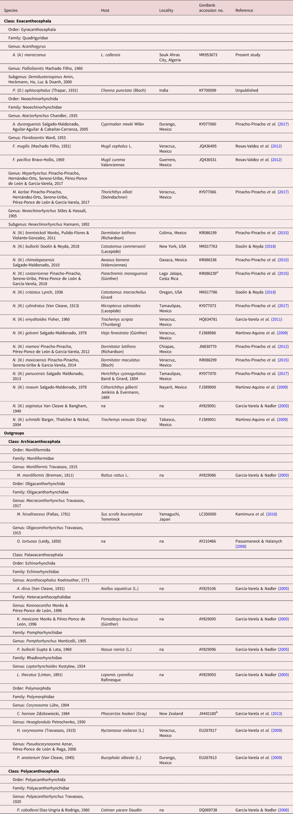
Newly generated sequences were aligned together with sequence data for species included in the class Eoacanthocephala in order to assess the phylogenetic relationships of the genus Acanthogyrus within this class. The dataset for the Eoacanthocephala comprised sequences for four genera and 18 species included in the order Neoechinorhynchida, and a single sequence of Palliolisentis (Demidueterospinus) ophiocephalus (Thapar, 1931) of the order Gyracanthocephala, as currently available in the GenBank database (see table 1 for details). The partial sequences of the 28S rRNA gene of Acanthogyrus (Acanthosentis) tilapiae Baylis, 1947 (nec 1948; see Baylis, Reference Baylis1947) available in the GenBank database (ATU53000) generated by Chenuil et al. (Reference Chenuil, Solignac and Bernard1997) were not included in the analysed dataset because of the very short length of the sequence (311 bp). Based on previous molecular phylogenies, sequences of species included in the classes Archiacanthocephala, Palaeacanthocephala and Polyacanthocephala were used as external groups (table 1). Contiguous sequences were assembled and inspected for errors in platform Geneious® v10.1.3 (Kearse et al., Reference Kearse, Moir and Wilson2012), and gene alignments were built by the program ClustalW (Thompson et al., Reference Thompson, Higgins and Gibson1994), which is implemented in the website http://www.genome.jp/tools/clustalw/, with the approach ‘SLOW/ACCURATE’ and weight matrix ‘CLUSTALW (FOR DNA)’. Maximum likelihood analysis was performed by RAxML v8.2.11 (Stamatakis, Reference Stamatakis2014) with 1000 bootstrap replicates. A Bayesian inference tree was generated by MrBayes v3.2.2 (Huelsenbeck & Ronquist, Reference Huelsenbeck and Ronquist2001), in which four independent MC3 runs of ten million generations each were realized, and for each run, two chains were used; tree topologies were sampled every 1000 generations, the heating parameter value was 0.2 and ‘Burn-in’ was set to 25%. The evolution model GTR + G for both RAxML and MrBayes was estimated with the program jModelTest v2.1.10 (Darriba et al., Reference Darriba, Taboada and Doallo2012) using corrected Akaike Information Criterion. Genetic distances (uncorrected p-distance) and the number of parsimony informative sites in the alignment were calculated with Geneious® and PAUP 4.0a165, respectively (Swofford, Reference Swofford2002).
Acanthocephalans included in the phylogenetic analyses with data on the host, locality and GenBank accession number (28S rDNA).

a Identified as Neoechinorhynchus (N.) golvani by Pinacho-Pinacho et al. (Reference Pinacho-Pinacho, Sereno-Uribe and Pérez-Ponce de León2015) (see Pinacho-Pinacho et al., Reference Pinacho-Pinacho, Sereno-Uribe and García-Varela2019).
b Identified as Corynosoma australe Johnston, 1937 by García-Varela et al. (Reference García-Varela, Pérez-Ponce de León, Aznar and Nadler2013) (see Hernández-Orts et al., Reference Hernández-Orts, Smales and Pinacho-Pinacho2017).
Results
Acanthogyrus (Acanthosentis) maroccanus (Dollfus, 1951)
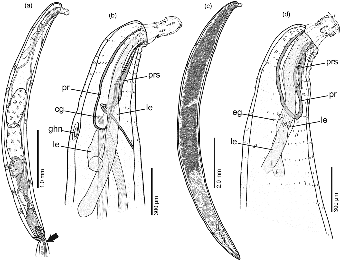
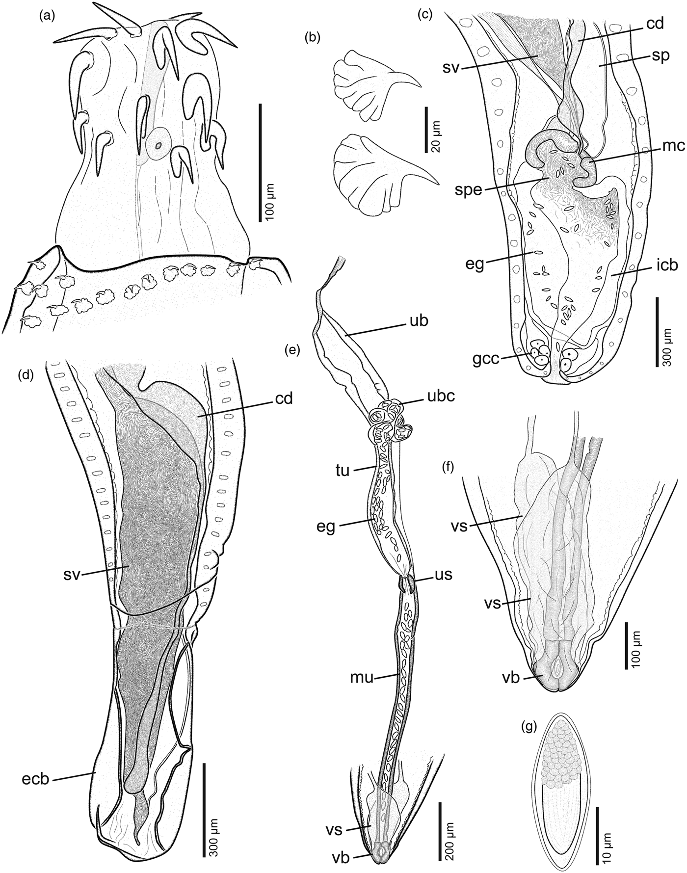
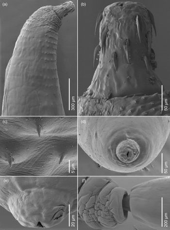
(figs 1–4; supplementary table S1)
Acanthogyrus (Acanthosentis) maroccanus (Dollfus, 1951) ex Luciobarbus callensis (Valenciennes) from Oued Charef dam lake, Algeria. (a) Male, whole mount, lateral view. Arrow indicates the posterior end of female inserted through the genital pore of the male. (b) Anterior end of male, lateral view. (c) Female, whole mount, lateral view. (d) Anterior end of female, lateral view. Abbreviations: cg, cephalic ganglion; eg, egg; ghn, giant hypodermal nucleus; le, lemniscus; pr, proboscis receptacle; prs, para-receptacle structure.

Acanthogyrus (Acanthosentis) maroccanus (Dollfus, 1951) ex Luciobarbus callensis (Valenciennes) from Oued Charef dam lake, Algeria. (a) Proboscis armature of female, lateral view. (b) Trunk spines of female, lateral view. (c) Posterior end of male with an inverted bursa, showing sperm, mature eggs and glandular cellular cluster at the posterior end of the body. (d) Posterior end of male with detail of reproductive structures extending into the copulatory bursa. (e) Female reproductive system, lateral view. (f) Posterior end of female showing a paired vaginal sleeve. (g) Mature egg. Abbreviations: cd, cement duct; eg, egg; gcc, glandular cellular cluster; ecb, everted copulatory bursa; icb, inverted copulatory bursa; mc, muscular cap of copulatory bursa; mu, muscular uterus; sp, Saefftigen's pouch; spe, sperm; sv, seminal vesicle; tu, tubular uterus; ub uterine bell; ubc, uterine bell cells; us, uterine sphincter; vb, vagina bulb; vs, vaginal sleeve.

Scanning electron micrographs of adult males of Acanthogyrus (Acanthosentis) maroccanus (Dollfus, 1951) ex Luciobarbus callensis (Valenciennes) from Oued Charef dam lake, Algeria. (a) Anterior end, lateral view. (b) Proboscis armature, lateral view. (c) Proboscis, apical view. Arrow indicates the lateral anterior hook displaced posteriorly. (d) Posterior hooks near the base of proboscis, ventral view. (e) Copulatory bursa, lateral view. (f) Copulatory cap, ventral view.

Scanning electron micrographs of adult females of Acanthogyrus (Acanthosentis) maroccanus (Dollfus, 1951) ex Luciobarbus callensis (Valenciennes) from Oued Charef dam lake, Algeria. (a) Anterior end, lateral view. (b) Proboscis armature, lateral view. (c) Trunk spines, ventral view. (d) Posterior end, sub-apical view. (e) Genital pore, ventral view. (f) Male and female in copulation. Note that the posterior end of the female (right) is inserted into the posterior end of the male (left).

Redescription
General. Quadrigyridae, with characters of Acanthogyrus and subgenus Acanthosentis. Sexual dimorphism evident, females larger than males (fig. 1a, c). Proboscis short, cylindrical, with small apical organ, pump in middle, armed with three irregularly arranged circles (especially anterior and middle circle) of six rooted hooks each, and one mononucleated large cell (figs 1b, d, 2a, 3a, b and 4a, b). Two lateral proboscis hooks in anterior and middle circle displaced posteriorly, but do not differ in size from two dorsal and two ventral hooks of each circle (figs 2a, 3b, c and 4b). Anterior hooks of similar length as middle hooks (fig. 2a). Posterior hooks shorter than anterior and middle hooks (figs 3b, d and 4b). Roots simple, without manubria, shorter than blades (fig. 2a). Neck present, unarmed (figs 3b and 4b). Trunk short, cylindrical (fig. 1a, c). Giant hypodermal nuclei 9–12; 2–3 ventral, 6–9 dorsal (fig. 1a), not observed in most specimens (especially in females). Complete circles of spines on anterior trunk scare, usually fewer spines dorsally (figs 1b, d, 3a and 4a). Spines become widely spaced with no or fewer dorsal spines posteriorly (figs 1b, d, 3a and 4a). Proboscis receptacle single-walled (fig. 1b, d), 2.0–5.5 (3.9; n = 4) times longer than proboscis in males and 2.9–5.7 (4.1; n = 3) times longer in females, with small, ellipsoidal cerebral ganglion close to its base (fig. 1b). Para-receptacle structure evident in both sexes, contiguous with ventral side of receptacle (fig. 1b, d). Lemnisci subequal, elongate, longer than proboscis receptacle. Gonopore terminal in both sexes (figs 1a, c and 4d, e). Genital spines absent.
Male. Based on 18 mounted specimens and four for SEM. Trunk 3.4–7.2 mm (5.3 mm; n = 18) × 0.44–1.04 mm (0.7 mm; n = 18). Anterior trunk with 14–19 circles of rosethorn-shaped cuticular spines, covering about 14–15% of trunk, reaching to posterior level proboscis receptacle (figs 1b and 3a). Proboscis 130–158 (141; n = 5) × 81–107 (96; n = 5). Anterior, middle and posterior hooks 49–63 (58; n = 15), 43–61 (55; n = 12), 34–42 (38; n = 9) long, respectively. Roots of anterior, middle and posterior hooks 28–38 (33; n = 14), 28–38 (32; n = 11), 22–35 (27; n = 9) long, respectively. Neck 42–93 (55; n = 4) × 127–176 (144; n = 4). Proboscis receptacle 341–984 (646; n = 10) × 59–121 (93; n = 11). Lemnisci 0.90–1.47 mm (1.13 mm; n = 8) long. Reproductive system occupying approximately 43–63% (54%; n = 11) of trunk length (fig. 1a). Testes equatorial, ovoid, in tandem, often overlapped (fig. 1a). Anterior testis 549–1347 (850; n = 11) × 264–625 (430; n = 11). Posterior testis 597–1209 (851; n = 10) × 266–658 (439; n = 10). Cement gland subspherical, 278–612 (454; n = 10) × 234–443 (339; n = 9), with 4–6 large giant nuclei (fig. 1a). Sperm ducts join seminal vesicle at approximately posterior level of cement gland (fig. 1a). Saefftigen's pouch dorsal to cement duct and seminal vesicle (fig. 1a). Cement duct joins with seminal vesicle duct and Saefftigen's pouch anterior to copulatory bursa. Glandular cellular cluster observed at posterior end of body (fig. 2c). Sperm and mature eggs sometimes observed inside inverted copulatory bursa (fig. 2c). Posterior male reproductive structures extending into fully everted bursa (fig. 2d). Everted copulatory bursa 360–720 (484; n = 5) × 210–336 (275; n = 5) (figs 2d and 3e). Cement plug observed in some specimens (fig. 3f).
Female. Based on 19 gravid mounted specimens and four for SEM. Trunk 7.7–12.9 mm (10.5 mm; n = 14) × 0.4–1.2 mm (0.9 mm; n = 14). Anterior trunk with 16–19 circles rosethorn-shaped spines (figs 2b and 4c), covering about 5–11% of trunk, reaching almost to posterior level of lemnisci (fig. 1d). Proboscis 114–198 (151; n = 7) × 92–134 (112; n = 6). Anterior, middle and posterior hooks 57–62 (59; n = 11), 53–60 (57; n = 7), 33–42 (39; n = 9) long, respectively. Roots of anterior, middle and posterior hooks 29–34 (32; n = 10), 28–41 (31; n = 6), 26–34 (30; n = 9) long, respectively. Neck 56–79 (67; n = 4) × 138–167 (150; n = 4). Proboscis receptacle 572–1031 (801; n = 10) × 102–149 (125; n = 9). Lemnisci 1.50–1.64 mm (1.56 mm; n = 3) long. Reproductive system 1.68–1.79 mm (1.73 mm; n = 2) long, post-equatorial, representing 15–16% of trunk length. Uterine bell attached to ventral body wall, with subspherical uterine bell cells (fig. 2e). Uterus differentiated to tubular and muscular part separated by sphincter (fig. 2e). Vaginal bulb, simple, globular, 35–106 (67; n = 13) long. Vagina bulb flanked with paired conical muscular jacket (vaginal sleeve), 204–501 (341; n = 5) long, extending anteriorly as reproductive ligaments and attaching posteriorly at posterior end of trunk (fig. 2e, f). Posterior end of female sometimes contracted after mating (fig. 4d, e). Mature eggs containing a fully developed acanthor fusiform (fig. 2g), elongate, 24–29 (26; n = 32) × 9–11 (10; n = 28).
Taxonomic summary
Type host. Luciobarbus setivimensis (Valenciennes) (Cypriniformes: Cyprinidae).
Additional host. Luciobarbus callensis (Valenciennes) (Cypriniformes: Cyprinidae), Algerian barb.
Type locality. Azrou (Middle Atlas), Morocco.
New localities. Oued Charef dam lake, Souk Ahras City and Zit Emba dam lake, Skikda City, Algeria (no acanthocephalans were collected from Beni Haroun dam lake).
Site of infection: Intestine.
Infection parameters. Oued Charef dam lake: prevalence 32% (n = 289); abundance = 0.9; mean intensity = 2.7; intensity range = 1–12. Zit Emba dam lake: prevalence 36% (n = 36); abundance = 1.4; mean intensity = 3.9; intensity range = 1–12.
Voucher material. Four males and three females (IPCAS A-109); three males and two females (NHMUK 2019.8.12.1-6); one male and three females (HCMLP-He 7541).
Representative sequences. MK953673 (28S rRNA, partial sequence).
Remarks
The newly collected specimens from L. callensis in Algeria are morphologically similar to those described by Dollfus (Reference Dollfus1951) and Meddour et al. (Reference Meddour, Meddour-Bouderda and Brahim-Tazi2010) in having a similar body length, proboscis size, length of the anterior and middle hooks (which are larger than the posterior hooks), shape of roots of the hooks (simple without manubria) and the number of circles of tegumental spines (supplementary table S1). The eggs are also similar in size, although the eggs illustrated in Fig. 6 by Meddour et al. (Reference Meddour, Meddour-Bouderda and Brahim-Tazi2010) are smaller (28–30 µm × 10–12 µm, n = 6) than mentioned in their text (34–38 µm × 9–15 µm).
The newly collected material enlarges the range of intraspecific variability for the number of dorsal hypodermal nuclei, the length of the trunk and the anterior hooks of males, the size of the testis and the size of the eggs, all values being lower than those previously reported in the literature (see supplementary table S1). In contrast, our study also revealed the higher upper limits for the number of circles of cuticular spines, the size of the proboscis of females, the length of the middle hooks for males and females, the length of the posterior hooks of females, the length of the proboscis receptacle of males and the length of lemniscus for males and females (supplementary table S1).
Our morphological observations revealed that the proboscis hooks of A. (A.) maroccanus are not arranged in perfect (regular), or nearly perfect, circles reported for this species in the key to the species of the subgenus Acanthosentis provided by Amin (Reference Amin2005). Our study has shown that two lateral hooks in the anterior and middle circle are displaced posteriorly. This arrangement of the hooks of A. (A.) maroccanus was already observed by Dollfus (Reference Dollfus1951) and Meddour et al. (Reference Meddour, Meddour-Bouderda and Brahim-Tazi2010).
The present study also shows that the posterior male reproductive structures extend into the everted bursa in A. (A.) maroccanus (fig. 2d). This feature was previously described only in A. (A.) parareceptaclis Amin, 2005 from Japan by Amin (Reference Amin2005), although this characteristic may also be present in A. (A.) phillipi (Mashego, 1988) from South Africa (see Fig. 4 in Mashego, Reference Mashego1988). However, as noted by Amin (Reference Amin2005), it is uncertain if this feature appears in other species of the subgenus Acanthosentis, mainly because the everted bursa has not been described for some species.
Examination of the newly collected specimens of A. (A.) maroccanus has also shown the presence of two structures, which were not reported previously, and which are uncommon among species of the subgenus Acanthosentis, i.e. the para-receptacle structure and the vaginal sleeve. The para-receptacle structure has also been reported in species of Neoechinorhynchus, Paratenuisentis Bullock & Samuel, 1975 and Tenuisentis Van Cleave, 1936 (see Amin et al., Reference Amin, Heckmann and Standing2007, Reference Amin, Evans and Boungou2016, Reference Amin, Heckmann and Ha2018; Herlyn & Taraschewski, Reference Herlyn and Taraschewski2017). To our knowledge, the para-receptacle structure has been observed in both sexes only in A. (A.) parareceptaclis, A. (A.) barmeshoori Amin, Gholami, Akhlaghi & Heckmann, 2013 and A. (A.) kashmirensis Amin, Heckmann & Zargar, 2017. However, A. (A.) parareceptaclis and A. (A.) barmeshoori are characterized by having two para-receptacle structures, one contiguous with the ventral side of the proboscis receptacle and the other at the posterior end of the trunk (see Amin, Reference Amin2005; Amin et al., Reference Amin, Gholami and Akhlaghi2013), whereas only the para-receptacle structure contiguous with the ventral side of receptacle is evident in A. (A.) kashmirensis (Amin et al., 2017). In A. (A.) maroccanus, only the antero-ventral para-receptacle structure was observed (fig. 1b, d), whereas the similar posterior structure is apparently absent (fig. 2c, d), especially in females (fig. 2e, f). A vaginal sleeve was described only in A. (A.) parareceptaclis by Amin (Reference Amin2005), but this structure could also be present in A. (A.) tripathi Rai, 1967 from India, as indicated by Rai (Reference Rai1967).
During the parasitological examination of Algerian barbs in our study, numerous copulating pairs of A. (A.) maroccanus were collected. Both individuals were found firmly attached to the intestinal wall of the fish. The fully everted copulatory bursa of male attaches to the posterior end of the female. Then, the bursa is withdrawn into the body without releasing the female, and the attached portion of the female is inserted through the genital pore of the male (fig. 1a). Inside the invaginated bursa, the muscular cap of the bursa is contracted anteriorly (fig. 1a; see also Fig. 61 in Dollfus, Reference Dollfus1951), and the female is firmly trapped by the genital pore of the male (figs 1a and 4f), suggesting that insemination may occur before the female is lodged inside the male. In some copulating females, ovarian balls and unripe eggs were observed, indicating that copulation occurs more than once. Moreover, in one copulating pair, pieces of the copulatory cap of females were observed in the invaginated bursa of the male. When the copulating pair is separated, the posterior end of the female is strongly contracted (fig. 4d, e), but the posterior end returns to its normal shape after some time (figs 1c and 2e, f). In some separated males, sperm and unripe eggs were observed in the invaginated bursa (fig. 2c). A similar copulatory behaviour was observed in A. (A.) holospinus (Sen, Reference Sen1938), A. (A.) dattai (Podder, Reference Podder1938) and A. (A.) tilapiae (see Podder, Reference Podder1938; Sen, Reference Sen1938; Baylis, Reference Baylis1947).
Molecular analyses
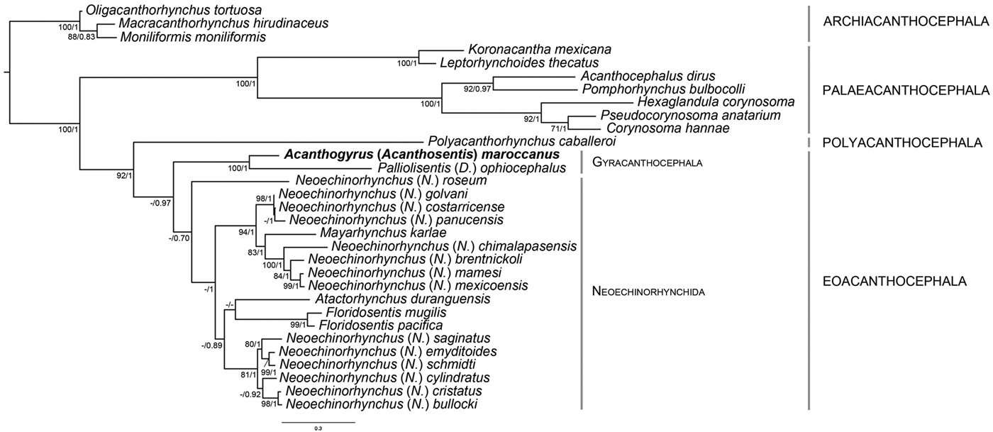
Three identical sequences for the D1–D3 region of the 28S rDNA (991–1085 nt long) were generated from isolates of A. (A.) maroccanus collected from L. callensis from Algeria. The alignment for the 28S rDNA dataset comprised 1618 nt positions, of which 599 (37%) were parsimony-informative. Phylogenetic relations estimated by maximum likelihood and Bayesian inference methods resulted in consensus trees with identical topologies (Fig. 5).
Phylogenetic tree resulting from maximum likelihood analysis of partial (D1–D3 regions) sequences of 28S rDNA of Acanthogyrus (Acanthosentis) maroccanus from Algeria. Nodal supports are depicted as bootstraps values (≥70% shown only) followed by posterior probabilities (≥0.70 shown only). The branch-length scale bar indicates the expected number of substitutions per site. The newly generated sequence is indicated in bold.

The phylogenetic relationships at the class level were very similar to the phylogenetic hypotheses obtained in previous studies (e.g. García-Varela & Nadler, Reference García-Varela and Nadler2005; Verweyen et al., Reference Verweyen, Klimpel and Palm2011), where the Polyacanthocephala was the sister clade of the Eoacanthocephala. Within the Eoacanthocephala, which includes members of the Gyracanthocephala and the Neoechinorhynchida, our sequence of A. (A.) maroccanus was placed in a well-supported clade with P. (D.) ophiocephalus, both species included in the Quadrigyridae, the only family within the Gyracanthocephala. The clade formed by the members of the Gyracanthocephala was found to be sister of a clade including all members of the Neoechinorhynchida, but with low support. Within the Neoechinorhynchida, the phylogenetic analyses showed Neoechinorhynchus (N.) roseum (GenBank accession no. FJ389000) as an earliest diverging lineage and two further groups. The first well-supported group was formed by two clades of Neoechinorhynchus spp. (the isolates of three and four species, respectively) and Mayarhynchus karlae (GenBank accession no. KY077066). The second group, having low support, was formed by a clade of Floridosentis mugilis (GenBank accession no. JQ436495), F. pacifica (GenBank accession no. JQ436531) and A. duranguensis (GenBank accession no. KY077080), and its sister clade of six species of Neoechinorhynchus. Under the current taxon sampling, Neochinorhynchus was resolved as a polyphyletic group with at least three separate clades and the lineage of N. (N.) roseum. The present phylogenetic analysis is largely consistent with previous phylogenetic assessments for species of Neoechinorhynchus (e.g. García-Varela & Pinacho-Pinacho, Reference García-Varela and Pinacho-Pinacho2019).
The genetic divergence value between A. (A.) maroccanus and P. (D.) ophiocephalus (the other available member of the Quadrigyridae) was 39.2%. Variation of the interspecific genetic divergence between A. (A.) maroccanus and other members of the Eoacanthocephala ranged between 33.9% (Neoechinorhynchus (Neoechinorhynchus) golvani) and 43.6% (F. pacifica).
Discussion
Acanthogyrus (A.) maroccanus has been registered only in L. setivimensis and L. callensis from Algeria and Morocco (Dollfus, Reference Dollfus1951; Meddour, Reference Meddour2009; Meddour et al., Reference Meddour, Meddour-Bouderda and Brahim-Tazi2010). This acanthocephalan has been reported in freshwater cyprinids collected in Oubeira Lake and several dam lakes (i.e. Ain Dalia, Bouhamdane, Oued Charef and Zit Emba) or streams (i.e. Bounamoussa, El Kebir and Seybouse) from East Algeria (Meddour et al., Reference Meddour, Meddour-Bouderda and Brahim-Tazi2010; present study).
The interspecific relationships and phylogenetic position of Acanthogyrus remain poorly explored. According to the GenBank dataset, molecular data for mitochondrial and nuclear genes have been provided only for two of the 47 species of the genus. Chenuil et al. (Reference Chenuil, Solignac and Bernard1997) provided a partial fragment of the 28S rRNA gene of A. (A.) tilapiae; however, this is extremely short (311 bp) and, thus, could hardly be compared with our newly generated sequences, which are much longer (991–1085 bp). Even though our phylogenetic analysis showed that our isolate of A. (A.) maroccanus forms a poorly supported clade within the Eoacanthocephala, the topology recovered from different analyses was always identical. The phylogenetic signal detected in our study shows the limitation of the currently available molecular data to understand the interrelations of species of the Quadrigyridae.
Supplementary material
To view supplementary material for this article, please visit https://doi.org/10.1017/S0022149X19000737.
Acknowledgements
The authors are indebted to two anonymous reviewers for their helpful suggestions, and to M. Borovková and R. Kuchta, IPCAS, České Budějovice for their technical help during the examination of the acanthocephalans. Thanks are also due to D. Hernández-Mena, Centro de Investigación y de Estudios Avanzados del Instituto Politécnico Nacional – Unidad Mérida, Yucatán, Mexico for his valuable comments and assistance with the phylogenetic analyses.
Financial support
This study was partly supported by the Faculty of Natural and Life Sciences, University of Mohamed Cherif Messaadia of Algeria (CNEPRU: 410120150001), Institute of Parasitology, Biology Centre of the Czech Academy of Sciences (RVO: 60077344), Czech Science Foundation (project No. P505/12/G112), Grant Agency VEGA (project No. 2/0159/16), and Agencia Nacional de Promoción Científica y Tecnológica of Argentina (PICT No. 2017-3232).
Conflicts of interest
None.
Ethical standards
The authors assert that all procedures contributing to this work comply with the ethical standards of the relevant national and institutional guides on the care and use of animals.