Introduction
Acute ST-segment elevation myocardial infarction is a well-recognised medical emergency in adults and commonly results from atherosclerotic coronary artery disease and plaque rupture. Conversely, myocardial infarction is rare in paediatric patients and often linked to structural coronary artery abnormalities. Reference Towbin, Garson, Bricker and McNamara1 The diagnostic criteria for myocardial infarction, including electrocardiographic, echocardiographic, and enzymatic data, are well defined in adults but present challenges in paediatric cases. Reference Gazit, Avari, Balzer and Rhee2,Reference Towbin, Bricker and Garson3 Electrocardiographic diagnostic criteria of ischaemia are especially difficult to define in children as the cardiac structure changes with age. Additionally, non-ischaemic conditions such as pericarditis may present with similar symptoms as myocardial infarction and ST-segment changes.
ST-elevation myocardial infarction is classified into two types based on its underlying mechanism. Reference Thygesen, Alpert and Jaffe4 Type I occurs secondary to acute coronary thrombosis from plaque rupture, ulceration, or erosion, leading to complete occlusion of the coronary artery, and is the most common aetiology of myocardial infarction in adults. Reference Towbin, Garson, Bricker and McNamara1 Treatment of Type I ST-elevation myocardial infarction includes immediate revascularization with percutaneous coronary intervention. Type II ST-elevation myocardial infarction occurs secondary to a mismatched myocardial oxygen supply and demand and is caused by non-thrombotic aetiologies such as congenital coronary anomalies, poor cardiac function, sepsis, severe anaemia, or coronary vasospasm. Reference Thygesen, Alpert and Jaffe4 Electrocardiogram findings may show transient ST-segment elevation. Patients presenting with Type II ST-elevation myocardial infarction may have underlying coronary artery disease, but the primary cause is not plaque rupture. The treatment differs from Type I ST-elevation myocardial infarction, as percutaneous coronary intervention is not indicated, and the underlying cause should be treated. Reference Thygesen, Alpert and Jaffe4
Paediatric myocardial infarction is a rare condition, with poorly defined diagnostic criteria and limited guidance on prompt treatment and intervention, resulting in clinicians frequently relying on adult algorithms due to the absence of paediatric-specific evidence. This study is the first to explore the aetiology and outcomes of paediatric patients presenting with concerns for ST-elevation myocardial infarction. It aims to review the incidence, diagnostic findings, interventions, treatment, and outcomes of paediatric patients presenting with ST-elevation myocardial infarction concerns at a paediatric institution and an affiliated adult hospital.
Materials and method
Study design and participants
A retrospective chart review was conducted over a ten-year period, from January 2013 to December 2023, at both a freestanding children’s hospital and an affiliated adult hospital designated as a Level I Comprehensive Heart Attack Center by the American Heart Association. The study aimed to identify patients diagnosed with ST-elevation myocardial infarction. Patients reviewed included those with ICD-10 codes containing ST-elevation myocardial infarction, non- ST-elevation myocardial infarction, elevated troponin level, chest pain, and cardiac arrest to ensure all patients presenting with ST-elevation myocardial infarction were included. All electrocardiograms were reviewed and confirmed by the electrophysiology team. Institutional Review Board approval was obtained at each participating hospital prior to the study.
Inclusion criteria
Patients aged 1 day to 21 years who met all the following criteria were included:
-
i. Symptoms consistent with myocardial infarction, such as chest pain, shoulder pain, shortness of breath, diaphoresis, lightheadedness, syncope, nausea, or palpitations.
-
ii. Documented elevation in one or more cardiac enzymes (e.g., troponin, creatine kinase-MB).
-
iii. Electrocardiogram findings of ST-segment abnormalities or changes from baseline electrocardiogram.
Exclusion criteria
Patients excluded were those who did not exhibit electrocardiogram changes consistent with myocardial infarction or those without documented elevated cardiac enzymes. Additionally, patients with incomplete records or missing critical data were excluded to ensure comprehensive and accurate analysis. Duplicate records of patients who were treated at both hospitals during their care were combined.
Data collection
Data were collected from electronic medical records, including patient demographics, clinical presentation, diagnostic findings, interventions, treatments, and outcomes. The specific demographic variables collected were patient age at presentation, sex, and race. Medical histories, including a history of CHD, were evaluated as well as symptoms at presentation (chest pain, dyspnoea, shortness of breath, shoulder pain, palpitations, nausea, syncope, cardiac arrest). Results from diagnostic testing were evaluated, including electrocardiogram changes, echocardiographic findings, cardiac enzymatic marker levels, CT, and/or MRI results, and results of cardiac stress testing. The collected data included interventions with pharmacological treatments, surgical procedures, and interventional cardiology procedures. Short- and long-term outcomes were evaluated including transfer of patients between facilities, survival rates, and complications of procedures. Study data were collected and managed using REDCap electronic data capture tools hosted at Children’s Mercy Hospital, Kansas City. Reference Harris, Taylor, Thielke, Payne, Gonzalez and Conde5,Reference Harris, Taylor and Minor6 REDCap (Research Electronic Data Capture) is a secure, web-based software platform designed to support data capture for research studies, providing 1) an intuitive interface for validated data capture; 2) audit trails for tracking data manipulation and export procedures; 3) automated export procedures for seamless data downloads to common statistical packages; and 4) procedures for data integration and interoperability with external sources. Reference Harris, Taylor, Thielke, Payne, Gonzalez and Conde5,Reference Harris, Taylor and Minor6
Analysis methods
Descriptive statistics were used to summarise patient demographics, clinical presentation, diagnostic findings, interventions, and outcomes. Continuous variables were expressed as means ± standard deviations or medians and ranges as appropriate, and categorical variables as frequencies and percentages. SAS version 9.4 (SAS Institute Inc., Cary, NC) was used for all analyses.
Results
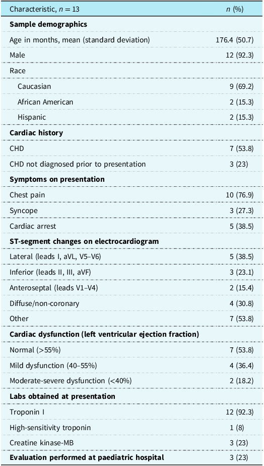
Out of 965 patients screened, 13 patients met the inclusion criteria for this study. As described in Table 1, the cohort had a mean age of 15 years (standard deviation ± 4 years), with the youngest patient being 11 months old. Most of the patients were male (12 males, 92%), and the remaining patient was female (1 female, 8%). Ethnic distribution was predominantly Caucasian (69%), followed by African American (15%) and Hispanic (15%). Chest pain was the most common presenting symptom (77%), while less frequently reported symptoms included shoulder pain (23%), shortness of breath (31%), diaphoresis (8%), lightheadedness (25%), syncope (27%), nausea (15%), and palpitations (31%). All patients exhibited ST-segment changes on ECG consistent with myocardial infarction, with ST-segment elevations observed in the inferior (leads II, III, and aVF), lateral (leads I, aVL, V5–V6), anteroseptal (leads V1–V4), and noncoronary regions. Echocardiography was performed in all patients and revealed a normal ejection fraction in 54% of patients, while regional wall motion abnormalities were seen in five patients (38%) in the left ventricle (n = 2), septal wall (n = 1), and diffuse motion abnormalities (n = 2), Troponin levels were elevated in all patients, with evaluation of troponin I levels in 12 patients (range 0.05 to 22.9ng/mL, reference range 0.00–0.04 ng/mL) and high-sensitivity troponin in the remaining patient (22,900 ng/L, reference range 0.00–20 ng/L). Creatine kinase-MB levels were not routinely collected in patients at either institution, with the level being obtained in 3 patients (23%) and elevated in 1 patient. Additional diagnostic testing included CT angiography (31%), cardiac MRI (38%), and exercise stress testing (15%), although these diagnostic tests were not performed during the initial evaluation.
Summary of patient descriptive data

Most patients in this cohort (54%) had CHD. Four patients (31%) had previously identified CHD and included a patient with a previously repaired anomalous right coronary artery, a patient with Williams syndrome and a single left coronary artery with known stenosis, a patient with repaired congenitally corrected transposition of the great arteries, and a patient with subaortic stenosis who had previously undergone a Ross/Konno procedure. There were 3 patients (23%) with unidentified CHD in the cohort, two of whom presented following cardiac arrest and the third presented with chest pain following a syncopal event. All three newly diagnosed congenital defects involved structural coronary abnormalities including a patient with a myocardial bridge, a patient with an anomalous left coronary arising from the pulmonary artery, and a patient with an anomalous origin of the circumflex artery.
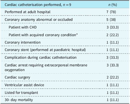
As shown in Table 2, 10 patients (77%) were either transferred to or remained at an adult hospital for emergent ST-elevation myocardial infarction evaluation, with seven (54%) undergoing cardiac catheterisation outside of the paediatric hospital. No patients received coronary intervention in the cardiac catheterisation lab at the adult hospital. Four patients (31%) had cardiac arrest, with three of these events occurring at the adult hospital. Three patients (23%) required initiation of extracorporeal membrane oxygenation support in the cardiac catheterisation lab, all of whom had congenital cardiac anomalies. There was one death in a patient with chromosome duplication in 16q24 and diffuse vasospastic disease who underwent cardiac catheterisation at the adult hospital with findings of profoundly vasospastic coronary arteries and collaterals suggestive of chronicity. There were ultimately no interventions performed in the cardiac catheterisation lab for this patient, and the patient died in the ICU at the adult hospital the following day. Three patients (23%) were discharged without undergoing a cardiac catheterisation despite meeting adult criteria for myocardial infarction of ST-segment changes, clinical symptoms of myocardial infarction, and elevated cardiac biomarkers. These patients were found to have pericarditis, myocarditis, and significant drug use, with resolution of symptoms and ST-segment changes with supportive care.
Cardiac catheterisation data and outcomes

* Previous Kawasaki disease, chromosomal duplication in 16q24/associated diffuse chronic vasospastic disease.
Two patients (15%) underwent cardiac catheterisation at the paediatric hospital with one patient who presented with chest pain undergoing coronary stent placement for known coronary ostial scarring and left coronary artery stenosis following a Ross/Konno procedure for subaortic stenosis. This patient had a cardiac arrest during coronary evaluation and was cannulated onto extracorporeal membrane oxygenation prior to coronary stent placement. The patient was subsequently listed for cardiac transplant. The second patient presented following a cardiac arrest with a previous diagnosis of Kawasaki disease. Coronary evaluation demonstrated adequate coronary collaterals despite right coronary occlusion, so did not receive a coronary intervention. One patient with a diagnosis of Williams syndrome and known single left coronary stenosis was electively transferred to an adult hospital following cannulation to extracorporeal membrane oxygenation support for surgical coronary artery bypass grafting after presenting to the paediatric hospital with chest pain and subsequent cardiac arrest. (Supplemental Figure 1).
Discussion
Acute myocardial infarction is a rare cause of morbidity and mortality in childhood, with limited published data on guidelines, evaluation, and treatment. This study reviewed data on patients presenting to a large paediatric hospital and affiliated adult Level I Comprehensive Heart Attack Center with concerns of ST-elevation myocardial infarction. Clinical identification of paediatric myocardial infarction can be difficult, with atypical presentations often complicating the diagnosis. Reference Celermajer, Sholler, Howman-Giles and Celermajer7 Chest pain, the hallmark symptom of myocardial ischaemia and infarction in adults, is rarely secondary to cardiac ischaemia in children and adolescents which makes the diagnosis of myocardial infarction in this population particularly challenging. This is evident in our study as over a ten-year period, less than 2% of patients with ICD-10 codes relating to myocardial infarction met the widely used adult criteria of ST-segment changes and elevated cardiac biomarkers.
No patients within our cohort were found to have a Type I ST-elevation myocardial infarction, as the ischaemic events were not a result of plaque rupture but were instead linked to CHD or structural coronary issues. In the paediatric literature, the most common coronary defects associated with myocardial ischaemia include an anomalous left coronary artery originating from the pulmonary artery and coronary abnormalities following Kawasaki disease. Reference Celermajer, Sholler, Howman-Giles and Celermajer7,Reference Reich and Campbell8 Both diagnoses were present in this patient cohort. A total of 3 patients (23%) were found to have previously undiagnosed abnormal coronary anatomy, all of whom presented following cardiac arrest or syncope. Two of these patients required cardiac surgery as one patient was found to have an anomalous left coronary artery arising from the pulmonary artery and another required surgical unroofing of a myocardial bridge on the left anterior descending coronary artery. The third patient with previously undiagnosed CHD did not undergo surgical intervention on the discovered anomalous circumflex artery as it was not deemed to be the cause of cardiac arrest, but ultimately had an implanted cardiac defibrillator placed for idiopathic ventricular tachycardia. All patients requiring extracorporeal membrane oxygenation had CHD, thereby reinforcing that this cohort represents a fragile patient population during general anaesthesia. However, mortality was low in this cohort (7%), with only one death occurring in a child with known chromosome duplication in 16q24, leading to diffuse vasospastic disease and chronic severe coronary stenosis.
The twelve-lead electrocardiogram is a critical tool in diagnosing myocardial infarction and provides valuable insight into the acute phase of myocardial injury. In adults, myocardial infarction can be diagnosed with a high specificity through evaluation of ST-segment elevation in a coronary distribution pattern on electrocardiogram.Reference Michael, Masry, Khan and Das 9 In this cohort, ST-segment changes were observed in all patients, with the most common distributions being lateral (leads I, aVL, V5–V6) (42%) and diffuse/non-coronary (33%) regions. Three patients in our study had a final diagnosis of pericarditis or myocarditis with two of these patients undergoing cardiac catheterisation without the need for coronary intervention. These results emphasise the importance of recognising electrocardiogram changes as part of the diagnostic process, but also consider non-ischaemic causes of ST-segment changes in paediatric patients.
Considerable variability in medical management was observed, highlighting the need for the development of evidence-based guidelines that account for the unique pathophysiology in the paediatric population. No coronary interventions were required in patients who underwent coronary evaluation at the adult facility, suggesting the aetiology of coronary ischaemia in children is rarely a Type I ST-elevation myocardial infarction and time-sensitive percutaneous coronary intervention may not be indicated. CHD was more prevalent among patients who suffered cardiac arrest during or following cardiac catheterisation, underscoring the fragility of these patients and the importance of addressing this condition early in the diagnostic process at a paediatric facility with expertise in CHD.
Current facility protocols and considerations
Previously, the studied paediatric hospital followed an ST-elevation myocardial infarction/acute coronary syndrome activation protocol that included urgent notification of the associated adult hospital when there was suspicion for myocardial infarction by the paediatric cardiologist involved in the patient’s care. If ST-elevation myocardial infarction was confirmed, the patient was promptly transferred to the adult hospital, where they proceeded directly to the cardiac catheterisation lab under the care of an adult interventional cardiologist. The diagnostic criteria for myocardial infarction in this protocol were aligned with established adult guidelines, which include ST-elevation greater than 1 mm and elevated levels of cardiac biomarkers such as troponin or creatine kinase-MB.
Given these study findings, the previous protocol was modified. While the adult cardiology team continues to be involved in decision-making, one key change was made: conducting additional diagnostic evaluations, including cardiac catheterisation and/or coronary CT angiography at the paediatric hospital prior to patient transfer. Based on the data findings, this will allow for a more informed, case-by-case determination of the need for transfer, helping to assess potential CHD, congenital coronary artery anomalies, and coronary occlusion before making the decision to transfer care to the adult hospital. Such an approach would help address the unique complexities associated with paediatric myocardial infarction, ensuring that congenital anomalies are adequately considered, and appropriate interventions are carried out in a timely manner at a paediatric facility with CHD expertise.
Study limitations and future multicenter collaboration
One of the primary limitations of this study is the small sample size, with only 13 patients meeting the inclusion criteria over a ten-year period. This limited sample size restricts the generalizability of the findings and highlights the rarity of acute myocardial infarction in paediatric patients. The small cohort also validates the challenges in studying such a rare condition and the need for larger, multicentre studies to validate these findings and develop robust clinical guidelines.
To address these limitations, collaboration with other paediatric centres is essential. By pooling data from multiple institutions, we can increase the sample size and enhance the statistical power of future studies. Such collaboration would facilitate a more comprehensive understanding of paediatric myocardial infarction, including its incidence, clinical presentation, management strategies, and outcomes.
This study underscores the rarity of acute myocardial infarction in paediatric patients despite the common presentation of ST-segment changes on electrocardiogram. The findings reveal significant variability in medical management and emphasise the importance of initial diagnostic evaluations at paediatric centres, particularly to rule out CHD. The lack of coronary intervention during cardiac catheterisation at the adult facility suggests that paediatric cases may be effectively managed within paediatric settings. The study calls for the development of standardised guidelines to ensure consistent and effective management of acute myocardial infarction in paediatric patients, ultimately aiming to improve clinical outcomes for this unique population.
Supplementary material
The supplementary material for this article can be found at https://doi.org/10.1017/S1047951126111366.
Acknowledgements
We would like to express our sincere gratitude to Children’s Mercy Hospital and The University of Kansas Health System for their invaluable collaboration and support in this study. Their contributions were essential in facilitating data collection and providing the necessary resources for this research. We also extend our appreciation to the medical staff and researchers at both institutions for their dedication and expertise, which greatly enhanced the quality and scope of this study.
Financial support
This research received no specific grant from any funding agency, commercial, or not-for-profit sectors.
Competing interests
Competing interests: The author(s) declare none.
Ethical standard
This study (IRB Protocol #STUDY00002612) was reviewed and determined to be exempt from Institutional Review Board oversight under 45 CFR 46.104(d)(4)(iii). This exemption applies to secondary research involving identifiable private health information when the use is regulated under the Health Insurance Portability and Accountability Act for purposes of health care operations, research, or public health activities. As such, informed consent was not required, and no contact with subjects occurred. All data were handled in accordance with applicable privacy and confidentiality standards.