Introduction
Osteoporosis is a progressive, systemic metabolic bone disorder which results in lowered bone mineral density (BMD) thus increasing an individual's risk of fracture. It is a serious public health issue affecting one in two women and one in five men aged over 50 years (Keen, Reference Keen2007), and with increasing life expectancy its incidence is rising. Osteoporosis is asymptomatic in the absence of fractures and often remains undiagnosed; hence it has been termed the “silent disease.” In the U.K. osteoporotic fractures result in an annual cost to the National Health Service (NHS) of £1.8 billion (Dolan and Torgerson Reference Dolan and Torgerson1998). Hip fractures are particularly serious, causing much mortality and reduced quality of life, with only 30% of patients returning to their pre-morbid level of function (Chrischilles et al., Reference Chrischilles, Butler, Davis and Wallace1991).
A variety of risk factors are associated with osteoporosis (Cummings et al., Reference Cummings1995; Johnell, Reference Johnell1996). These include age, low body mass index (BMI), immobility, smoking, previous fragility fractures and certain medicines and medical conditions. Patients with mental illnesses, such as schizophrenia andaffective disorders, are also at increased risk of osteoporosis, partly because of their psychotropic medication (Misra et al., Reference Misra, Papakostas and Klibanski2004). Those with learning disabilities are also at higher risk (Aspray et al., Reference Aspray, Francis, Thompson, Quilliam, Rawlings and Tyrer1998; Tyler et al., Reference Tyler, Snyder and Zyzanski2000). Relatively little is known about the incidence of osteoporosis in elderly hospitalized patients with severe mental illness, although these patients would be expected to be at particular risk.
Central dual energy X-ray absorptiometry (DEXA) scans are the definitive means for measuring BMD. Because these are not easily accessed by residents of institutions, peripheral scanners are an acceptable alternative, although in general, positive findings should be confirmed by central scan (Blake et al., Reference Blake, Patel and Fogelman1998; Kirk et al., Reference Kirk, Nichols and Spangler2002).
As part of a service development at our psychiatric hospital, we screened older inpatients for risk factors for osteoporosis and used a peripheral DEXA scanner to determine the proportion with osteoporosis. We looked for associations between patient variables and fragility fractures and osteoporosis as found on DEXA scans. We also assessed patient acceptability of the scanning technique and followed up patients to determine how many were subsequently receiving medication for osteoporosis.
Method
The survey was conducted in Townsend Division, a 102-bedded unit mainly for older adults with complex psychiatric needs within a large charitable tertiary referral centre. Townsend Division consists of five wards for older adults with severe mental illness and challenging and/or offending behavior and one ward for patients with Huntington's disease. Local Research Ethics Committee approval was not necessary since the survey was part of a service development for ongoing screening and treatment of osteoporosis.
Between March and July 2007, BS interviewed each patient's consultant psychiatrist about the following osteoporosis risk factors:
• age ≥75 years
• post-menopausal status
• previous diagnosis of osteoporosis
• weight loss of >5% during the past year and/or BMI <22
• thoracic kyphosis
• fragility fracture(s) recorded in past medical records
• predisposing medical disorders e.g. rheumatoid arthritis, malabsorption.
• predisposing medication, e.g. antipsychotics, anticonvulsants, systemic corticosteroids.
Additional details were obtained from case notes and medication charts, including the number of falls within the past year. BS also measured each patient's weight and height.
All patients were invited to undergo scanning of the wrist and calcaneum using a DEXA scanner (EXA-3000 model; OsteoSys Co Ltd, 2001). We used the World Health Organization's definition of osteoporosis (T-score ≤ −2.5) and osteopenia (T-score between −1.0 and −2.5) (WHO, 1994). We also reported Z-scores (the number of standard deviations the patient's bone density is above or below that expected for their age). Where results differed between wrist and calcanium, the lower BMD score was adopted.
Patients judged by their psychiatrist to have the mental capacity to consent to scanning were afterwards questioned about the experience and acceptability of being scanned and their understanding of the procedure.
Patients were followed up in February 2008 to see if action had been taken in the light of the survey's findings.
Statistical analysis
Results were analyzed using SPSS version 14.0 (SPSS Inc., 2006). The χ2, Fisher Exact and Mann-Whitney tests were used to examine patient variables and the presence or absence of fragility fractures and osteoporosis on the DEXA scan. Pearson's correlation was used to examine the relationship between age and length of stay, and also the number of falls, body mass index, length of stay and T- and Z-scores. The independent samples T-test was used to compare mean Z-scores associated with patient variables.
Results
A total of 89 patients were screened for osteoporosis risk factors, of whom 72 (57 males and 15 females) underwent DEXA scans (wrist = 68; calcaneum = 70). Thirteen declined scanning and four were too agitated to be scanned.
Demographic and clinical details and risk factors for osteoporosis of patient population
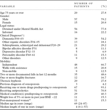
The demographic and clinical details are given in Table 1. Dementia (54%) and schizophrenia (29%) were the most common diagnoses. Seventeen patients were aged under 65 years, ten of whom had Huntington's disease. Regarding risk factors for osteoporosis, over a quarter were aged 75 years or more, over a third had a predisposing medical condition, and a fifth had a thoracic kyphosis. Almost all were receiving osteoporosis predisposing drugs: antipsychotics (N = 51), anticonvulsants (36), corticosteroids (4), and others (4). Only three of the 12 patients with a previous fragility fracture were receiving medication for osteoporosis.
Demographic and clinical characteristics of patients who underwent peripheral DEXA scanning (N = 72)

*ICD-10 Clinical Diagnoses (WHO, 1992).
**Of the 15 females, nine (60%) were post-menopausal.
†Patients could have more than one diagnosis.
Results of peripheral DEXA scanning
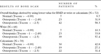
According to WHO criteria, 58.3% of patients were osteoporotic (males 56.1%; females 66.7%). Only 9.7% had a normal scan result. Of the ten patients with Huntington's disease, three had osteoporosis and three osteopenia. Osteoporosis was detected more frequently at the wrist than the calcanium (57.4% vs 31.4%; see Table 2).
Numbers of patients with osteopenia and osteoporosis according to peripheral DEXA scan (N = 72)

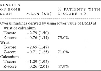
The overall mean T-score was −2.79 and 75% of patients had a negative Z-score. Mean calcaneal Z-score (0.26) was above zero and higher than that for the wrist (−0.71) (see Table 3).
T-score and Z-score results (N = 72)

Patient variables associated with fragility fractures
Patients aged 75 years and over were more likely to have had a fragility fracture than younger patients (8/20 (40.0%) vs 4/52 (7.7%) Fisher Exact 2-tailed P < 0.005), as were patients with thoracic kyphosis (10/20 (50.0%) vs 2/52 (3.9%) χ2 = 22.2, P < 0.0001). The number of falls in the past year, immobility, female gender, drugs or medical conditions predisposing to osteoporosis, recent weight loss and/or BMI < 22 were not associated with fragility fractures.
Patient variables associated with osteoporosis on DEXA scan
Significantly more patients with a history of fragility fracture had osteoporosis on DEXA scan than those without (12/12 (100.0%) vs 30/60 (50.0%), χ2 = 10.3, P = 0.01), as did those aged 75 years or over (16/20 (80.0%) vs 26/52 (50.0%), χ2 = 5.35, P < 0.05) compared with younger patients. More patients with thoracic kyphosis had osteoporosis than those without (16/20 (80.0%) vs 26/52 (50.0%), χ2 = 5.35, P < 0.05), as did those receiving drug(s) predisposing to osteoporosis (40/42 (95.2%) vs 23/30 (76.7%), Fisher Exact2-tailed P < 0.005). Patients with osteoporosis had a higher median length of stay (four years vs three years, Mann-Whitney Z = 2.14, P < 0.05). There was no significant correlation between patient age and length of hospital stay or between number of falls and T-scores. There were no significant associations between osteoporosis and gender, recent weight loss or BMI < 22, mobility status, medical conditions that predispose to osteoporosis or a diagnosis of schizophrenia.
Relationship of patient variables to Z-scores
Patients receiving one or more osteoporosis predisposing drugs had a lower mean Z-score than those not receiving such drugs (T = 2.35, P < 0.05) but no association was found specifically for antipsychotics or anticonvulsants. Those with thoracic kyphosis showed a trend towards a lower mean Z-score but the result was not quite statistically significant (T = 2.00, P = 0.05). Length of hospital stay and Z-score were negatively correlated (Pearson's correlation = −0.31, P < 0.01). No other significant associations were found between patient variables and Z-scores.
Patients' experience of undergoing a bone scan
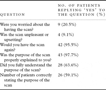
Prior to scanning, patients were given a standardized verbal explanation of the purpose of the scan and what they were likely to experience. A total of 44/72 (61.1%) patients were judged to have the capacity to consent to the scan and were questioned about it (Table 4).
Patients' responses to questions about the DEXA scanning process (N = 44)

A fifth of patients reported being worried about having the scan, though few said it was unpleasant or upsetting and most said they would be willing to have another. Only half of those with capacity correctly described the scan's purpose.
Management of patients with osteoporosis and osteopenia
Patients with osteoporosis or osteopenia on DEXA scan were managed according to the osteoporosis guidelines published by the National Institute for Health and Clinical Excellence (2005). Those with osteopenia were referred for a specialist opinion (consultant physician or radiologist) and for a central DEXA scan and those with osteoporosis for drug therapy, e.g. calcium and vitamin D, bisphosphonates, etc. Patients of Townsend Division routinely undergo falls risk assessment and formulation of a management plan to minimize identified risks. Falls are monitored and physiotherapists provide exercise programs. A dietician monitors BMI and patients' food intake.
At follow-up, 68/72 of the scanned patients were still inpatients. For 16 a central DEXA scan was indicated but nine (56.3%) were considered too behaviorally disturbed. Bisphosphonates were indicated for 40 patients but only nine (22.5%) were receiving them, either because of dyphagia or because they could not remain vertical for 30 minutes after drug administration.
Discussion
In this survey of mainly male older adults, 58% had osteoporosis and 32% osteopenia. For those patients aged over 65 years, 70% had osteoporosis on peripheral DEXA scan. Interestingly, three of the ten Huntington's disease patients also had osteoporosis. Mean Z-scores were substantially higher than T-scores, indicating that much of the osteoporosis was age related rather than having a secondary cause. A history of fragility fractures was fairly common (17%) but only a quarter of these patients were receiving drug treatment for osteoporosis. Most of the variables we found associated with osteoporosis – age of 75 years or over, thoracic kyphosis (Ensrud et al., Reference Ensrud1997) and fragility fractures (Center et al., Reference Center, Nguyen, Schneider, Sambrook and Eisman1999; Boonen et al., Reference Boonen, Vanderschueren, Haentjens and Lips2006) – have been described by previous authors. We also found that greater length of hospital stay was associated with osteoporosis. This finding could not be explained by length of stay being confounded by patient age. It may relate to reduced physical activity and limited access to sunlight in our patients. Analysis of variables associated with Z-scores showed length of stay to be negatively correlated with bone mineral density, as were osteoporosis-inducing drugs.
Comparing our findings with those of others is difficult, given the use of different types of scanners, different definitions of osteoporosis and differing age and gender characteristics of study populations. Compared with the findings of a large U.K. general population sample of similar age, our patients' Z-scores were substantially lower (Holt et al., Reference Holt2002). The prevalence of osteoporosis in institutionalized populations is much higher but subjects are older and few studies report Z-scores. Aspray et al. (Reference Aspray, Stevenson, Abdy, Rawlings, Holland and Francis2006) studied care home residents with dementia (mean age 85 years) and found 69% to be osteoporotic. Among elderly (mean age 82 years) institutionalized Canadian patients with dementia, 95% had osteoporosis (Juby, Reference Juby2004). Both institutionalized and community patients with learning disabilities have also been found to have markedly reduced BMD (Aspray et al., Reference Aspray, Francis, Thompson, Quilliam, Rawlings and Tyrer1998; Tyler et al., Reference Tyler, Snyder and Zyzanski2000). Risk factors include vitamin D deficiency, anticonvulsant drugs, hypogonadism and reduced physical activity. Many of our patients had schizophrenia and most were male. Older males with schizophrenia are at risk of developing secondary osteoporosis from long-term antipsychotics and have greater mortality from hip fractures than females (Meyer and Lehman, Reference Meyer and Lehman2006). We found no association between schizophrenia or antipsychotics and osteoporosis but patient numbers were small.
Despite high levels of positive symptoms and disturbed behavior, it was possible to scan over 80% of our patient population. Only 20% of patients said they were anxious about having the scan and 95% said they would be willing to be scanned again. Other authors have reported successful peripheral scanning rates of 74–100% in nursing home residents (Juby, Reference Juby2004; Aspray et al., Reference Aspray, Stevenson, Abdy, Rawlings, Holland and Francis2006) but these populations are likely to be less disturbed than ours.
In the follow-up study, less than half the patients were deemed suitable to undergo a central scan and less than a quarter of patients were receiving the indicated drug treatment for osteoporosis, mainly due to problems with bisphosphonate administration. For these patients other drugs for osteoporosis should be considered, such as strontium ranelate and calcitonin nasal spray.
The study has a number of important limitations. There was no control group with which to compare patients because the screening of patients was a service development rather than a research study. The number of patients scanned was relatively small with few women, and so separate gender analysis was not possible. With larger patient numbers more associations with low BMD would probably have emerged. We did not use a standard risk factor tool, such as FRAX (Kanis et al., Reference Kanis, Johnell, Oden and McCloskey2008) to identify those patients at highest risk of osteoporosis, nor did we measure vitamin D levels, or assess dietary calcium. Fragility fractures are likely to have been under-reported since we relied on case note information. The study was conducted in a tertiary referral centre, so the findings lack generalizability to other institutional populations. Mean T-scores were lower at the wrist than calcanium. In our analysis we used the lower of the two scores as indicating BMD but this discordance made interpretation of our results in relation to other studies difficult. Discordance between peripheral and central scan results is problematic (Moayyeri et al., Reference Moayyeri, Soltani, Tabri, Sadatsafavi, Hossein-neghad and Larijani2005; Melton et al., Reference Melton2005).
Despite the high prevalence of osteoporosis and fragility fractures in residents of long-term care facilities, several studies have reported low levels of treatment (Duque et al., Reference Duque2006; Wright, Reference Wright2007). In our survey, the low proportion of patients deemed suitable to undergo a central scan or treatment with bisphosphonates suggests that the management of this patient group is not straightforward. Difficulties with using oral bisphosphonates to treat osteoporosis can now be circumvented by their once-yearly administration as an intravenous infusion (Black et al., Reference Black2007). However, a proportion of our patients would also have been unsuitable for an intravenous infusion due to agitation and aggression. For such patients other antiresorptive therapies, such as strontium ranelate, could be used. The prevention of osteoporosis and fragility fractures also involves ongoing monitoring of risk factors with risk reduction where possible, falls risk assessment and prevention, exercise programs, assessment of calcium intake and vitamin D levels, and calcium and vitamin D supplementation. Detection and treatment of causes of secondary osteoporosis is important and may require referral to a specialist bone clinic.
Conflict of interest
None.
Description of authors' roles
Mr. Stubbs collected the data and contributed to the manuscript. Dr. Haw analyzed the data and wrote the paper. Dr Zapata-Bravo designed the study and contributed to the paper.