INTRODUCTION
Sea anemones (order Actiniaria) are among the benthic invertebrate groups inhabiting the coral reefs environments along the Caribbean Sea. Although actiniarians have been studied since the 18th Century in the Caribbean Sea, thorough taxonomic research of sea anemones meeting modern standards in the Mexican Caribbean began very recently (González-Muñoz et al., Reference González-Muñoz, Simões, Sánchez-Rodríguez, Rodríguez and Segura-Puertas2012). Fourteen species have been previously recorded for the Mexican Caribbean (INE, 1998a, b, 2000; Jordán-Dahlgren, Reference Jordán-Dahlgren and Mejía2008; González-Muñoz et al., Reference González-Muñoz, Simões, Sánchez-Rodríguez, Rodríguez and Segura-Puertas2012), but only ten of them have been taxonomically diagnosed (González-Muñoz et al., Reference González-Muñoz, Simões, Sánchez-Rodríguez, Rodríguez and Segura-Puertas2012). In the present study, we document the first records of Homostichanthus duerdeni (Carlgren, Reference Carlgren1900) and Anemonia sargassensis Hargitt, Reference Hargitt1908 in the Mexican Caribbean. Homostichanthus duerdeni is taxonomically diagnosed and images of living specimens, internal anatomy and cnidae are provided. Anemonia sargassensis was taxonomically diagnosed in a recent study of sea anemones of coral reefs in the southern Gulf of Mexico (González-Muñoz et al., Reference González-Muñoz, Simões, Tello-Musi and Rodríguez2013), but here we extended its geographical distribution range to the Mexican Caribbean. In addition, the taxonomic status of previous records of actiniarians in the Mexican Caribbean is discussed and updated.
MATERIALS AND METHODS
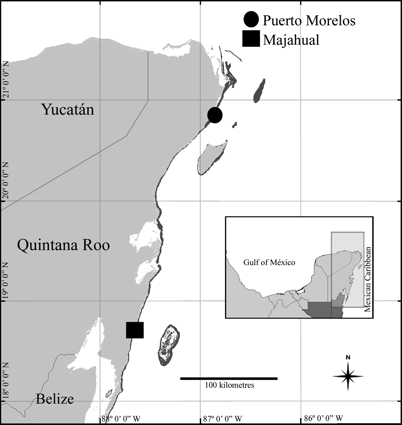
Observations and collections of specimens were made in Puerto Morelos and the Majahual reefs (Figure 1) during March 2013. Specimens were collected during day, by hand and snorkelling, using a hammer and chisel. Collected specimens were transferred to the laboratory and maintained in an aquarium to photograph their colour in life. Specimens were relaxed in 5% MgCl2 seawater solution and fixed in 10% seawater formalin. Squash preparations of small amounts of tissue from tentacles, actinopharynx, filaments, column and marginal projections were made from two preserved specimens of Homostichanthus duerdeni to study the cnidom. Cnidae were examined using a Nikon Labophot-2 light microscope (1000× oil immersion), photographed and haphazardly measured. Cnida terminology follows Mariscal (Reference Mariscal, Muscatine and Lenhoff1974) and Östman (Reference Östman2000). Measurements of specimens were obtained from living and relaxed specimens. Histological sections of 8 μm were made from two specimens of H. duerdeni, and stained with hematoxylin and eosin (Estrada et al., Reference Estrada-Flores, Peralta and Rivas1982). External anatomy and dissections were examined from specimens of Anemonia sargassensis for identification. Specimens were deposited in the Collection of the Gulf of Mexico and Mexican Caribbean Sea (Registration code: YUC-CC-254-11) of Unidad Multidisciplinaria de Docencia e Investigación en Sisal (UMDI-Sisal) at the Universidad Nacional Autónoma de México (UNAM). Taxonomic classification follows Rodríguez et al. (Reference Rodríguez, Barbeitos, Brugler, Crowley, Grajales, Gusmão, Häussermann, Reft and Daly2014).
Map of the Mexican Caribbean, indicating locations of specimens reported in this account.

RESULTS
SYSTEMATICS
Order ACTINIARIA Hertwig, Reference Hertwig1882
Suborder ENTHEMONAE Rodríguez & Daly in Rodríguez et al. (Reference Rodríguez, Barbeitos, Brugler, Crowley, Grajales, Gusmão, Häussermann, Reft and Daly2014)
Superfamily ACTINIOIDEA Rafinesque, Reference Rafinesque1815
Family ACTINIIDAE Rafinesque, Reference Rafinesque1815
Genus Anemonia Risso, Reference Risso1826
Anemonia sargassensis Hargitt, Reference Hargitt1908
(Figure 2A–C)
Anemonia sargassensis Hargitt, Reference Hargitt1908, pp. 117–118.
Anemonia sargassensis Hargitt, Reference Hargitt1908: (A) oral view; (B) pedal disc view; (C) lateral view. Abbreviations: (t) tentacles; (od) oral disc; (pd) pedal disc; (mp) marginal projections; (c) column. Scale bar = 10 mm.
Anemonia antillensis Pax, Reference Pax1924, p. 94, pp. 99–100, p. 119.
Anemonia sargassiensis [sic]: Carlgren, Reference Carlgren1949, p. 50.
MATERIAL EXAMINED
Puerto Morelos reef (20°52.09′N 86°52.02′W; three specimens).
DIAGNOSIS
Oral disc 8–25 mm in diameter, smooth, wider than column, reddish to dark orange with whitish or yellow radial stripes from the periphery of the mouth to the base of the tentacles (Figure 2A). Tentacles irregularly arranged in four or five cycles, inner cycles longer than outer ones, relatively long, smooth, slender, tapering distally, reddish to dark-orange with yellowish tips and sometimes with pink flashes (Figure 2A–C). Pedal disc 10–20 mm in diameter, reddish or bright to dark orange (Figure 2B). Column cylindrical, short, smooth, reddish to dark orange, with marginal projections poorly marked (Figure 2C) forming acrorhagi with holotrichs and basitrichs.
For further information on external and internal anatomical details and cnidae see González-Muñoz et al. (Reference González-Muñoz, Simões, Tello-Musi and Rodríguez2013).
REMARKS ON GEOGRAPHICAL DISTRIBUTION
Anemonia sargassensis has been reported in the Northern Atlantic coast of the United States (Hargitt, Reference Hargitt1908; Field, Reference Field1949), the North-western and Southern Gulf of Mexico (Carlgren & Hedgpeth, Reference Carlgren and Hedgpeth1952; González-Muñoz et al., Reference González-Muñoz, Simões, Tello-Musi and Rodríguez2013), Cuba (Varela, Reference Varela2002), Jamaica (Fisher, Reference Fisher1976), Curaçao (Pax, Reference Pax1924), Venezuela (Agudo, Reference Agudo1987) and in Brazil (Zamponi et al., Reference Zamponi, Belém, Schlenz and Acuña1998). This contribution represents the first record of A. sargassensis for the Mexican Caribbean. The nearest previous record of this species in the Caribbean Sea is in Caleta de los Piojos, Cuba (Varela, Reference Varela2002), about 230 km away from Puerto Morelos reef. This species inhabits the underside of rocks and coral rubble of the lagoon reef zone, between 0.5 and 1.5 m.
Family HOMOSTICHANTHIDAE Duerden, Reference Duerden1900
Genus Homostichanthus Duerden, Reference Duerden1900
Homostichanthus duerdeni (Carlgren, Reference Carlgren1900)
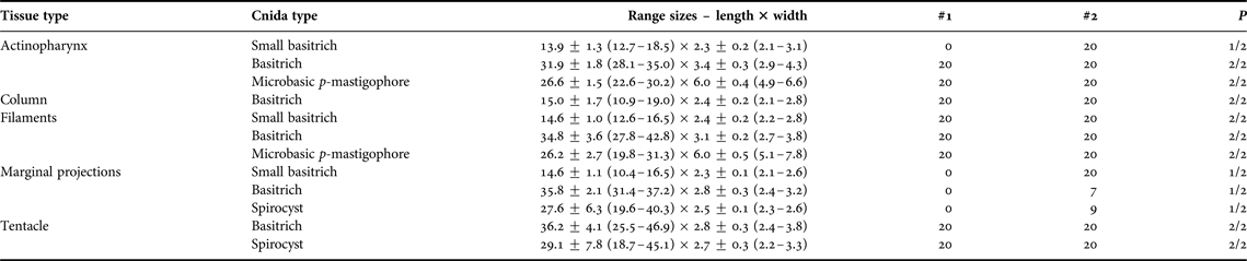
(Figure 3A–U; Tables 1 & 2)
Homostichanthus Duerdeni [sic] Carlgren, Reference Carlgren1900, pp. 117–118.
Homostichanthus duerdeni (Carlgren, Reference Carlgren1900): (A) oral view showing the endocoelic and exocoelic rows of tentacles; (B) lateral view, detail of margin and marginal projections; (C) lateral view; (D) cross-section at the actinopharynx level; (E) cross-section through proximal column showing the retractor and parietobasilar muscles; (F) cross-section through proximal column, detail of oocysts; (G) longitudinal section of proximal column, detail of basilar muscles; (H) longitudinal section through margin, detail of marginal projection; (I) longitudinal section through tentacle. Cnidom – Actinopharynx: (J) small basitrich; (K) basitrich; (L) microbasic p-mastigophore. Column: (M) small basitrich. Filament: (N) small basitrich; (O) basitrich; (P) microbasic p-mastigophore. Marginal projections: (Q) small basitrich; (R) basitrich; (S) spirocyst. Tentacle: (T) basitrich; (U) spirocyst. Abbreviations: (a) actinopharynx; (bm) basilar muscles; (c) column; (dm) directive mesenteries; (e) epidermis; (fo) fosse; (g) gastrodermis; (li) limbus; (lm) longitudinal muscles; (m) mesoglea; (mp) marginal projections; (o) oocysts; (od) oral disc; (pm) parietobasilar muscles; (rm) retractor muscles; (s) siphonoglyph; (t) tentacles. Scale: A–C = 10 mm; D–I = 200 μm; J–U = 25 μm.
Size and distribution of preserved cnidae from examined specimens of Homostichanthus duerdeni. Range size data are arranged as follows: Length mean ± SD (length min–max) × width mean ± SD (width min–max); all measurements are in μm; ‘#1’ and ‘#2’ is the number of capsules measured per specimen examined; ‘P’ is the ratio of number of specimens in which cnidae are found to number of specimens examined.
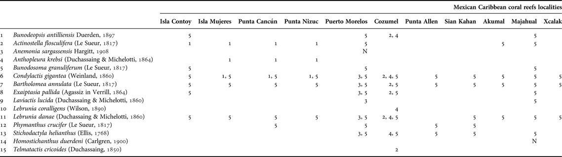
Distribution of sea anemones recorded in the coral reefs of the Mexican Caribbean. Citations: (1) INE (1998a); (2) INE (1998b); (3) INE (2000); (4) Jordán-Dahlgren (Reference Jordán-Dahlgren and Mejía2008); (5) González-Muñoz et al. (Reference González-Muñoz, Simões, Sánchez-Rodríguez, Rodríguez and Segura-Puertas2012); (N) new records.
Homostichanthus anemone: Duerden, Reference Duerden1900, p. 142, pp. 167–173; Pl. X, Figure 8; pl. XII, Figures 4–6; pl. XIV, Figure 2; pl. XV, Figure 1.
Homostichanthus duerdeni: Duerden, Reference Duerden1902, pp. 365–367.
Homosticanthus duerdeni: Herrera-Moreno, Reference Herrera-Moreno1981, p. 2.
Stichodactyla duerdeni: Zamponi et al., Reference Zamponi, Belém, Schlenz and Acuña1998, p. 94, 38, 40.
Actinoporus elegans: Human & Deloach, Reference Human and Deloach2002, pp. 92–93.
Stychodactyla duerdeni: Wirtz et al., Reference Wirtz, Melo and Grave2009, p. 6.
MATERIAL EXAMINED
Majahual reef (18°41.01′N 87°43.15′W; two specimens).
DIAGNOSIS
Oral disc 54–72 mm in diameter, wider than column, grey or white with brown and bright green spots (Figure 3A). Margin crenulated, with a row of very reduced marginal projections (Figure 3B); fossa poorly marked. Tentacles very numerous (>1000), very short, blunt, digitiform, slightly capitated, arranged in radial endocoelic and exocoelic rows covering almost the entire oral disc surface; each endocoelic row with 14–22 tentacles, exocoelic rows with 4–6 tentacles, restricted to outermost portion of oral disc (Figure 3A). Tentacles white or grey distally, beige proximally (Figure 3B), some tentacles with bright green spots. Column cup-shape, long, 22–30 mm in diameter and 18–58 mm in height; smooth, pale brown distally, pale orange in the mid part and bright orange proximally (Figure 3C). Pedal disc well-developed, 22–32 mm in diameter, bright orange. Mesenteries hexamerously arranged in 4–5 cycles: first and second cycle perfect, others imperfect; more mesenteries distally than proximally. Two pairs of directives, each attached to a well-developed siphonoglyph (Figure 3D). Retractor muscles strong and restricted (Figure 3E); parietobasilar muscles well-developed with free mesogleal pennon (Figure 3E). Gonochoric (?): oocysts 0.28–0.44 mm in diameter in strongest mesenteries of first cycle (Figure 3F). Basilar muscles well-developed (Figure 3G). Marginal sphincter muscle not observed in specimens examined (Figure 3H). Longitudinal muscles of tentacles ectodermal (Figure 3I). Zooxanthellae present. Cnidom: basitrichs, microbasic p-mastigophores and spirocysts (Figure 3J–U; Table 1).
NATURAL HISTORY
Homostichanthus duerdeni inhabits seagrass patches formed by Thalassia testudinum, often between 0.5 and 2 m, in the lagoon reef zone. It is usually found with the column completely burrowed in the sand, but with the pedal disc actually adhered to burrowed rocks or shells. It has been reported in symbiotic association with Periclimenes rathbunae Schmitt, Reference Schmitt1924, Thor amboinensis (De Man, Reference De Man1888) and Thor manningi Chace, Reference Chace1972 (Mercado & Capriles, Reference Mercado and Capriles1982; Ritson-Williams & Paul, Reference Ritson-Williams and Paul2007).
DISTRIBUTION
Jamaica (Duerden, Reference Duerden1898; Carlgren, Reference Carlgren1900, Reference Carlgren1949), Brazil (Corrêa, Reference Corrêa1973; Belém, Reference Belém1980; Zamponi et al., Reference Zamponi, Belém, Schlenz and Acuña1998; Wirtz et al., Reference Wirtz, Melo and Grave2009), Cuba (Herrera-Moreno, Reference Herrera-Moreno1981), Curaçao (Human & Deloach, Reference Human and Deloach2002) and Venezuela (Agudo, Reference Agudo1987). This is the first record for Mexico, found in the Majahual reef.
REMARKS
Although we did not observe a marginal sphincter muscle in the specimens histologically examined, a very feeble endodermal marginal sphincter has been reported in Homostichanthus duerdeni (Duerden, Reference Duerden1900; Belém, Reference Belém1980; Dunn, Reference Dunn and Parker1982); Belém (Reference Belém1980) even pointed out that the marginal sphincter muscle of H. duerdeni is comparable in size with that present in Anemonia sargassensis (about 400 μm). In addition, Belém (Reference Belém1980) reported a series of three small, translucent and flattened marginal spherules corresponding with each exocoelic marginal space and containing basitrichs, spirocysts and probably atrichs; because they are very close to each other they are only distinguishable when specimens are fully expanded (Belém, Reference Belém1980). We observed the marginal projections as single reduced marginal spherules on each exocoelic marginal space (Figure 3B), containing spirocysts and two size categories of basitrichs (small and large). The measurements of cnidae fit well otherwise with those previously reported for H. duerdeni (Carlgren, Reference Carlgren1945; Belém, Reference Belém1980). Specimens reported by Belém (Reference Belém1980) present gametogenic tissue in all well-developed mesenteries from first cycle onwards, and have the oral disc olive to pale green, mouth with yellow or scarlet lips, column pink proximally and silver-grey distally, tentacles green to yellow or brownish, and pedal disc pale pink and translucent. Similarly, larger specimens than those in this study (oral disc of about 150–200 mm) have been reported in Brazil (Belém, Reference Belém1980; Wirtz et al., Reference Wirtz, Melo and Grave2009). We agree with Wirtz et al. (Reference Wirtz, Melo and Grave2009) in that the photograph in Human & Deloach (Reference Human and Deloach2002) labelled as Actinoporus elegans Duchassaing, Reference Duchassaing1850, actually depicts a specimen of H. duerdeni. According to Belém (Reference Belém1980), the internal anatomy of H. duerdeni may be irregular in having more than two siphonoglyphs and directive mesenteries due to longitudinal fission (Dunn, Reference Dunn and Parker1982).
DISCUSSION
In addition to Anemonia sargassensis and Homostichanthus duerdeni, 14 other species of sea anemones have been reported for the Mexican Caribbean (González-Muñoz et al., Reference González-Muñoz, Simões, Sánchez-Rodríguez, Rodríguez and Segura-Puertas2012). However, the taxonomic status of some of these previous records has been recently changed after the taxonomic revision of the family Aiptasiidae (Grajales & Rodríguez, Reference Grajales and Rodríguez2014). The species Aiptasia tagetes (Duchassaing & Michelotti, Reference Duchassaing and Michelotti1864) has been synonymized with Exaiptasia pallida (Agassiz in Verrill, Reference Verrill1864) (= Aiptasia pallida), and the genus Laviactis Grajales & Rodríguez, Reference Grajales and Rodríguez2014 has been erected to place the species Laviactis lucida (Duchassaing & Michelotti, Reference Duchassaing and Michelotti1860) (= Ragactis lucida) (Grajales & Rodríguez, Reference Grajales and Rodríguez2014). Thus, 15 species are currently reported for the Mexican Caribbean (Table 2), of which only 13 species have been taxonomically diagnosed in previous studies of sea anemones from coral reefs in the Mexican Atlantic (González-Muñoz et al., Reference González-Muñoz, Simões, Sánchez-Rodríguez, Rodríguez and Segura-Puertas2012, Reference González-Muñoz, Simões, Tello-Musi and Rodríguez2013). Although Anthopleura krebsi (Duchassaing & Michelotti, Reference Duchassaing and Michelotti1864) and Telmatactis cricoides (Duchassaing, Reference Duchassaing1850) (= Telmatactis americana) have been recorded in Parque Marino de Isla Mujeres-Punta Cancún-Punta Nizuc and Parque Marino de Cozumel, respectively (INE, 1998a, b), formal identification and taxonomic diagnosis of these two species are needed to confirm these records for the Mexican Caribbean. Further detailed studies of actiniarians along coral reefs and coastal environments are still needed to complete the distribution range of sea anemones in the Mexican Caribbean.
ACKNOWLEDGEMENTS
We thank the Comisión Nacional de Áreas Naturales Protegidas (CONANP) and the staff at the Parque Nacional Arrecife de Puerto Morelos for their helpful assistance. Maribel Badillo-Alemán, Gemma Martínez-Moreno and Patricia Guadarrama-Chávez (UMDI-Sisal) provided access and support to histological facilities and the microscopy lab; Alfredo Gallardo-Torres (UMDI-Sisal) and Alejandro Córdova (FESI-UNAM) helped in the field. All specimens were collected under consent of Mexican law, collecting permit approved by Comisión Nacional de Acuacultura y Pesca (ref. 07332.250810.4060). The first author is grateful to the Programa de Becas Postdoctorales of the Comisión Nacional de Ciencia y Tecnología (CONACyT; grant 232318). This work was partially supported by the project CONACyT–SEMARNAT (to N. Simões, grant 108285) and DGAPA–PAPIME–UNAM (to N. Simões, grant PE207210). Comments from Dr Fabián H. Acuña, Dr Agustín Garese and one anonymous referee improved this manuscript.