Introduction
Pneumocephalus is a common consequence of both trauma and skull surgery. Spontaneous pneumocephalus, however, is a rare disorder that occurs as a result of skull base defects, and a pressure gradient causing air to enter the cranium. These patients commonly present with headache, although they may also complain of cerebrospinal fluid (CSF) rhinorrhoea, weakness, sensory disturbance, vomiting, aphasia or visual disturbances.Reference Pennings, Liauw and Cremers1
Alien limb syndrome is a motor disorder defined by involuntary movements that occur in addition to, or instead of, a planned or willed movement; the patient is usually aware of these involuntary movements but is unable to control them.Reference Josephs and Rossor2 This is an uncommon phenomenon found in patients with lesions typically in the frontal lobe or the corpus callosum. These lesions are most commonly vascular or neoplastic, although alien limb syndrome has been reported in neurodegenerative conditions such as corticobasal degeneration.
We report a case of spontaneous pneumocephalus in which the presenting complaint was alien limb syndrome. This is, to our knowledge, the first reported case of alien limb syndrome due to pneumocephalus of any aetiology.
Case report
A 27-year-old woman presented with a three day history of being unaware of her left arm performing actions. For example, she described finding her left hand holding a cup she had previously put down. She subsequently developed reduced coordination in her left leg. She was otherwise well, with no other neurological, otological or rhinological symptoms. She had a history of a right-sided sinus operation as an 11-year-old, for sinusitis, which had a good effect and was without complication. She had no other past medical history. She had never Scuba-dived.
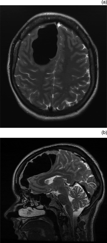
Neurological examination was normal apart from an upgoing plantar reflex on the left side and difficulty with tandem gait. She occasionally lifted her left arm up but then put it down when asked whether she had become aware of her actions. Magnetic resonance imaging demonstrated a 4 × 4 × 7 cm lesion with markedly low signal on T1- and T2-weighted sequences (Figure 1).
(a) Axial and (b) sagittal, T2-weighted magnetic resonance imaging scans demonstrating a large, balloon-like collection of intracranial air extending posterosuperiorly from the subfrontal region to cause marked distortion of the right frontal lobe.

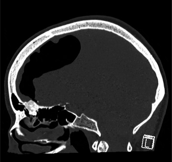
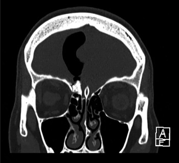
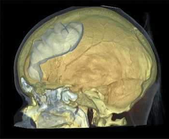
Computed tomography scanning demonstrated a large, gas-filled lesion in the right frontal lobe. The lesion measured 4 × 4 × 7 cm and was markedly hypodense. It appeared to originate from an enostosis and defect in the anterior skull base at the cribriform plate (Figures 2 to 4). The appearances were in keeping with an intraparenchymal pneumocephalus.
Subsequent sagittal computed tomography scan showing a well circumscribed, bony mass projecting from the junction of the right frontal and anterior ethmoid sinuses into the anterior cranial fossa. L = left

Coronal computed tomography scan showing the inferior ‘neck’ of the intracranial air collection converging on a small skull base defect lying along the lateral border of a bony mass, and communicating with the aerated sinuses. A = anterior

Volume-rendered, three-dimensional computed tomography representation demonstrating the intracranial gas collection.

The patient underwent right frontal craniotomy, at which stage a bony mass was excised, the pneumocephalus was aspirated, and a defect in the cribriform plate was repaired.
She made an excellent post-operative recovery. Her neurological symptoms were completely resolved by the first day after the operation. She was discharged one week after presentation.
Histopathological examination of the excised enostosis demonstrated normal lamellar bone.
Discussion
Pathophysiology
Pneumocephalus is caused by a defect in the integrity of the skull in combination with a pressure gradient that allows air to flow into the skull vault. These factors are commonly seen in traumatic skull fractures or during cranial surgery when the skull is deliberately opened. Markham et al. Reference Markham3 published a 1967 review of pneumocephalus which described 295 cases, for which trauma and surgery were frequent aetiological factors associated with 73.9 and 3.7 per cent, respectively.
Spontaneous pneumocephalus, however, occurs much less commonly. It is a rare disorder that arises due to defects of either the anterior or the lateral skull base, in combination with a state in which air flows into the cranium down a pressure gradient. There have been prior reports of spontaneous pneumocephalus in patients with osteomata eroding into the frontalReference Hamid, Huneidi and Afshar4 or ethmoidal sinus.Reference Bartlett5 Furthermore, pneumocephalus has been reported with other skull base and sinus disorders such as a pneumocoeleReference Añorbe, Aisa and Saenz de Ormijana6, Reference Park and Kim7 and pneumosinus dilatans.Reference Lee, Park, Kwon and Suk8 Non-traumatic pneumocephalus has also been described in cases of barotrauma,Reference Hermans, Bracke and Demeyer9, Reference Jarjour and Wilson10 including use of the Valsalva manouevre.Reference Babl, Arnett, Barnett, Brancato, Kharasch and Janecka11
In the presented case, it is difficult to assess the pathophysiological importance of the patient's prior sinusitis and sinus surgery. It is possible that surgical injury may have initiated an osteoblastic response, resulting in enostosis and a skull base defect.
Presentation
Pneumocephalus most commonly presents with headache,Reference Markham3 although symptoms may include weakness,Reference Hamid, Huneidi and Afshar4 meningitis, papilloedema and cranial nerve palsies.Reference Babl, Arnett, Barnett, Brancato, Kharasch and Janecka11 Due to the condition's rarity and variety of presentations, it may not initially be suspected, but may be readily identified on imaging.Reference Andrews and Canalis12
In the reported case, the presentation involved involuntary motor phenomena. These are most commonly seen in patients with frontal and callosal pathology such as strokes, tumours or neurodegenerative lesions.Reference Scepkowski and Cronin-Golomb13, Reference Spector, Freeman and Cheshire14 These reports correlate well with the current case, in which the mass effect exerted by the pneumocephalus had morphological features more commonly associated with a frontal intraparenchymal mass, rather than those associated with conventional sub- or extradural collections (as seen in the majority of pneumocephalus cases). It may well be the case that the location of our patient's pneumocephalus led to her dramatic presentation. Furthermore, the lack of headache, lack of a single identifiable trigger, and unusual manner of presentation may indicate a subacute collection of intracranial air.
Management
Due to the comparative rarity of cases, there is no clear consensus on the management of spontaneous pneumocephalus. Reported cases have generally had good outcomes, whether they were managed conservatively or invasively. The management of these cases varied depending on the volume and location of gas, the underlying aetiology, and the clinical presentation.
Examples of cases in which conservative management produced a good outcome include patients with an identifiable trigger which can be reversed, for example, a patient who repeatedly performed the Valsalva manoeuvreReference Schrijver and Berendse15 and a patient who repeatedly blew his nose.Reference Lee, Park, Kwon and Suk8 In these cases, there was no suspicion of meningism or an ongoing dural defect. However, in some patients with a reversible trigger, such as nose blowing, conservative treatment is insufficient to relieve symptoms, and craniotomy and repair may eventually be required.Reference Richards, Saeed, Laitt and Ramsden16, Reference Tucker, Miyake, Tsuji, Ukita, Nishihara and Ito17 Among the reported cases, craniotomy and repair were frequently necessary.
In addition to craniotomy for repair of these defects, there is some evidence to support alternative surgical approaches. In a series of 10 cases of pneumocephalus and CSF leakage following endoscopic sinus and microscopic skull base surgery, those patients who underwent conservative or endoscopic repair were examined.Reference DelGaudio and Ingley18 Those patients with larger deficits failed conservative treatment. Eight of the 10 patients were successfully treated endoscopically. One patient with otogenic pneumocephalus and a temporal bone pneumatocoele required antrostomy and sealing of the aditus,Reference Añorbe, Aisa and Saenz de Ormijana6 although otogenic pneumocephalus is more commonly managed with a middle fossa craniotomy.Reference Wilkinson, Meyer and Rubinstein19
• A case of intraparenchymal spontaneous pneumocephalus presented with alien limb syndrome
• Spontaneous pneumocephalus results from a skull base defect with consequent air pressure gradient
• Skull base defects can present in many unusual ways
• They should be part of the differential diagnosis of neurological symptoms
• Pneumocephalus symptomatology depends on adjacent cranial structures
In the current case, the clinical state of the patient and the intraparenchymal location of the collection precluded a conservative approach. In this patient, the choice of an endoscopic versus cranial approach could be debated. However, it was decided to employ a cranial approach, in order to fully relieve the air collection and to clearly excise the enostosis for histological analysis.
Conclusion
This patient had a dramatic presentation of pneumocephalus which required urgent treatment. This condition should be considered in the differential diagnosis of neurological phenomena reminiscent of stroke or space-occupying lesions. The management of pneumocephalus depends upon a number of factors, and should be decided on a case by case basis.
Acknowledgement
We would like to acknowledge the assistance of Drs Raj Kapoor and Arani Nitkunan.