Introduction
Antarctica is the coldest and most extreme region on Earth with an area of ~14 million km2, lacking surface liquid freshwater and with only ~0.3% ice-free lands (Kimble Reference Kimble2004). The temperature usually remains < 0°C except for during the short ‘summer’ period, when in some places the soil partially melts. Antarctica and its biomes are constantly exposed to extreme climatic conditions including low temperatures with wide fluctuations, high periodic radiation fluxes, desiccation, hypersalinity and low nutrient status (Cowan & Tow Reference Cowan and Tow2004). Due to these harsh conditions, microorganisms inhabiting this area must have extraordinary systems of adaptation for surviving in such an environment (O'Brien et al. Reference O'Brien, Sharp, Russell and Roller2004).
Glacier surfaces can be colonized by bacteria and algae (Massalski et al. Reference Massalski, Kostikov, Olech and Hoffmann2009, Rybalka et al. Reference Rybalka, Andersen, Kostikov, Mohr, Massalski, Olech and Friedl2009), as well as more complex organisms such as mosses and even higher plants, which in turn can be inhabited by the Tardigrada, Nematoda, Enchytraeidae and Arachnida (Foreman et al. Reference Foreman, Sattler, Mikucki, Porazinska and Priscu2007, Cameron et al. Reference Cameron, Hodson and Osborn2012). Early surveys of Antarctic wildlife using serological approaches showed the occurrence of Birnaviridae, Flaviviridae, Herpesviridae, Orthomyxoviridae and Paramyxoviridae viruses circulating in seals (Phocidae), penguins (Spheniscidae), petrels (Procellariidae), etc. These data were also recently confirmed and expanded using modern metagenomics techniques demonstrating sequences of adeno-, anello-, orthomyxo-, papilloma-, paramyxo-, polyoma- and togaviruses associated with many Antarctic animals (Smeele et al. Reference Smeele, Ainley and Varsani2018). Several experimental works also prove the occurrence of different plant viruses in Antarctica (Pearce & Wilson Reference Pearce and Wilson2003, Polischuk et al. Reference Polischuk, Budzanivska, Shevchenko and Oliynik2007).
Viruses are the most abundant and genetically diverse acellular species in the ocean. Virus detection is associated with bacterial abundance and/or activity (Weinbauer Reference Weinbauer2004). The presence of viable microflora in Antarctic ecosystems implies the possibility of phage preservation/survival and also suggests that many, if not most, viruses found in such environment are usually phages. It is well known that bacteriophages are the most numerous creatures on Earth, and they play an unrivalled ecological role in water and terrestrial ecosystems. They are recognized as an important factor in regulating the structure and population of microorganisms (Säwström et al. Reference Säwström, Lisle, Anesio, Priscu and Laybourn-Parry2008, López-Bueno et al. Reference López-Bueno, Tamames, Velázquez, Moya, Quesada and Alcamí2009). Recently, a total of 22,222 viral populations' epipelagic and mesopelagic double-stranded DNA (dsDNA), representing 867 viral clusters (in groups at approximately the genus level) have been isolated (Brum et al. Reference Brum, Hurwitz, Schofield, Ducklow and Sullivan2016). Metagenomic studies have revealed high viral diversity. Diverse single-stranded DNA (ssDNA) and dsDNA viruses have been found in Limnopolar Lake located on Livingston Island, West Antarctica. Limnopolar Lake recorded phenomenally high species richness, including 5130 viral genotypes in spring and as many as 9730 viral genotypes in summer (López-Bueno et al. Reference López-Bueno, Tamames, Velázquez, Moya, Quesada and Alcamí2009, Reference López-Bueno, Rastrojo, Peiró, Arenas and Alcamí2015). In the Sargasso Sea, 5140 genotypes were estimated, and in the Gulf of Mexico this number was 15,400; 106–108 virus particles ml-1 were found in the floating ice of the Ross Sea; 0.6–5.0 × 10⁵ virus particles ml-1 have been found in sea-ice samples in Prydz Bay, East Antarctica (Gowing et al. Reference Gowing, Riggs, Garrison, Gibson and Jeffries2002, Angly et al. Reference Angly, Felts, Breitbart, Salamon, Edwards and Carlson2006, López-Bueno et al. Reference López-Bueno, Tamames, Velázquez, Moya, Quesada and Alcamí2009, Paterson & Laybourn-Parry Reference Paterson and Laybourn-Parry2012). Importantly, the first cultivated phage-host systems have been recently isolated from 1 year and 2 year winter sea ice in the Weddell Sea (Luhtanen et al. Reference Luhtanen, Eronen-Rasimus, Oksanen, Tison, Delille and Dieckmann2018). The detection of RNA viruses in the Antarctic aquatic environment has been reported in Limnopolar Lake (López-Bueno et al. Reference López-Bueno, Tamames, Velázquez, Moya, Quesada and Alcamí2009). RNA viruses remain poorly understood at the poles. Polar water regions offer exciting opportunities for discovery and can indicate the role of viruses in evolution (Culley et al. Reference Culley, Lang and Suttle2006). Examination of the phages of the studied polar ecosystems, including soil, showed that they are mostly inhabited by bacteriophages of the Siphoviridae, Myoviridae and Podoviridae families (Zablocki et al. Reference Zablocki, van Zyl, Adriaenssens, Rubagotti, Tuffin, Cary and Cowan2014, Gong et al. Reference Gong, Liang, Wang, Jiang, Yang and Xia2018). However, most research is focused on water ecosystems, while our knowledge on soil biotopes remains very limited.
Currently, there is a growing interest in research of this kind. The predicted global warming makes it relevant to study the environmental effects of permafrost thawing. As a result of Antarctic glaciers melting and the thawing of permafrost ice, the environment is ‘contaminated’ with their microflora. Therefore, there is a possibility of dangerous infections of an unknown nature appearing due to the activation of ancient microflora, and also the transfer by bacteriophages of ancient virulence and pathogenicity factors stored in their genomes (Lavysh et al. Reference Lavysh, Sokolova, Minakhin, Yakunina, Artamonova and Kozyavkin2016). Monitoring the structure and condition of the phage component of microbial communities is also needed in order to assess the potential risks to the biosecurity of the Antarctic region (Paterson & Laybourn-Parry Reference Paterson and Laybourn-Parry2012).
In polar regions characterized by limited food chains, the role of the diversity of viruses in the functioning of the ecosystem is still relatively little studied. As in all studies of metaviromes, a large number of sequences cannot be taxonomically attributed - usually, viral sequences obtained from metagenomes do not correspond to sequences from known databases (López-Bueno et al. Reference López-Bueno, Tamames, Velázquez, Moya, Quesada and Alcamí2009). This number was noted to reach 91%, due to which this enormous genetic diversity has been named viral dark matter and, therefore, the mysterious virosphere (Siddharth & Krishnamurthy Reference Siddharth and Krishnamurthy2017). The study of phage-host systems provides valuable information about the microbial communities of Antarctica. To our knowledge, to date, only three virus-host systems have been isolated from the Arctic, three from Antarctica and seven from ice from the Baltic Sea (Borriss et al. Reference Borriss, Helmke, Hanschke and Schweder2003, Luhtanen et al. Reference Luhtanen, Eronen-Rasimus, Kaartokallio, Rintala, Autio and Roine2014, Yu et al. Reference Yu, Chen, Shen, Zhao, Tang and Su2015). Most research on the marine environment is carried out in the water column, while knowledge of viruses and their functions in sea ice and soil is still very limited (Luhtanen et al. Reference Luhtanen, Eronen-Rasimus, Oksanen, Tison, Delille and Dieckmann2018, Yau & Seth-Pasricha Reference Yau and Seth-Pasricha2019). The question remains unresolved as to whether polar viral species differ significantly from low-latitude ones or whether they differ between the poles. The notion of the great importance of viruses as the main ‘predators’ in various biomes, especially in polar lakes, where multicellular animals are absent, is of significant interest.
Here, we report on the isolation of cultured phage-host systems from soil samples of terrestrial biotopes collected from several islands in the vicinity of Ukrainian Antarctic Station (UAS) ‘Academician Vernadsky’. The study of phage-host systems provides valuable information regarding the interaction of individual microorganisms with bacteriophages in frozen soil and their potential role in the ecosystem.
Materials and methods
Sampling
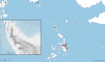
Moss and soil samples were collected during the 23rd Ukrainian Antarctic Expedition (2019) in the vicinity of UAS ‘Academician Vernadsky’ (former UK ‘Faraday’ station) (Fig. 1). Sampling was carried out at a fixed number of mapped reference sampling points common for the biological units of the expeditions, which allowed for a more accurate and controlled collection of lichen, moss and soil material, as well as the following timeline comparison of the results. The sampling sites and types of samples are listed in Table I.
a. Location of Ukrainian Antarctic Station (UAS) ‘Academician Vernadsky’ (indicated with arrow) on Argentina Islands and b. sampling locations on Irizar Island, Pitermann Island and Galindez Island (S1–S6 - sample numbers). For geographical coordinates, please see Table I. Original maps are generated using the Scientific Committee on Antarctic Research (SCAR) Antarctic Digital Database and Antarctic Digital Database map viewer resource (https://www.add.scar.org).

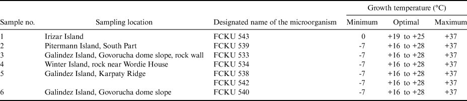
Location and type of collected Antarctic samples.

The samples were transferred into sterile Falcon tubes (Chongqing New World Trading Haimen, China) and stored at < 0°C in the field and during transport, and then at -80°C in the laboratory.
Bacteria cultivation and identification
All contamination-sensitive procedures were performed inside a laminar flow hood. For isolation of the microorganisms, 2 g of soil/substrate were suspended in 10 ml of sterile 0.9% NaCl solution and vortexed for 30 s.
The supernatant was serially diluted (10-1–10-6) and added to 100 ml of sterile 1/100 diluted (0.08 g l-1) Difco Nutrient Broth (Becton Dickinson, USA) and thoroughly mixed (Connon & Giovannoni Reference Connon and Giovannoni2002).
Afterwards, 1 ml of the resulting mixture was spread on plates with previously solidified 1.5% Difco Reasoner's 2A agar (R2A agar; Becton Dickinson, USA), beef extract agar (BEA; HiVeg), trypticase-soy agar (TSA; Sigma), nutrient agar (NA; Sigma), malt extract agar (MEA; Merck KGaA, Germany), MacConkey agar (Sigma), potato dextrose agar (PDA; Sigma), King A medium (KA medium; HiMedia Laboratories Pvt. Ltd, India) or King B medium (KB medium; HiMedia Laboratories Pvt. Ltd, India). Nutrient compositions were based on media previously used for the isolation of bacteria (Cho & Giovannoni Reference Cho and Giovannoni2004, Pulschen et al. Reference Pulschen, Bendia, Fricker, Pellizari, Galante and Rodrigues2017). Cultivation of Antarctic microorganisms was carried out at different temperatures from -7°C to +40°C (-10°C, -7°C, -4°C, 0°C, +4°C, +10°C, +16°C, +19°C, +25°C, +28°C, +37°C, +40°C) in the dark for up to 5 weeks. To avoid desiccation, plates were incubated inside polyethylene bags (Antibus et al. Reference Antibus, Leff, Hall, Baeseman and Blackwood2012, Orellana-Saez et al. Reference Orellana-Saez, Pacheco, Costa, Mendez, Miossec and Meneses2019). Bacterial colonies were chosen based on their varying morphological characteristics to avoid picking similar colonies that developed on the plates at the same time. For isolation, previously picked colonies were then streaked on new sterile 1/100 solid media plates. Then these plates were incubated at +20°C in the dark for 24 h (these incubation conditions were experimentally proven to be optimal). After isolation, cells were re-streaked on sterile 1/100 solid media plates and 1/10 solid media plates (10 times less diluted) to evaluate growth preferences. Where possible, cells were cultivated in liquid 1/10 and 1/100 broth to generate the biomass necessary for the following cryostock preparation. However, many isolates failed to grow in liquid media. In such cases, the colonies were recovered from the Petri dishes (Pulschen et al. Reference Pulschen, Bendia, Fricker, Pellizari, Galante and Rodrigues2017).
Cytological examination of the obtained bacterial cultures was performed by smearing with subsequent staining by Gram using a set of Gram dyes (Merck KGaA, Germany) following the manufacturer's instructions (Gregersen Reference Gregersen1978, Silhavy et al. Reference Silhavy, Kahne and Walker2010). The morphology of bacterial cells and spores was studied using a Primo Star (Germany) binocular microscope at low (×40) and high (×100) magnification. For high magnification, immersion oil was used (Bauman Reference Bauman2017). Photography of Gram-stained microorganisms and measurements of required parameters (cell and spore diameter, cell spacing, etc.) were performed using an Axio Vision 4.8 morphometric computer program and a PrimoStar Carl Zeiss binocular microscope (Germany).
Determination of physiological and biochemical characteristics and identification of pure cultures of isolated Antarctic bacteria were carried out using the microbiological analyser ‘Vitek-2’ (bioMérieux, France) with corresponding ID cards for Gram-positive, Gram-negative and spore-forming bacteria, as well as with MIKRO-LA-TEST diagnostic strips (LaChema, Czech Republic) following the manufacturer's instructions as described previously (Spanu et al. Reference Spanu, Sanguinetti, Ciccaglione, D'Inzeo, Romano, Leone and Fadda2003).
Determination of the aerobic respiration or fermentation ability of isolated Antarctic microorganisms was performed with the oxidation-fermentation (OF) test using Hugh Leifson OF-glucose medium (Cowan & Steel Reference Cowan and Steel1973). The presence of terminal cytochrome c-oxidase in the respiratory chain of bacteria (oxidase test) was determined using the Bactident Oxidase test system (Merck KGaA, Germany) as described previously (Hemraj et al. Reference Hemraj, Sharma and Gupta2013) with the addition of appropriate reagents (tetra-methyl-p-phenylenediamine dihydrochloride (TMPD) and N,N-dimethyl-p-phenylene diamine hydrochloride (DMPD)) to the medium.
Determination of the nitrate respiration ability of isolated Antarctic microorganisms (nitrate reduction test) was performed using nitrate broth and Grissom reagent (Cook Reference Cook1950).
The effect of molecular oxygen on bacteria was studied using thioglycol medium (Leijh et al. Reference Leijh, van Zwet, ter Kuile and van Furth1984).
To test Antarctic microorganisms for catalase production, a 3% solution of hydrogen peroxide was used (Taylor & Achanzar Reference Taylor and Achanzar1972).
Bacterial motility was determined using Motility Test Medium (HiMedia Laboratories Pvt. Ltd, India) (MacFaddin Reference MacFaddin1985).
The ability of studied bacteria to form soluble pigments was determined using KA medium (Pseudomonas agar (for fluorescein)) and KB medium (Pseudomonas agar (for pyocyanin)) (HiMedia Laboratories Pvt. Ltd, India) (Wasserman Reference Wasserman1965).
The bacterial ability to hydrolyse gelatin was determined using M9 saline medium with 0.4% gelatin. Gelatin hydrolysis was determined via the development of cleared zones around bacterial colonies (Miller Reference Miller1972).
Bacteriophage isolation
Samples were homogenized in sterile mortars with autoclaved 0.1 M phosphate-buffered saline, pH 7.4 (1:2, w/v), at 4°C (Sambrook & Russell Reference Sambrook and Russell2001). To isolate the viruses, bacterial strains were used that could form a bacterial lawn. For further characterization, the phages were individually plaque-purified three times on agar plates (using viruses from only one plaque at a time). The plaques were placed in individual sterile tubes and filled with buffer. Next, 100 ml of each sample (biologically ‘clean’ line) was mixed with 100 ml of a nutrient broth (meat peptone broth; Uglich, Russia) of a four-fold concentration and with 8 ml of broth with host bacterial culture (previously isolated from this sample), and then incubated for 6 h at 20°C with constant aeration (Spencer Reference Spencer1960).
The resulting supernatant was centrifuged at 1600 g for 10 min using an OPTIMA L-90K centrifuge (Beckman Coulter, Japan) and then passed through a 0.22 μm membrane filter (Chongqing New World Trading Haimen, China). Virus particles were recovered from the resulting filtrate by centrifugation at 43,667 g for 6 h in autoclaved 30 ml Nalgene polypropylene copolymer tubes (Thermo Fisher Scientific, MA, USA) using an OPTIMA L-90K centrifuge. The sediments from individual tubes were further successively re-suspended as follows: each sediment was re-suspended in 3 ml of Tris-EDTA (TE) buffer. The resulting mixture was then transferred to the next sterile tube and the sediment was again thoroughly re-suspended in 1 ml of ТЕ buffer (Hansen et al. Reference Hansen, Tolker-Nielsen, Givskov and Molin1998, Adriaenssens et al. Reference Adriaenssens, Van Zyl, De Maayer, Rubagotti, Rybicki, Tuffin and Cowan2015). After this procedure, the presence of any phages active against particular bacteria was detected by plating and plaque formation, as described below.
Bacteriophage titration
The titre of each phage isolated from collected samples, expressed as plaque-forming units (pfu), was determined using the double-layer plaque (DLA) technique as described by Sambrook & Russell (Reference Sambrook and Russell2001). Briefly, phage stock (100 μl) was mixed with 200 μl of respective host bacterial culture (3 × 10⁷–1 × 10⁸ colony-forming units (cfu)/ml). The resulting mixture was added to 4 ml of top 0.7% R2A agar (melted and cooled to 35°C), gently homogenized and poured into a sterile 90 mm Petri dish (Plastiques-Gosselin, France) previously filled with 10 ml of solidified bottom 1.5% R2A agar. The plates were gently swirled, dried for 10 min at room temperature in sterile conditions, inverted and incubated at 20°C in the dark for up to 24 h. Phage titre was expressed as pfu/ml and determined by following formula (Adams Reference Adams1959):
 $${\rm pfu}/{\rm ml\ } = {\rm Number\ of\ plaques\ } \times {\rm Dilution\ factor}$$
Evaluation of the spectrum of lytic activity of bacteriophages
To determine the spectrum of lytic activity of isolated bacteriophages towards various strains of microorganisms, we used Antarctic bacteria isolated in this study (six isolates) and the following laboratory bacterial isolates: Clavibacter michiganensis, Enterobacter cloacae, Erwinia carotovora, Escherichia coli, Pseudomonas fluorescens 8573, Pseudomonas syringae pv. lachrymans 7591, Pseudomonas savastanoi pv. phaseolicola 4013, Serratia marcescens and Xanthomonas campestris (nine isolates). Phage stock (100 μl) was mixed with 200 μl of bacterial culture (3 × 10⁷–1 × 10⁸ cfu/ml). The resulting mixture was added to 4 ml of top 0.7% R2A agar (melted and cooled to 35°C), gently homogenized and poured into a sterile 90 mm Petri dish (Plastiques-Gosselin, France) previously filled with 10 ml of solidified bottom 1.5% R2A agar. Sterile (phage- and bacteria-free) R2A medium was used as a negative control. The dishes were incubated at 20°C for 1–14 days. All positive results were further confirmed with a plaque assay, as described above (Kutter Reference Kutter2009).
Transmission electron microscopy of virus-like particles
Transmission electron microscopy (TEM) for studying the morphology of virus-like particles (VLPs) was conducted employing standard techniques of negative contrasting for clarified virus preparations.
Briefly, copper grids (Sigma, USA) were coated with chloroform-dissolved 0.2% polyvinyl formaldehyde (Serva, Germany) and dried overnight on filter paper at room temperature. Then, the coated grids were deposited in the clarified homogenate of the final sample filtrates, incubated for 2–15 min at room temperature and dried on filter paper for 1 min. The samples deposited onto grids were stained with 2.5% uranyl acetate (Serva, Germany) and examined using a JEM 1400 (JEOL, Japan) transmission electron microscope. The samples were photographed at a magnification of 5000–60,000× (Ackermann Reference Ackermann2009).
Results
This work was focused on the isolation and characterization of certain microorganisms and their tentative viruses from the samples collected in the vicinity of UAS on the Argentine islands in Antarctica (see Table I). The paper describes bacteria recovered from the collected samples and experimentally established optimal conditions for their cultivation, along with the bacteriophages that have been isolated and particularly the interactions of the counterparts in the phage-host system.
The cultivation experiments resulted in the successful growth of several bacterial cultures that were subsequently designated as FCKU 543, FCKU 539, FCKU 533, FCKU 534, FCKU 538, FCKU 542 and FCKU 540 (Table II). ‘FCKU’ historically refers to Fungi Collection of Kyiv University, which was later expanded to include bacteria as well (see details in Table III). The growth temperatures (minimal, optimal and maximal) were determined for each of the isolated microorganisms. All cultures were cultivable at 0°C. Seven cultures showed obvious signs of growth when incubated at -7°C (the lowest temperature tested in this study) for 60 days. Optimal growth temperatures were determined by judging the fastest cell growth when incubated for 1–3 days. For most of these bacteria, the optimal temperature was in the range of 16–28°C.
Extreme and optimal temperatures for the cultivation of bacteria isolated from the Antarctic samples.

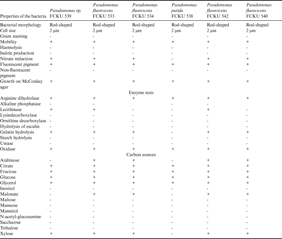
Cultural, morphological and biochemical properties of the bacteria isolated from the Antarctic samples.

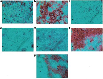
The cultural, morphological and biochemical features of these microorganisms were further established. The culture of FCKU 543 (from sample 1; Table II) was isolated, propagated and stored on BEA medium. After 24 h of cultivation at 20°C under aerobic conditions, microorganisms formed round colonies with even edges ~2–3 mm in diameter. The colonies had raised profiles with a soft pasty texture, homogeneous structure and smooth matt surface. FCKU 543 produced an insoluble lemon-yellow pigment, forming a kind of halo in the agar surrounding the bacterial colonies. Microscopy studies showed spherical Gram-positive cells without spores or capsules (Fig. 2a). The cells were arranged separately or formed cubic aggregates. Due to the cultivation in BEA medium, the bacterial culture was uniformly cloudy with a slightly flocculent precipitate. The bacteria were also able to grow on NA, TSA and PDA media. FCKU 543 bacteria were catalase-positive, aerobic and could not ferment glucose or mannitol. Their optimum temperature was in the range of 19–25°C; however, bacterial growth was also observed at 0–37°C. Based on the experimentally established cultural, morphological and biochemical properties, and according to Bergey's Manual of Systematic Bacteriology, the isolated FCKU 543 bacterium can be attributed to the Micrococcae family. However, the FCKU 543 isolate was subsequently lost due to technical reasons, and hence was not used further in this research.
Morphology of bacterial cells. a. Micricoccus sp. FCKU 543, b. Pseudomonas sp. FCKU 539, c. Pseudomonas fluorescens FCKU 533, d. Pseudomonas fluorescens FCKU 534, e. Pseudomonas putida FCKU 538, f. Pseudomonas fluorescens FCKU 542 and g. Pseudomonas fluorescens FCKU 540 (Gram staining, magnification 1600×).

The cultures of FCKU 539, FCKU 533, FCKU 534, FCKU 538, FCKU 542 and FCKU 540 (from samples 2–6; Table II) were isolated and cultured on NA medium at 20°C for 1 day. All of these microorganisms formed similar round superficial transparent greyish colonies with uneven edges reaching ~1–2 mm (24 h incubation) and 3–6 mm (48 h incubation) in diameter. The colonies had flat profiles with a homogeneous structure, a smooth shiny surface and a mucous texture. After 18 h of cultivation, the colonies became opalescent in transient light. Microscopy investigations revealed motile Gram-negative rod-shaped cells with rounded ends, reaching ~1.5–3.0 μm in length (Fig. 2b–g). Such cells were arranged separately or in pairs and had no capsules or spores. Cultivation of these bacteria in meat peptone broth produced sediment-free, uniformly cloudy solutions with thin smooth films. The bacteria were also able to grow on potato agar, TSA and M9 media.
When cultivated on KA medium, FCKU 539, FCKU 533, FCKU 534, FCKU 538, FCKU 542 and FCKU 540 isolates did not produce soluble pigments, whereas soluble yellow-green fluorescent pigments were produced if grown on KB medium for 3–6 days at 20°C. All of these bacteria were aerobic, catalase- and oxidase-positive, able to utilize citrate and did not produce either indole or hydrogen sulphide. In contrast to FCKU 538, bacterial isolates FCKU 539, FCKU 533, FCKU 534, FCKU 542 and FCKU 540 were able to hydrolyse gelatin and to reduce nitrates. The optimum temperature was in the range of 16–28°C; however, bacterial growth was also observed at 4–10°C. Furthermore, the detailed biochemical characteristics of the FCKU 539, FCKU 533, FCKU 534, FCKU 538, FCKU 542 and FCKU 540 isolates were established and are summarized in Table III.
Based on the experimentally established cultural, morphological and biochemical properties and according to Bergey's Manual of Systematic Bacteriology, all of these isolated bacteria can be attributed to the Pseudomonas genus. Identification of these strains using the Mikro-La-Test (Erba Lachema, Czech Republic) test system confirmed that the biochemical profiles of the FCKU 533, FCKU 534, FCKU 542 and FCKU 540 bacteria corresponds to that of Pseudomonas fluorescens with a 99% probability, whereas the FCKU 538 bacterium can be attributed to Pseudomonas putida with a probability of 99%. Specific identification of the Pseudomonas sp. FCKU 539 strain needs further investigation.
The FCKU 533, FCKU 534 and FCKU 542 isolates were similar in both morphological and biochemical parameters. However, the FCKU 539, FCKU 538 and FCKU 540 isolates slightly differed in their biochemical profiles. For example, the FCKU 539 isolate assimilated citrate fructose, glucose and glycerol but did not assimilate arabinose, unlike other isolates. The FCKU 538 isolate was also distinct in its ability to assimilate substances, and it did not reduce nitrate. The FCKU 540 isolate was shown to be negative for lecithinase (Table III).
Pure cultures of microorganisms that were isolated from the Antarctic samples listed in Table I and successfully cultivated (Pseudomonas sp. FCKU 539, P. fluorescens FCKU 533, P. fluorescens FCKU 534, P. putida FCKU 538, P. fluorescens FCKU 542 and P. fluorescens FCKU 540) were then deposited in the collection of the Educational and Scientific Center ‘Institute of Biology and Medicine’ of Taras Shevchenko National University of Kyiv. This collection forms a part of the ‘Culture Collection of Fungi at Kyiv University’ (FCKU) registered in the World Data Center for Microorganisms (WDCM 1000).
From the samples collected during previous 18th–20th expeditions to the UAS (2014–2016) we also isolated Bacillus subtilis FCKU 537 and described its biochemical and morphological properties. This isolate was also used in further research on phage-host interactions.
Here, we successfully recovered, retained and characterized six bacterial isolates from Antarctic samples, all belonging to the Pseudomonas genus. The line of evidence suggests that isolated bacteria were not truly psychrophilic but rather psychrotolerant microorganisms able to survive at extremely low temperatures while preferring warmer (moderate) conditions for their efficient growth.
In this work, we found that the identified Pseudomonas isolates (Tables II & III) were metabolically versatile and capable of metabolizing various carbon compounds, as well as using them as unique sources of carbon and energy.
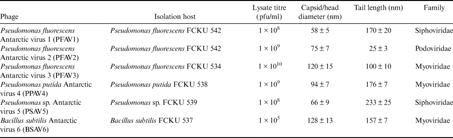
Seven bacterial strains that formed a bacterial lawn on the plates were further used for phage isolation from the samples (Table IV). In total, six phages were successfully isolated, cultured, purified and described in the laboratory. These phages were assigned tentative names reflecting the susceptible bacterial organism, isolation area and unique laboratory number. Here, we used following virus names and abbreviations: P. fluorescens Antarctic virus 1 (PFAV1), P. fluorescens Antarctic virus 2 (PFAV2), P. fluorescens Antarctic virus 3 (PFAV3), P. putida Antarctic virus 4 (PPAV4), Pseudomonas sp. Antarctic virus 5 (PSAV5) and B. subtilis Antarctic virus 6 (BSAV6). All six of these phages proved to be lytic (at least against their natural bacterial hosts) and produced clear plaques of different diameters: 2–3 mm for PFAV1, 1–2 mm for PFAV2, 0.5–1.0 mm for PFAV3, 2–3 mm for PPAV4, 1–2 mm for PSAV5 and 0.4–0.5 mm for BSAV6. Depending on the phage, the phage lysate titres varied from ~1 × 10⁵ to ~1 × 10¹⁰ pfu/ml (Table IV). The infectivity of the lysates was retained for several months when stored at 4°C.
Antarctic phages isolated from the soil samples in this study.

pfu = plaque-forming unit.
To characterize the phages, virus preparations were purified by sterilizing filtration through 0.22 μm membrane filters with subsequent ultracentrifugation, as described above. This method has proven to be the most effective for virus purification as confirmed by TEM.
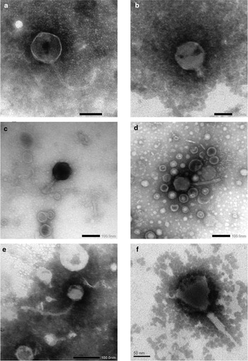
TEM of such soil eluates revealed particles, mostly with clearly visible tails. Only particles having spherical or icosahedral shapes resembling phage heads were considered to be VLPs, as opposed to other rod-shaped or filamentous morphotypes that were not taken into account due to the difficulties with their unambiguous detection against the background of organic matter.
According to the TEM data, these phage isolates had no lipid-containing envelope. In all of these cases, accurate morphological identification of virus tails allowed us to attribute these phages to the Caudovirales order (Fig. 3). The diameter of the virus capsid varied from 50 to 130 nm (Table IV). TEM of the purified virus preparations showed that the PFAV1 and PSAV5 phage isolates (Fig. 3a & e) had a typical siphovirus morphology with thin and flexible non-contractile tails. Specifically, the PFAV1 phage (Fig. 3a) had a ~58 nm head with a ~170 nm non-contractile tail, while the PSAV5 phage isolate (Fig. 3e) had a ~66 nm head and a ~233 nm non-contractile tail. The PFAV2 phage (Fig. 3b) was isolated from the same host as PFAV1 (P. fluorescens FCKU 542) but had a ~75 nm head with a very short rigid ~25 nm tail characteristic of podoviruses. The PFAV3, PРAV4 and BSAV6 phage isolates (Fig. 3c, d & f) varied in their size and had rigid contractile tails typical of myoviruses. The PFAV3 phage (Fig. 3c) had a ~120 nm head with a ~100 nm tail. The PPAV4 phage (Fig. 3d) had a ~94 nm head with a ~176 nm tail, whereas the BSAV6 phage isolate (Fig. 3f) had a ~128 nm head with a ~157 nm tail.
Morphology of viruses isolated from Antarctic soil samples as detected by transmission electron microscopy. a. Pseudomonas fluorescens Antarctic virus 1 (PFAV1), b. P. fluorescens Antarctic virus 2 (PFAV2), c. P. fluorescens Antarctic virus 3 (PFAV3), d. Pseudomonas putida Antarctic virus 4 (PPAV4), e. Pseudomonas sp. Antarctic virus 5 (PSAV5) and f. Bacillus subtilis Antarctic virus 6 (BSAV6). Scale bars are indicated on the images.

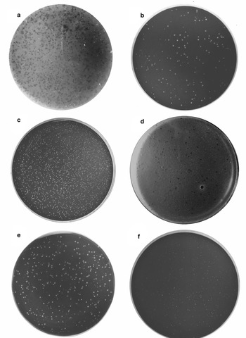
Furthermore, we proceeded with the determination of the optimal temperature for bacterial infection by the isolated phages. As is shown in Table II, all strains of isolated host bacteria formed colonies at temperatures from -7°C to +37°C. The temperature effect on phage replication (i.e. plaque formation) was tested at temperatures supporting host growth. All phages were able to infect their original hosts only at moderate temperatures from 4°C to 20°C, but not at higher temperatures. The PFAV1, PFAV2, PFAV3, PPAV4 and PSAV5 phages produced plaques in 3 days, whereas the BSAV6 phage produced plaques in 7 days at 16°C (Fig. 4a–f).
Lysis plaques observed using the double agar layer method. a. Pseudomonas fluorescens Antarctic virus 1 (PFAV1), b. P. fluorescens Antarctic virus 2 (PFAV2), c. P. fluorescens Antarctic virus 3 (PFAV3), d. Pseudomonas putida Antarctic virus 4 (PPAV4), e. Pseudomonas sp. Antarctic virus 5 (PSAV5) and f. Bacillus subtilis Antarctic virus 6 (BSAV6). Images were converted to black/white mode for higher contrast.

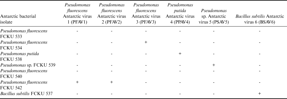
To determine the biological properties of the isolated phages, we cross-inoculated their natural bacterial hosts recovered from the same Antarctic samples (Table V).
Natural host ranges of the Antarctic phages.

Our results demonstrated that the PFAV1 and PFAV2 phages infected the same host (P. fluorescens FCKU 542) from which these viruses were recovered, but they were not able to replicate in other Antarctic bacterial isolates. Similarly, the PFAV3, PPAV4, PSAV5 and BSAV6 phages experimentally infected only those bacterial hosts from which these viruses were initially isolated: P. fluorescens FCKU 534, P. putida FCKU 538, Pseudomonas sp. FCKU 539 and B. subtilis FCKU 537, respectively. The obtained data show a high specificity of Antarctic phages towards their natural hosts and their inability to infect even closely related bacteria belonging to the same species P. fluorescens.
Here, we used a limited range of laboratory bacterial strains to determine whether the isolated phages were capable of infecting them (Table VI).
Sensitivity of laboratory bacterial strains to the isolated Antarctic phages.

C. michiganensis, E. cloacae, E. carotovora, E. coli, P. fluorescens 8573, P. syringae pv. lachrymans 7591, P. savastanoi pv. phaseolicola 4013, S. marcescens and X. campestris laboratory strains were inoculated with the PFAV1, PFAV2, PFAV3, PPAV4, PSAV5 and BSAV6 phages. According to the obtained data, only two such bacterial strains were susceptible to at least one of the phages. From these bacteria, P. savastanoi pv. phaseolicola 4013 was sensitive to the PFAV2 phage, whereas P. fluorescens 8573 supported infection by two phages, PFAV2 and PFAV3, isolated from different though related hosts (P. fluorescens FCKU 542 and P. fluorescens FCKU 534, respectively). Surprisingly, other phages isolated from Antarctic samples of Pseudomonas (PFAV1, PPAV4 and PSAV5) were not capable of inducing successful infection in these laboratory hosts, as well as the BSAV6 phage (Table VI). Hence, our results confirm the high specificity of phages isolated from Antarctic hosts towards bacteria from temperate climatic conditions.
In this study, we have recovered and characterized six phage isolates, all belonging to the order Caudovirales.
The bacteriophages PFAV1 and PSAV5 with long, non-contractile tails had a morphology that was characteristic of the Siphoviridae family. PFAV3, PPAV4 and BSAV6, with contractile tails consisting of a sheath and a central tube, had a morphology that was characteristic of the Myoviridae family. The PFAV2 phage had short tails, a morphology that was characteristic of the Podoviridae family. According to our results, none of the tested bacterial strains supported detectable lytic growth of either the PFAV, PPAV4, PSAV5 or BSAV6 phages (except for their natural hosts). However, PFAV2 podovirus and PFAV3 myovirus were initially isolated from closely related hosts (P. fluorescens FCKU 542 and P. fluorescens FCKU 534, respectively) and also had similar host ranges, whereas PFAV1 siphovirus was unable to infect closely related hosts (laboratory strains of Pseudomonas). A limited list of laboratory bacteria used in this study complements our data on natural virus host ranges (Table V) and demonstrates the ability of Antarctic phages to infect and lyse bacteria common in the temperate climate of Eurasia.
Discussion
Antarctic ecosystems, and especially their terrestrial biotopes, were previously shown to have surprisingly high levels of biodiversity and microorganism variability (Fierer & Jackson Reference Fierer and Jackson2006). Viruses are powerful factors driving the diversity of their hosts and the evolution of ecosystems as a whole (Suttle Reference Suttle2007).
The bacteria isolated in our study had an optimal growth temperature in the range of 16–28°C. From the samples collected during the previous 18th–20th expeditions to the UAS (2014–2016), we also isolated B. subtilis FCKU 537 and described its biochemical and morphological properties (Kondratiuk et al. Reference Kondratiuk, Beregova and Ostapchenko2016). Typing was based on experimentally established cultural, morphological and biochemical properties and according to Bergey's Manual of Systematic Bacteriology (Vos et al. Reference Vos, Garrity, Dorothy, Krieg, Ludwig and Rainey2009). These data are in agreement with similar previous studies (Straka & Stokes Reference Straka and Stokes1960, Margesin Reference Margesin2000). In line with our data, cold-tolerant or cold-adapted bacteria have been previously reported to exhibit a wider growth temperature range (from -5°C to +40°C) with an optimal growth temperature typically > 20°C (Cavicchioli et al. Reference Cavicchioli, Charlton, Ertan, Mohd Omar, Siddiqui and Williams2011). However, there is a study that proved the presence of actively respiring bacterial cells at incubation temperatures of -20°C (Junge et al. Reference Junge, Eicken and Deming2004). In addition, bacteria of the Pseudomonas genus are well known for their ability to colonize various ecological niches, to withstand unfavourable environmental conditions (Poblete-Castro et al. Reference Poblete-Castro, Becker, Dohnt, dos Santos and Wittmann2012) and to grow at nearly the same rate over a wide temperature range while producing comparable amounts of biomass (Pacheco et al. Reference Pacheco, Orellana-Saez, Pepczynska, Enrione, Bassas-Galia and Borrero-de Acuña2019).
Today, the genomes of nearly 200 psychrotolerant bacteria have been sequenced (Bowman Reference Bowman and Margesin2017). Bacterial communities of Antarctic soils are known to be highly localized and heterogeneous (Cary et al. Reference Cary, McDonald, Barrett and Cowan2010, Bottos et al. Reference Bottos, Scarrow, Archer, McDonald, Cary and Cowan2014). As of today, the following higher taxa of microorganisms have been reported in the land ecosystems of Antarctica: Proteobacteria, Actinobacteria, Firmicutes, Acidobacteria, Chloroflexi, Verrucomicrobia, Bacteroidetes, Armatimonadetes, Gemmatimonadetes and Planctomycetes (Teixeira et al. Reference Teixeira, Peixoto, Cury, Sul, Pellizari, Tiedje and Rosado2010, Van Horn et al. Reference Van Horn, Van Horn, Barrett, Gooseff, Altrichter and Geyer2013, Bakermans et al. Reference Bakermans, Skidmore, Douglas and McKay2014). Several major bacterial types are consistently found across the continent, but Proteobacteria represent the most common soil-type bacteria worldwide, even when diversity is analysed using a wide range of methods (clone libraries, pyrosequencing and microarrays) (Spain et al. Reference Spain, Krumholz and Elshahed2009). Pseudomonas is one of the typical representatives of Proteobacteria. Studies using a culture-dependent approach showed that Pseudomonas spp. are (sub)dominant among cultivated bacteria in the Arctic and Antarctic (Chauhan et al. Reference Chauhan, Bharti, Goyal, Varma and Jindal2015, Romaniuk et al. Reference Romaniuk, Ciok, Decewicz, Uhrynowski, Budzik and Nieckarz2018), which is in line with our data. Pseudomonads well tolerate various environmental conditions due to their high metabolic versatility (Ozen & Ussery Reference Ozen and Ussery2012). Representatives of Actinobacteria (Micrococcus) and Firmicutes (Bacillus) are no less common. The diversity of cultured bacteria in our studies was similar to those from ice cores from Antarctica, Greenland and other non-polar ice regions (Zhang et al. Reference Zhang, Yao, Tian, Xu and An2008).
However, other known Pseudomonas isolates collected far from Antarctica or from other extreme regions have not shown such metabolic capabilities, unlike our results, and none of them were able to catabolize xylose or arabinose despite being able to catabolize various C6 carbon sugars (Lee et al. Reference Lee, Cho, Yang, Jung, Hong and Kim2017, Orellana-Saez et al. Reference Orellana-Saez, Pacheco, Costa, Mendez, Miossec and Meneses2019). Previously, it was hypothesized that some bacteria, specifically Pseudomonas, might have developed a genetic repertoire enabling the metabolism of such sugars (C6 and C5), glycerol, organic acids and aromatics, partly thanks to an established ‘reservoir’ of Antarctic flowering plants such as Deschampsia antarctica and Colobanthus quitensis (Polischuk et al. Reference Polischuk, Budzanivska, Shevchenko and Oliynik2007) sourcing such carbon substrates.
There are quite a few articles devoted to the biological characteristics of viruses and describing their isolation from the polar regions. Studies of the Antarctic aquatic environment have shown that viruses are diverse and numerous, and they are able to change their lifestyles similarly to their microbial hosts, from pseudolysogenic to lytic, when climatic conditions favour microbial growth (Säwström et al. Reference Säwström, Lisle, Anesio, Priscu and Laybourn-Parry2008, Anesio & Bellas Reference Anesio and Bellas2011). Recent research in Antarctica and the Arctic reveals a wide variety of DNA viruses, including bacteriophages, viruses with circular ssDNA, algal phycodnaviruses and virophages, adapted to such extreme conditions. The first discovery of RNA viruses in Antarctica was reported in Limnopolar Lake. Most of the identified viruses were positive-sense ssRNA viruses later assigned to the Picornavirales order. Dicistroviridae viruses were most represented in the samples analysed from 2006 to 2010, while the Bacillarnavirus genus and Picornavirales representatives were found in the summer samples from 2007 and 2010 (López-Bueno et al. Reference López-Bueno, Tamames, Velázquez, Moya, Quesada and Alcamí2009, Lauro et al. Reference Lauro, DeMaere, Yau, Brown, Ng, Wilkins and Raftery2010, Yau et al. Reference Yau, Lauro, DeMaere, Brown, Thomas and Raftery2011, Brum et al. Reference Brum, Hurwitz, Schofield, Ducklow and Sullivan2016, Miranda et al. Reference Miranda, Culley, Schvarcz and Steward2016, Rastrojo & Alcamí Reference Rastrojo and Alcamí2018). In addition, virus diseases of Antarctic birds, including penguins and seagulls, were described, as well as those of seals (Pearce & Wilson Reference Pearce and Wilson2003). Furthermore, there are limited data on plant virus diversity in Antarctica (Pearce & Wilson Reference Pearce and Wilson2003, Polischuk et al. Reference Polischuk, Budzanivska, Shevchenko and Oliynik2007).
As for the bacteriophages, it was shown that the hypolytic communities of the Antarctic deserts host significant virus populations, where caudate phages of the Caudovirales order predominate (Williamson et al. Reference Williamson, Radosevich, Smith and Wommack2007, Zablocki et al. Reference Zablocki, Adriaenssens and Cowan2016, Adriaenssens et al. Reference Adriaenssens, Kramer, Van Goethem, Makhalanyane, Hogg and Cowan2017). The presence of RNA bacteriophages of the Leviviridae family, ssDNA phages of the Microviridae family and non-tail Tectiviridae (dsDNA) was also shown (Wei et al. Reference Wei, Lacap-Bugler, Lau, Caruso, Rao and de Los Rios2016).
Phages have also been previously found in Antarctic water and sea-ice samples, where the authors identified a sensitive bacterial host capable of supporting viral replication in the laboratory (Luhtanen et al. Reference Luhtanen, Eronen-Rasimus, Oksanen, Tison, Delille and Dieckmann2018). To our knowledge, to date, only three virus-host systems have been isolated from the Arctic and seven such systems have been isolated from the ice of the Baltic Sea (Borriss et al. Reference Borriss, Helmke, Hanschke and Schweder2003, Luhtanen et al. Reference Luhtanen, Eronen-Rasimus, Kaartokallio, Rintala, Autio and Roine2014, Yu et al. Reference Yu, Chen, Shen, Zhao, Tang and Su2015). However, no such work has been done on viruses and their tentative hosts isolated from the superficial soil layer in Antarctica. Therefore, the current work was focused on the isolation of phages from soil samples, their subsequent analysis and the search for a sensitive bacterial host from the same samples.
Nevertheless, much remains to be learned about virus-host interactions at low temperatures. The reproduction of viruses is dependent on the activity of the protein-synthesizing systems of bacteria, while DNA replication, protein synthesis, etc., in bacteria obviously slows down at low temperatures. Consequently, this should negatively affect the reproduction of viruses. Despite this, there is a growing body of evidence of virus abundance in Antarctica and other regions characterized by low/extremely low temperatures for most of the year (Rastrojo & Alcamí Reference Rastrojo and Alcamí2018, Sommers et al. Reference Sommers, Fontenele, Kringen, Kraberger, Porazinska and Darcy2019, Yau & Seth-Pasricha Reference Yau and Seth-Pasricha2019).
It should be noted that the capsid sizes of some phages isolated in this study differed from common VLP size groups (50–70 nm or < 110 nm) reported previously for Antarctic and Arctic sea-ice samples (Wei et al. Reference Wei, Lacap-Bugler, Lau, Caruso, Rao and de Los Rios2016, Adriaenssens et al. Reference Adriaenssens, Kramer, Van Goethem, Makhalanyane, Hogg and Cowan2017).
Our results contradict the hypothesis suggesting the predominance of polyvalent bacteriophages due to low levels of bacterial plankton in aquatic systems, probably reflecting differences in either the biology of virus-host interactions or their mutual availability in these conditions (Breitbart & Rohwer Reference Breitbart and Rohwer2005). However, our data are in agreement with the biological patterns previously observed in terrestrial biotopes. It was shown that most bacterial viruses were highly specific to a single type of microorganism (or even a bacterial strain) when tested in the laboratory (Koskella & Brockhurst Reference Koskella and Brockhurst2014). Most probably, Antarctic bacteria may have developed some virus-specific defence mechanisms resulting from their coevolution with Antarctic phages.
It was previously suggested that bacteriophages in biotopes with high host specificity serve as the mechanism that more frequently ‘kills’ hosts with higher growth rates (i.e. the most successful and numerous bacteria) occupying the top of the ecological niche (an ecological ‘behaviour’ of bacteriophages known as ‘kill the winner’; Thingstad & Lignell Reference Thingstad and Lignell1997). It follows that only viruses of dominant bacteria can be detected in the majority of samples. We assumed that if such bacteriophages do not infect laboratory cultures of bacteria, then this possibly indicates the absence of these and/or similar strains in the respective samples. Therefore, it was decided to determine whether Antarctic phages isolated in this study infect bacteria recovered from the soil samples previously collected from a temperate Eurasian climate in Ukraine.
Most of the phages isolated from the Antarctic region are characterized by high specificity. This pattern has been demonstrated in similar works (Borriss et al. Reference Borriss, Helmke, Hanschke and Schweder2003, Luhtanen et al. Reference Luhtanen, Eronen-Rasimus, Kaartokallio, Rintala, Autio and Roine2014). Our data on the PFAV1, PPAV4, PSAV5, and BSAV6 phages are in line with this evidence. However, the ability of the Antarctic phages PFAV2 and PFAV3 to infect bacterial isolates from temperate latitudes is a unique feature. We assume that some phages isolated in Antarctica may successfully replicate in certain European bacterial isolates and, possibly, can also shape the composition of the bacterial communities in temperate climates.
The ecology of bacteriophages as a key component of many bacterial communities has been studied primarily in temperate ecosystems, but little attention has been paid to the role of viruses in Antarctic ecosystems (Murray & Eldridge Reference Murray and Eldridge1994, Wommack et al. Reference Wommack, Ravel, Hill and Colwell1999a). Data on the spread of bacteriophages in the polar continental latitudes are very limited. However, they indicate that phages play an important role in these ecosystems dominated by microbial communities (Säwström et al. Reference Säwström, Lisle, Anesio, Priscu and Laybourn-Parry2008).
A number of reviews have described the role of viruses in marine ecosystems (Wommack et al. Reference Wommack, Ravel, Hill, Chun and Colwell1999b), showing that viruses play an important role in the structure and composition of water reservoir food (Murray & Eldridge Reference Murray and Eldridge1994, Suttle et al. Reference Suttle2007).
Since the first marine viral metagenomic study in 2008 (Säwström et al. Reference Säwström, Lisle, Anesio, Priscu and Laybourn-Parry2008), metagenomics has become a powerful tool enabling us to better understand viral diversity and variability, and it can also be used to directly address complex, uncultured environmental viral genomes (Angly et al. Reference Angly, Felts, Breitbart, Salamon, Edwards and Carlson2006). Metagenomics approaches in the study of natural populations of bacteria have probably led to an overestimation of the phage prevalence in the environment (Koskella & Brockhurst Reference Koskella and Brockhurst2014).
Currently, there are only two viral metagenomic studies of the Southern Ocean. These studies were conducted on the coastal waters of the Palmer Station Antarctica - Long Term Ecological Research (PAL-LTER) station and Station B of the Western Antarctic Peninsula (Brum et al. Reference Brum, Hurwitz, Schofield, Ducklow and Sullivan2016). It has even been hypothesized that the polar regions may be point sites of microbial evolution due to the higher degree of viral control in these microbial communities (Anesio & Bellas Reference Anesio and Bellas2011). Another recent study suggests that the waters of the Arctic Ocean are hotbeds of biodiversity for viruses (Gregory et al. Reference Gregory, Zayed, Conceição-Neto, Temperton, Bolduc and Alberti2019). These authors highlight the importance of these highly climate-damaged Arctic regions to global biodiversity.
However, the limited number of Antarctic viral metagenomic studies have focused exclusively on aquatic systems (Säwström et al. Reference Säwström, Lisle, Anesio, Priscu and Laybourn-Parry2008, López-Bueno et al. Reference López-Bueno, Tamames, Velázquez, Moya, Quesada and Alcamí2009), and recent meta-viral studies of saline meromictic lakes have revealed a high level of diversity of VLPs (mainly phages) and several virophages (Vos et al. Reference Vos, Garrity, Dorothy, Krieg, Ludwig and Rainey2009, Yau et al. Reference Yau, Lauro, DeMaere, Brown, Thomas and Raftery2011).
Nevertheless, terrestrial biotopes in Antarctica still mostly remained terra incognita in terms of virus research. To date, there are few studies on viruses isolated in Antarctica that follow a classical biological routine of phage isolation and cultivation on bacterial species (Williamson et al. Reference Williamson, Radosevich, Smith and Wommack2007, Swanson et al. Reference Swanson, Reavy, Makarova, Cock, Hopkins and Torranceet2012). In this article, we have described the isolation of a cultivated host-phage systems from terrestrial biotopes, as well as the diversity of bacterial viruses.
Acknowledgements
The authors wish to thank the staff of the National Antarctic Scientific Center (NASC) for providing the samples used in this research and the staff of the Centre of Collective Usage at the Zabolotny Institute of Microbiology and Virology of the National Academy of Sciences (NAS) of Ukraine for their invaluable help with the TEM studies. The use of the Scientific Committee on Antarctic Research (SCAR) Antarctic Digital Database and the Antarctic Digital Database map viewer resource (https://www.add.scar.org) supported by the British Antarctic Survey for generating sampling maps is also acknowledged. The authors wish to thank two anonymous reviewers for their helpful comments.
Financial support
This work was in part supported by the National Antarctic Scientific Center (NASC) of the Ministry of Education and Science of Ukraine (grant no. 17DF036-10).
Author contributions
OA, IB and TK designed the study. VH, OA and PZ conducted the experiments. VH, OA, TK and OS analysed the data. VH, OA, IB and OS wrote the manuscript. All authors reviewed the manuscript.