INTRODUCTION
The state of Santa Catarina (SC), in southern Brazil is the major Brazilian producer of the brown mussel Perna perna Linnaeus, 1758 and of the Pacific oyster Crassostrea gigas Thunberg, 1795; 10,600 and 3150 tons were produced during 2007, respectively. Particularly, the districts of Governador Celso Ramos and Penha contribute with most of the production, with 4900 and 2000 tons, respectively, in 2007. An episode of mortality of the mussel P. perna (almost 20%) was observed by the farmers from the district of Penha during August 2008. This event was notified to the Santa Catarina Institution for Agricultural Research and Rural Extension Services (EPAGRI) and then to the Santa Catarina Federal University (UFSC) in order to study the causes of this event.
Monstrilloids are protelean parasitic copepods infecting benthic polychaetes and prosobranch molluscs; recently, they have been recorded also in sponges (Huys et al., Reference Huys, Llewellyn-Hughes, Conroy-Dalton, Olson, Spinks and Johnston2007). The free-living adult stage is present in the water column and mature individuals are collected frequently by plankton nets in coastal waters from all latitudes (Davis, Reference Davis, Steidinger and Walter1984; Suárez-Morales, Reference Suárez-Morales2001). Very little is known about the development of these endoparasite crustaceans. The early works by Giard (Reference Giard1895, Reference Giard1896), Malaquin (Reference Malaquin1901), Caullery & Mesnil (Reference Caullery and Mesnil1914) and Pelseneer (Reference Pelseneer1914) were among the most relevant in describing the general patterns of larval development in Monstrilloida. There are some recent studies on the morphology of the free-swimming early naupliar stages (see Grygier & Ohtsuka, Reference Grygier and Ohtsuka1995). Most of their life cycle takes place in the body of the host after the last naupliar stage, which burrows into the host to complete its development. Shortly after the infection, a protective sheath is produced around the larva, which feeds from the host through a pair of ventral antenna-like processes (Davis, Reference Davis, Steidinger and Walter1984). The development takes place within the sheath until a pre-adult stage that emerges from the host as a free-living, non-feeding copepod. The free-living form rapidly becomes a reproductive adult in the water column.
In this paper we report the occurrence of different parasites in the brown mussel Perna perna collected from southern Brazil culture fields, particularly in reference to the finding of the endoparasitic monstrilloid copepods. We evaluate the intensity and prevalence of the infection by these crustacean parasites and their histopathological effects on the mantle tissues of the host.
MATERIALS AND METHODS
Specimens of the brown mussel P. perna (N = 90) were collected from a culture area at Penha, Santa Catarina State in August 2008 during a local episode of mussel mortality (almost 20%); the specimens were kept alive and analysed upon arrival at the laboratory. All animals were examined for external body and shell gross abnormalities. Ten specimens were randomly selected to determine the occurrence and extent of pathological conditions by histological examination, as follows: mussels were shucked and a sagittal (5 mm thick) section containing gills and visceral mass was excised, fixed in Davidson's solution, and embedded in a paraffin block. Serial tissue sections (5 µm) were stained with Harris haematoxylin and eosin. A piece of mantle with nodules from five animals was also submitted to histological examination as described above.
The mantle from 5 other mussels bearing abnormal nodules was also excised. Some nodules were dissected under the stereoscope to extract the copepod larvae. Isolated copepod larvae and entire nodules were immediately fixed in 70% alcohol for further taxonomic and parasitological examination. In order to facilitate the observation process the copepods were transferred to a freshly prepared solution of glycerol (70%) in ethanol, and then into pure glycerine. Most of the juvenile specimens examined were strongly bent inwards; these were observed from different angles and illustrations were made from undissected specimens.
Prevalence was estimated as the percentage of infected mussels in the sample. The intensity of pathogenic organisms was scored as low, moderate, or heavy according to the macroscopic observations.
RESULTS
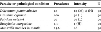
The mean values (±SD) for whole weight and shell length among the examined mussels were 30±7.9 g and 72±5.8 cm, respectively; these measurements are within the commercial size-range for this species. The prevalences of pathological conditions observed in P. perna mussels from the surveyed area are shown in Table 1.
Prevalence (%) of organisms or parasites on brown mussel Perna perna from the culture area of Penha, Santa Catarina State.

N, number of analysed animals; L, low; M, moderate; H, heavy; nd, not determined.
Macroscopic observations of the specimens examined showed the presence of the colonial tunicate Didemnum psammathodes Sluiter, 1895 covering the shell of 20% of the examined mussels. In some cases (9%) the tunicate covered more than half of the outer surface of the shell, which was considered as a heavy infestation. Ciliated turbellarian Urastoma cyprinae Graff, 1882 (Platyhelminthes) were observed moving freely throughout the gill lamellae in 100% of the mussels examined; this infestation occurred always at low intensity (few individuals). The polychaete Polydora websteri Hartman, 1943 occurred with 20% prevalence. The intensity was low; infested mussels had 1 or 2 small conchioline-covered tubes on the inner surface of the shells, but no observable damage was associated with the adjacent mantle. The trematode Bucephalus margaritae Ozaki & Ishibashi, 1934 (= varicus) (Buchephalidae) was detected macroscopically in one mussel (1.1%) (see Table 1). The infection by B. margaritae produced an orange pigmentation and a filamentous aspect of the mantle as a result of an intense parasite burden. The heavy intensity of infection probably caused host castration since the mantle was extremely thin with no apparent gonadal material, typically of red (female) or white (male) colour.
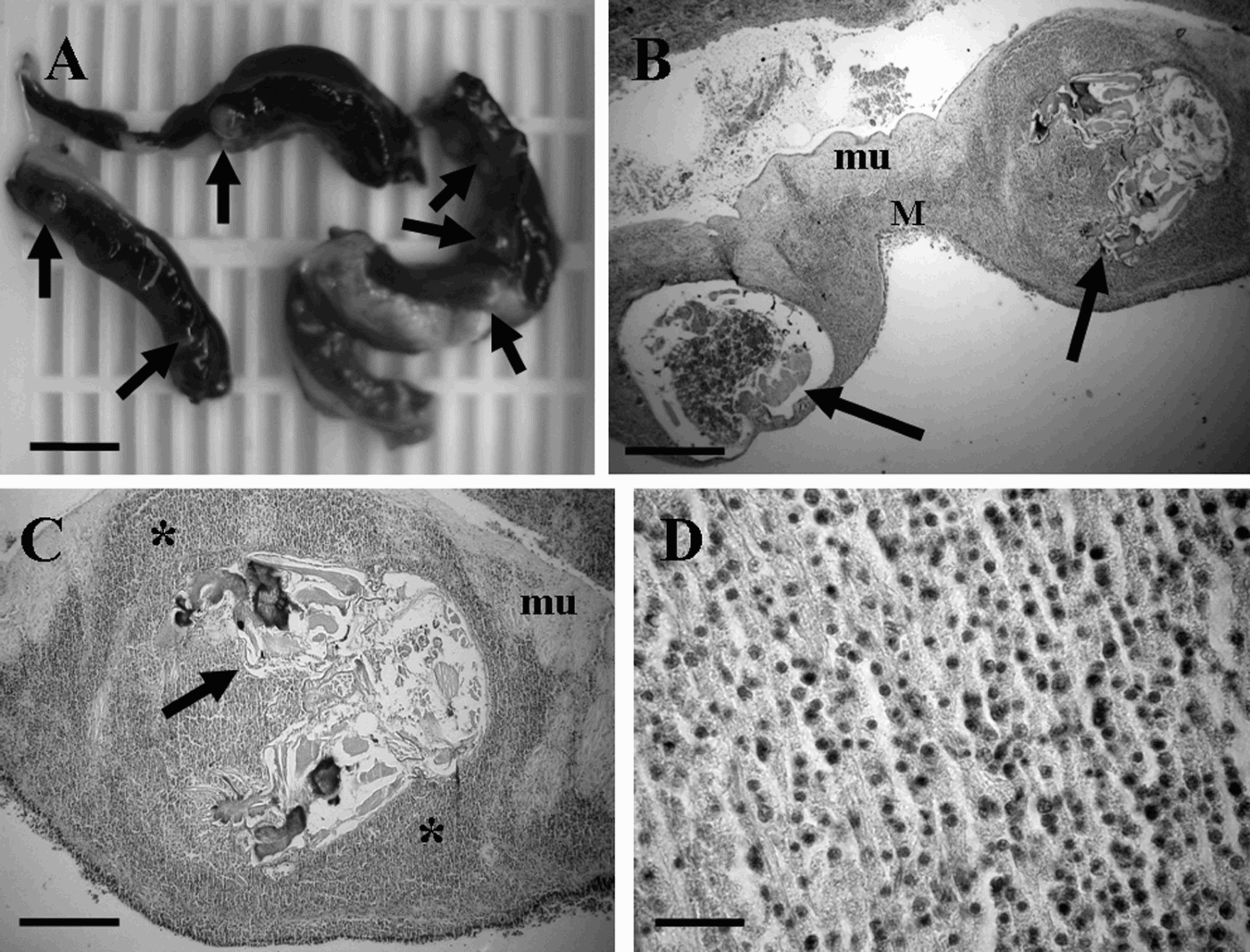
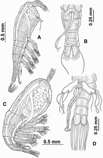
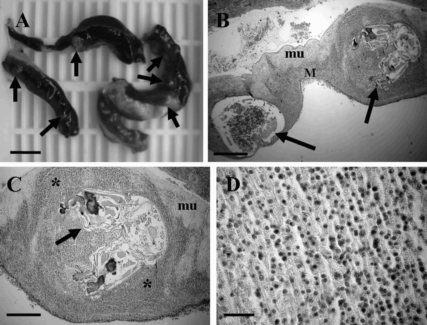
From all mussels examined, 23 had rose-coloured nodules near the border of the mantle, with sizes ranging from 1 to 4 mm (Table 1; Figure 1A); up to 10 nodules were detected on the mantle of the most infected animals. The nodules were opened under the stereomicroscope and the larval monstrilloid copepods recovered (Figure 2). The monstrilloid copepods were identified as belonging to the genus Monstrilla by the lack of eyes and by the presence of five caudal setae; all specimens examined were juvenile females, with 4-segmented antennules and a pair of structures on the genital somite that will become the ovigerous spines. At least two different developmental stages were observed in the specimens examined (see Figure 2).
Monstrilla sp. infecting the brown mussel Perna perna from Brazil. (A) Infected mussel's mantle with several nodules (arrows): scale bars = 10 mm; (B) general view of the mantle's border (M) showing two nodules containing larvae of copepods (arrows), and alteration of the architecture of mantle's tissue: scale bars = 100 µm. mu, muscle fibers of mantle's border; (C) copepod larvae induced-nodules formation and intense haemocytic infiltration (*): scale bars = 200 µm; (D) magnification of a connective tissue heavily infiltrated by haemocytes: scale bars = 20 µm.
General morphology of two juvenile specimens of Monstrilla sp. at different developmental stages recovered from the brown mussel Perna perna in Santa Catarina Island (southern Brazil). (A) Habitus of early female copepodite, lateral view; note the feeding tubes on the ventral surface, the genital somite is still 2-segmented; (B) urosome, ventral view showing genital spines and outer juvenile and inner cuticle of next stage; (C) habitus of juvenile late copepodite nearing its final moult, lateral view; (D) urosome, ventral view. Note that the total size of both specimens is equal, the early copepodite has bent caudal rami and antennules, and the late copepodite has the urosome bent inwards to fit in the chamber.

At the histological level, it was observed that the copepod larvae induced a strong alteration of the mantle's architecture. The copepod larvae were observed: (1) surrounded by a space; probably filled with the sheath (before histological procedure) produced by the larva after the infection; (2) encapsulated by a thick layer of infiltrated haemocytes resulting from the host response to the invasive process of larval development within the host tissue; and (3) invaded by those haemocytes which destroyed the parasite's wall and penetrated into their internal tissues (see Figure 1).
DISCUSSION
The brown mussel P. perna cultured at Penha, Santa Catarina State, was sampled after a mortality episode; the population was studied herein to determine possible links with parasitic organisms. Different commensal and parasitic organisms were recorded; most of them occurred at low levels of prevalence and intensity. A similar mortality event occurred in the summer of 2007 but no parasitological survey was carried out because the economic loss was relatively unimportant.
The ascidian Didemnum psammathodes was observed covering partially the mussels' outer shell surfaces, except for 8 individuals that had almost the whole shell inhabited by D. psammathodes; this condition might have compromised to some extent the bivalves' filtering process. The ascidians D. psammathodes and Didemnum perlucidum Monniot, 1983 are frequent fouling species inhabiting bivalve shells and aquaculture equipment, such as lantern nets or long-lines at Santa Catarina Island and Penha (A.R.M. Magalhães, Brazil, personal communication; Kremer, Reference Kremer2008). Didemnum sp. was reported as one of the most common fouling species covering the shells of pearl oysters in Australia (Guenther & De Nys, Reference Guenther and De Nys2006).
The turbellarian Urastoma cyprinae has long been considered as a commensal to different species of bivalve molluscs, including mussels; however, in some cases it can become a true parasite, causing some damage to the host, as reported for the oyster Crassostrea virginica (Brun et al., Reference Brun, Boghen and Allard1999) and the mussel Mytilus galloprovincialis (Robledo et al., Reference Robledo, Cáceres-Martínez, Sluys and Figueras1994). Cáceres-Martínez et al. (Reference Cáceres-Mártinez, Vásquez-Yeomans and Sluys1998) reported prevalences of U. cyprinae varying from 57 to 100% in the mussel M. galloprovincialis from a culture area in Baja California, Mexico and observed haemocytic infiltration around the turbellarian parasite. Although the occurrence of U. cyprinae in the examined mussels was widespread, it was present as a free-living organism moving around the gills. Our histopathological analysis did not reveal any lesions or host response to the turbellarian on the mussel's gills. Hence, there is no evidence that this turbellarian causes morbidity or is parasitic to the populations of P. perna from the surveyed area.
Burrowing forms of the polychaete genus Polydora are common parasites infesting the inner shell's surface of edible molluscs (Lauckner, Reference Lauckner and Kinne1983). There are several culture areas at Santa Catarina Island with high prevalences (up to 100%) of Polydora websteri Hartman, 1943 infesting the oysters Crassostrea gigas (Sabry & Magalhães, Reference Sabry and Magalhães2005); however, prevalences and intensities are low in the brown mussel P. perna (Magalhães & Ferreira, Reference Magalhães, Ferreira and Silva-Souza2006). Polydora can cause significant damage to the oyster C. gigas, including weakness of the shell and lesions to the adductor muscle and mantle (Sabry & Magalhaes, Reference Sabry and Magalhães2005). However, the main problem related to this parasite concerns the repulsive aspect of the shells resulting from the presence of abundant blisters and detrital tubes of Polydora; the infected specimens lose their commercial value and are rejected by potential consumers (Magalhães & Ferreira, Reference Magalhães, Ferreira and Silva-Souza2006).
Bucephalus margaritae is the foremost parasite infecting the brown mussel P. perna at Brazil and the Santa Catarina coast (Magalhães & Ferreira, Reference Magalhães, Ferreira and Silva-Souza2006; Cochôa & Magalhães, Reference Cochôa and Magalhães2008). Bucephalus sp. has been blamed for host (P. perna) castration and induction of haemocytic infiltration at connective tissue and consequent reduction in the number of circulating haemocytes (da Silva et al., Reference da Silva, Magalhães and Barracco2002). During this work, the local prevalence of this trematode (1.1%) was very low in comparison to culture and wild areas at Santa Catarina Island such as Ribeirão da Ilha (south) and Ponta das Canas (north) reaching 32.1 and 63.6%, respectively (da Costa, Reference da Costa2007), or even the same area at Penha with previously recorded prevalences higher than 20% (de Mesquita et al., Reference de Mesquita, Magalhães, Marenzi, Barbosa, Resgalla, Weber and da Conceição2008).
The copepod Monstrilla sp. was found infecting the mantle of P. perna mussel with a considerable prevalence in this cultured population. The work by Pelseneer (Reference Pelseneer1914) includes the only previous record of monstrilloid copepods in a mollusc host; he found a low prevalence (2%, 4 out of 200) of these parasites in a population of the prosobranch gastropod Odostomia rissoides (now considered as a junior synonym of O. scalaris) from the English Channel. These specimens were reared to adulthood and identified as Monstrilla helgolandica Claus, 1863. Hartman (Reference Hartman1964) reported a 1% prevalence of M. capitellicola Hartman, Reference Hartman1964 in the polychaete Capitella capitata oculata Hartman, 1961 from California. This species was described based on immature females only, as Hartman (Reference Hartman1964) indicates that they were still enclosed in the capsular sheath. Because of this, the species is not currently recognized as valid and its taxonomic status remains uncertain. Unfortunately, no adults of Monstrilla were collected from the cultures of P. perna in this survey, but the occurrence of the genus could be confirmed by the analysis of the morphology of the juveniles. The prevalence in the cultured mussels during our survey in Brazil is higher than that observed by Pelseneer (Reference Pelseneer1914) in a natural population of the prosobranch O. scalaris. The moderate prevalence (25.6%) of Monstrilla in the brown mussels from the surveyed area is very likely a result of the high culture density of potential hosts for larval monstrilloids. The availability of hosts is an essential factor in determining the adult populations of monstrilloids in the water column, as suggested by Suárez-Morales (Reference Suárez-Morales2001) who recorded a dense aggregation of monstrilloids in a reef environment, where high densities of polychaete hosts were expected.
The specimens examined were identified as juvenile females belonging to the genus Monstrilla by the presence of two urosomites posterior to the genital somite (Figure 2); however, this character alone does not differentiate Monstrilla from Monstrillopsis. The specimens obtained from P. perna have five caudal setae; Monstrilla is the only genus in which five caudal setae are found in females (Grygier, Reference Grygier1994; Suárez-Morales & Gasca, Reference Suárez-Morales and Gasca2004). The setal armament of the caudal rami remains unchanged during the juvenile and pre-adult stages of Mostrilloida; for instance, Caullery & Mesnil (Reference Caullery and Mesnil1914) described the juveniles of both Cymbasoma germanicus Timm, 1893 and C. malaquini Caullery & Mesnil, Reference Caullery and Mesnil1914 from polychaetes and found three caudal setae, a genus-related character, at different stages. The stability of this character through the ontogenetic development of the copepodid phase in the Monstrilloida is also confirmed by Huys et al. (Reference Huys, Llewellyn-Hughes, Conroy-Dalton, Olson, Spinks and Johnston2007). Another characteristic present in the juvenile specimens examined was the absence of eyes even in those specimens nearing their final moult; this is another feature exclusive to the genus Monstrilla (Grygier, Reference Grygier1994). In these specimens there were also remains of the feeding tubules connecting them to the host; these appendages remain almost entirely within the host after the pre-adult emerges (Caullery & Mesnil, Reference Caullery and Mesnil1914; Davis, Reference Davis, Steidinger and Walter1984). The armature and structure of the fifth leg is an important taxonomic characteristic within the group and it appears to remain constant during the last developmental stage as well as in the adult; the specimens examined clearly show a fifth leg with an inner lobe armed with a single seta and an outer lobe with three setae (Figure 2). Two species in the region are known to present such features: Monstrilla pustulata Suárez-Morales & Dias, Reference Suárez-Morales and Dias2001 and M. brasiliensis Suárez-Morales & Dias, Reference Suárez-Morales and Dias2000, both from Brazil. The taxonomy of the Monstrilloida is largely based on the morphology of the adult; hence, it is not possible to provide a reliable identification of the immature specimens until adult individuals can be recovered from the area or reared in laboratory. A complete morphological and taxonomic analysis of these specimens is still due.
The present work is the first study on the histopathological effects of the endoparasitic stages of the Monstrilloida on its hosts. We demonstrated the damage caused by the copepod larvae to the host at histological level, nodules containing copepods were already macroscopically evident on the mantle's border; some of them were large enough to potentially cause a deficiency in the open–close shell mechanism; however, this aspect needs further evaluation. Nevertheless, the presence of copepod larvae infecting the mantle of the mussel P. perna caused a severe alteration of the histological structure of the mantle. Accordingly, an important inflammatory response consisting of haemocyte migration into the copepod larvae tissue was observed, indicating that haemocytes were able to recognize the copepod larvae as a foreign invasive organism. In some cases the haemocytes were able to disrupt the copepod wall enabling tissue destruction. The present work provided interesting data to encourage further research on lesions and host response caused by a Monstrilla copepod larva.
After an anecdotal mention of a monstrilloid present in a bivalve in Hong Kong (Boxshall & Halsey, Reference Boxshall and Halsey2004), the finding of Monstrilla in P. perna is the first documented record of such crustacean parasites in bivalve molluscs; furthermore, there was no previous report of monstrilloids as parasites of commercial, edible molluscs.
ACKNOWLEDGEMENTS
The authors are grateful to the Santa Catarina Institution for Agricultural Research and Rural Extension Services (EPAGRI) for contacting Dr P.M. da Silva for mussel analyses, Dr A.R.M. Magalhães for taxonomic help and Dr Ju-shey Ho who encouraged the development of this study.