Introduction
Stegophorus macronectes (Johnston & Mawson, Reference Johnston and Mawson1942) (Nematoda, Acuariidae) is a gastrointestinal parasite found in Australian, Subantarctic and Antarctic bird species (Barbosa & Palacios, Reference Barbosa and Palacios2009; Vidal et al., Reference Vidal, Ortiz, Díaz, Ruiz de Ybañez, Amat, Palacios, Benzal, Valera, Dela Cruz, Motas and Barbosa2012; Diaz et al., Reference Diaz, Fusaro, Longarzo, Coria, Vidal, Jerez, Ortiz and Barbosa2013). The taxonomic classification of the species has changed since it was first described. Originally described as Paryseria macronectes in the southern giant petrel Macronectes giganteus (Gmelin) and the grey-headed albatross Thalassarche chrysostoma (Forster) in South Australia (Johnston & Mawson, Reference Johnston and Mawson1942), the species was later redescribed by Zdzitowiecki & Dróżdż (Reference Zdzitowiecki and Dróżdż1980) based on specimens found in the type host M. giganteus, the subantarctic skua Catharacta lonnbergi (Mathews) and the sheathbill Chionis alba (Gmelin), all collected on King George Island, South Shetland Islands, Antarctica. These authors considered Stegophorus paradeliae Johnston & Mawson, Reference Johnston and Mawson1945 and Stegophorus adeliae Johnston, Reference Johnston1938 sensu Petter Reference Petter1959, both collected from penguins (Pygoscelis adeliae (Hombron & Jacquinot) and P. papua, respectively), to be identical to S. macronectes (see Johnston & Mawson, Reference Johnston and Mawson1945; Mawson, Reference Mawson1953; Petter, Reference Petter1959; Zdzitowiecki & Dróżdż, Reference Zdzitowiecki and Dróżdż1980). Although most reports given under this name or its synonyms contribute to the morphological description of the species (Johnston & Mawson, Reference Johnston and Mawson1945; Mawson, Reference Mawson1953; Petter, Reference Petter1959; Zdzitowiecki & Dróżdż, Reference Zdzitowiecki and Dróżdż1980), many were made on the basis of badly preserved, fragmented and/or exclusively female specimens. Therefore, some of the more commonly used diagnostic characters may not be appropriate for species identification (e.g. the number of collarette teeth or the position of deirids or nerve ring). An updated morphological description of this species is therefore necessary.
Accurate identification of parasites at any point of the life cycle is crucial for diagnosing infection. However, parasite identification using morphological characters can be problematic when only larvae or small portions of an individual are available (Zhu et al., Reference Zhu, Gasser, Podolska and Chilton1998). Also, in some instances, preservation methods, such as freezing, can break the weak eggshell or cause morphological deformities, making identification difficult (Pritchard & Kruse, Reference Pritchard and Kruse1982). Molecular information from DNA sequences provides a high level of specificity for the diagnosis and identification of parasite species (Prichard & Tait, Reference Prichard and Tait2001). Thus, specific molecular probes for identification may provide a more reliable diagnosis compared with traditional techniques. With this in mind, we report the first molecular characterization of S. macronectes.
To date, only six of the over 150 currently described species of the family have been characterized molecularly, and only four species share a common molecular marker (Nadler et al., Reference Nadler, Carreno, Mejía-Madrid, Ullberg, Pagan, Houston and Hugot2007; Honisch & Krone, Reference Honisch and Krone2008; Perera et al., Reference Perera, Maia, Jorge and Harris2013). Furthermore, previous phylogenetic analyses have only been done at the order level, with uneven representation of the main families (e.g. Blaxter et al., Reference Blaxter, De Ley, Carey, Liu, Scheldeman, Vierstraete, Vanfleteren, Mackey, Dorris, Frisse, Vida and Thomas1998; Nadler et al., Reference Nadler, Carreno, Mejía-Madrid, Ullberg, Pagan, Houston and Hugot2007; Černotíková et al., Reference Černotíková, Horák and Moravec2011). Therefore, phylogenetic studies using molecular data from S. macronectes, among other species of the order, may reveal new evolutionary relationships for this understudied group of parasites.
The aims of this paper are as follows: (1) to describe the morphological features of S. macronectes, providing an updated description of the species; (2) evaluate the reliability of the morphological traits used for identification; (3) characterize the species molecularly; (4) develop primer pairs for molecular diagnoses; and (5) to determine the phylogenetic position of the species within Spirurida.
Materials and methods
Collection and examination of nematodes
Acuarioid nematodes (n= 1157) were collected from 64 gastrointestinal tracts of chinstrap penguins Pygoscelis antarctica recently deceased due to natural causes (61 chicks and 3 adults), from the Vapour Col breeding colony on Deception Island (63°00′S, 60°40′W), South Shetland Islands, Antarctica, during the austral summers (December–February) from 2005 to 2009. Gastrointestinal packages were extracted, placed in labelled plastic bags and frozen at − 20°C until analysis. In the laboratory, nematodes were recovered from the stomach and preserved in 70% ethanol. Parasite identification was based on morphometric features, following a specific bibliography (Johnston & Mawson, Reference Johnston and Mawson1942, Reference Johnston and Mawson1945; Petter, Reference Petter1959; Yamaguti, Reference Yamaguti and Yamaguti1961; Chabaud, Reference Chabaud, Anderson, Chabaud and Willmott1974; Zdzitowiecki & Dróżdż, Reference Zdzitowiecki and Dróżdż1980).
Ten male and ten female relaxed and well-preserved specimens were measured. Rigid and/or badly preserved specimens that had morphological alterations were also analysed for comparison. Nematodes were cleared with Amman lactophenol or 25% glycerin ethanol prior to observation under an optical microscope. Drawings were made with the aid of a camera lucida. Several specimens were dried using the critical point method (Bray, Reference Bray, Williams and Clifford2000), examined by scanning electron microscopy (JEOL-6100®; JEOL, Tokyo, Japan) and photographed. Measurements (in micrometres unless otherwise stated) are reported as means, with standard deviations in parentheses. In addition, collarette teeth in 815 individuals (556 females, 239 males and 20 immature specimens) were counted and differences analysed using the Kruskal–Wallis test.
Molecular analysis
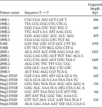
Several relaxed, well-preserved individuals with precise morphological identification were chosen for DNA isolation following the protocol by Floyd et al. (Reference Floyd, Abebe, Papert and Blaxter2002). Nuclear rDNA sequences (18S, 5.8S, 28S and internal transcribed spacer (ITS) regions) for S. macronectes were amplified using seven primer pairs (table 1), three for 18S, one spanning the ITS regions and 5.8S, and three for 28S. Polymerase chain reaction (PCR) amplifications were made using 50 μl of 1 × Ecogen Taq buffer (Ecogen, Madrid, Spain), 3 mmMgCl2, 0.2 mm of each deoxynucleotide triphosphate (dNTP), 10 μm of each primer and 1 U of Taq polymerase (Ecogen). Two microlitres of isolated DNA were used as the template for each reaction. The PCR conditions began with an initial denaturation step at 94°C for 5 min, followed by 36 cycles at 94°C for 1 min, 54°C for 1 min and 72°C for 2 min, and a final extension step at 72°C for 8 min. Five microlitres of each PCR product were checked in agarose gels stained with GelRedTM (Biotium, California, USA) and then purified with the GFXTM PCR DNA and Gel Band purification kit (GE Healthcare, Madrid, Spain). Fragments were then cloned into the pGEM® T-Easy vector (Promega, Madrid, Spain) and sequenced with M13 primers in an ‘ABI PRISMTM 310 Genetic Analyzer’ (Applied Biosystems, Madrid, Spain) automatic sequencer. The ‘ABI PRISMTM BigDye Terminator’ (Applied Biosystems) method was used with a 60-s injection time and 120-min run time. The ‘ABI PRISMTM 310 Collection v.1.1.2’ program was used for data acquisition, and ‘v.3.0 Sequencing Analysis’ (Applied Biosystems) for sequence analysis.
Primer pairs used for sequencing (molecular characterization) 18S, 5.8S, 28S and molecular diagnosis of ITS regions of Stegophorus macronectes.

For a diagnostic test for the presence of S. macronectes, species-specific primer pairs against the two ITS regions (ITS1 and ITS2), were designed. Primer pairs were validated in ten different worms by positive PCR amplification. Seven of the worms showed morphological alterations while the other three showed the morphology typically described for this species. PCR amplification conditions were as stated above. In addition, primer pairs were also tested using an egg solution prepared from gravid females.
Primer-pair specificity was assessed by testing these primers in another species of the Stegophorus genus, Stegophorus diomedeae (Johnston & Mawson, Reference Johnston and Mawson1942), isolated from Thalassarche melanophris Temminck (Chubut 2009), and in two other marine bird parasite genera of the family Acuariidae, Syncuaria sp., isolated from Phalacrocorax brasilianus Gmelin in Buenos Aires, Argentina in 2011, and Paracuaria adunca (Creplin, 1846) obtained from Larus dominicanus Lichtenstein in Chubut, Argentina in 2012. These specimens were collected, identified and provided by J.I.D. DNA was isolated using the QIAamp® DNA Mini Kit (Qiagen, Madrid, Spain), and PCR amplifications were performed using the aforementioned conditions.
Phylogenetic analyses
Sequences of 18S rDNA from Spirurida (n= 106) and from other orders (Strongylida, Oxyurida, Ascaridida and Rhabditida) were retrieved from GenBank (see supplementary table S1). Sequences, including S. macronectes, were aligned in ClustalX (Thompson et al., Reference Thompson, Gibson, Plewniak, Jeanmougin and Higgins1997) using default settings. The resulting alignment was checked and adjusted with Se-Al v2.0a11 (Rambaut, Reference Rambaut2002). A matrix with the final alignment was generated (available upon request from the corresponding author). The ITS regions, 5.8S and 28S sequences were not used due to the small number of Spirurida sequences available in GenBank. Gblocks (Castresana, Reference Castresana2000) was used to analyse the matrix. The complete and Gblocks matrices were compared.
The best-fit model for nucleotide substitution in the resulting matrix was GTR+I+G, determined by the Akaike information criterion (AIC) in a jModelTest (Posada, Reference Posada2008). Phylogenetic analyses were performed using PhyML v3.0 (Guindon & Gascuel, Reference Guindon and Gascuel2003) for maximum likelihood (ML), PAUP* v4.0b10 (Swofford, Reference Swofford2002) for maximum parsimony (MP), and MrBayes v3.1.2 (Ronquist & Huelsenbeck, Reference Ronquist and Huelsenbeck2003) for Bayesian inference (BI).
Supports for ML and MP analyses were determined by performing 1000 bootstrap replicates. For BI analyses, 5 million generations were performed in two parallel runs, sampling trees at 1000-generation intervals. The first 10% of sampled trees were discarded as burn-in, and the remaining trees were used to calculate the posterior probabilities. The maximum clade credibility tree was generated by TreeAnnotator (Drummond & Rambaut, Reference Drummond and Rambaut2007).
Results
Morphology
Stegophorus macronectes (Johnston & Mawson, Reference Johnston and Mawson1942). Spirurida, Acuariidae, Seuratinae.
Synonyms. Stegophorus paradeliae Johnston & Mawson, Reference Johnston and Mawson1945; Stegophorus adeliae Johnston, Reference Johnston1938 sensu Petter, Reference Petter1959.
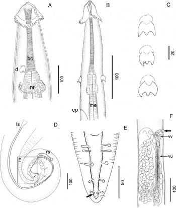
General morphology. Cuticle with fine transverse striations. Well-developed pseudolabia. Cephalic papillae at the same level as amphids and a short distance posterior to the oral opening. Pronounced apical process on each pseudolabium. Cephalic ornamentation appears as a collarette composed of two lateral lobes (hemi-collarettes) (fig. 1A, B). Each lobe emerges from the commissures of the buccal lips and has a continuous series of a varying number of teeth on its posterior border (fig. 1A, B). A short buccal capsule is lined with fine transverse striations. Large deirids, tridentate, with a sharp or blunt-ended middle denticle, sometimes bifid at the tip and shorter than the lateral denticle (fig. 1C). Deirids, almost always symmetrical, are usually located behind the junction of the buccal capsule and the oesophagus, but may sometimes occur at the same level (fig. 1A, B). Nerve ring usually located immediately posterior to buccal capsule–oesophagus junction, although occasionally located at the same level or less frequently anterior to it. Excretory pore posterior to nerve ring. Oesophagus straight, divided into muscular and glandular parts.
Stegophorus macronectes from Pygoscelis antarctica. (A) Female, anterior view; (B) male, anterior view; (C) deirids; (D) male, posterior end showing spiculae, papillae distribution and sessile papilla (black arrow); (E) male, detail of postcloacal papillae showing sessile papillae (black arrow) and phasmids (white arrow); (F) female, detail of vulva (black arrow), vagina vera, vagina uterine and eggs. bc, Buccal capsule; d, deirid; ep, excretory pore; ls, left spicule; me, muscular oesophagus; nr, nerve ring; rs, right spicule; vu, vagina uterine; vv, vagina vera.

Males: body length 7.15 ± 1.26 mm, maximum width 135 ± 25. Cephalic collarette length 56 ± 2.6. Deirids at 184 ± 30 from anterior end, 19 ± 2 long by 18 ± 1 wide. Nerve ring and excretory pore at 224 ± 57.4 and 250 from anterior end, respectively. Buccal capsule length 196 ± 55, muscular oesophagus length 602 ± 41 and glandular oesophagus length 1570 ± 30. Total oesophagus length 2170 ± 23. Long caudal alae present. Four pairs of precloacal papillae, first and third pairs smaller than second and fourth pairs (fig. 1D). Six pairs of postcloacal papillae, first and second pairs close to each other. Last three pedunculated pairs equally distant from each other. Inconspicuous sessile pair (sixth) of papillae at the base of fifth pedunculated pair (fig. 1E). Phasmids just behind the last pair of papillae. Left spicule very thin, 1034 ± 56 long, slightly dilated at distal end, ending in a sharply pointed tip. Right spicule 100 ± 17 long, slightly bent, ending in a crescent-like process pointing toward the anterior region. Tail 199 ± 20 in length (fig. 1D).
Females (all measurements are for specimens with uteri containing mature eggs): body length 17.7 ± 3 mm, maximum width 300 ± 71. Cephalic collarette length 87 ± 10. Deirids at 201 ± 30 from anterior end, 25.6 ± 1 long by 24 ± 3 wide. Nerve ring and excretory pore 239 ± 32.6 and 331 ± 71 from anterior end, respectively. Buccal capsule 201 ± 41 long. Muscular and glandular oesophagus 1500 ± 30 and 1350 ± 68 long, respectively. Vulva located at the end of the second third of body length, 9.96 mm from anterior end (45–64% of body length) on a small cuticular protuberance. Vagina divided into vagina vera and vagina uterine (fig. 1F). Mature larvated eggs (measured in the uterus near the ovejector) 42 ± 1 × 22 ± 1. Tail 159 ± 20 long.
Statistical analyses showed significant differences between the number of collarette teeth in males, females and immature specimens (H 2,813= 35.25, P < 0.0001). Immature specimens had fewer teeth on each hemi-collarette (mean = 15 ± 2.4) compared with adult males (mean = 18 ± 2.6) and females (mean = 17 ± 2.4).
Taxonomic summary
-
Type host. Macronectes giganteus (Gmelin).
-
Site of infection. Stomach.
-
Type locality. South Australia.
-
Other hosts. Thalassarche chrysostoma (Forster); Pygoscelis papua (Foster); Eudyptes chrysolophus (Brandt); Eudyptes chrysocome (Forster); Catharacta lonnbergi (Mathews); Chionis alba (Gmelin); Pygoscelis adeliae (Hombron & Jacquinot); Pygoscelis antarctica (Foster).
-
Other localities. Heard Island; King George Island.
-
Host and locality of present material. Pygoscelis antarctica Forster; Deception Island (South Shetland Islands).
-
Mean intensity ± SD and prevalence: 24.3 ± 28.9 and 72% in chicks (n= 61); 39.5 ± 43. 9 and 67% in adults (n= 3) (Vidal et al., Reference Vidal, Ortiz, Díaz, Ruiz de Ybañez, Amat, Palacios, Benzal, Valera, Dela Cruz, Motas and Barbosa2012).
-
Voucher specimens. Deposited in the Helminthological Collection, Museo de La Plata, La Plata, Argentina (MLP 6513) and in the Museo Nacional de Ciencias Naturales, Madrid, Spain (MNCN 11.01/403 and MNCN 11.01/404).
Remarks
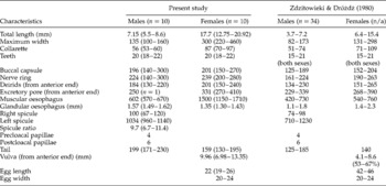
The general morphology and measurements of the specimens described here fully agree with those of S. macronectes provided by other authors (see Zdzitowieki & Dróżdż, 1980; table 2).
Measurements (means followed by range) of Stegophorus macronectes, given in the present study and by Zdzitowieki & Dróżdż (1980); measurements in micrometres unless otherwise stated; n, number of specimens examined; n/a, number not given.

According to the measurements given by other authors, the species is characterized as having a variable number of teeth, between 15 and 21, on the posterior border of each hemi-collarette (table 2). Most specimens isolated in this study were within this range; however, a few individuals had 11 teeth, while others had 27. Deirids are described as tridentate structures with the three cusps approximately equal in length, although the middle tooth is sometimes bifid (Johnston & Mawson, Reference Johnston and Mawson1942, 1945; Zdzitowiecki & Dróżdż, Reference Zdzitowiecki and Dróżdż1980). We observed some deirids with a bifid middle tooth, and some with one or two external bifid teeth. We also observed some deirids with two smaller protrusions between the main teeth (fig. 1C). In one specimen, we observed a deirid with a bifid middle tooth, while the deirid located on the opposite side had non-bifid teeth (fig. 1C).
Previous authors have described the deirid position at the buccal capsule–oesophagus junction, or posterior to it (Johnston & Mawson, Reference Johnston and Mawson1942, Reference Johnston and Mawson1945; Mawson, Reference Mawson1953; Zdzitowiecki & Dróżdż, Reference Zdzitowiecki and Dróżdż1980) and, in fact, have used this feature to distinguish the species (see Johnston & Mawson, Reference Johnston and Mawson1945; Zdzitowiecki & Dróżdż, Reference Zdzitowiecki and Dróżdż1980). However, some specimens may have been deformed, especially at the anterior end. In the present study, depending on the degree of contraction and specimen condition, we observed deirids appearing either before or after the buccal capsule–oesophagus junction.
Morphological features of S. macronectes match those of specimens studied by Petter (Reference Petter1959), who identified them as S. adeliae (Johnston, Reference Johnston1938). At present, the validity of S. adeliae appears doubtful. The original description was based on two distorted females, one of which was incomplete, consisting of only the posterior part (Johnston, Reference Johnston1938). Later, one of the two type specimens was assigned to a new species, S. paradeliae (Mawson, Reference Johnston and Mawson1945). Subsequently, other specimens, including males, were reported under this name (Mawson, Reference Mawson1953). Petter (Reference Petter1959), based on specimens from P. papua, considered S. paradeliae to be a synonym of S. adeliae. However, Zdzitowieki & Dróżdż (1980) considered S. paradeliae to be identical to S. macronectes. One author of the present study (J.I.D.) examined ten female specimens found in P. papua from the Petter nematode collection at the Musée d'Histoire Naturelle de Paris and found them to be identical to S. macronectes, supporting the finding of Zdzitowieki & Dróżdż (1980).
Molecular analysis
Nuclear rDNA sequences (18S, 5.8S, 28S and ITS regions) for S. macronectes were cloned and sequenced. Sequences from seven PCR products were assembled, resulting in a 6670-bp fragment (GenBank accession number HE793715) delimiting the 18S, 5.8S and 28S rDNA and ITS regions. Four species-specific primer pairs were then designed within the ITS regions for the molecular diagnosis of S. macronectes (table 1).
Adult S. macronectes having either the typical or altered morphologies resulted in positive PCR amplifications with the four primer pairs in all analyses. Positive amplifications were also obtained with a S. macronectes egg solution and in S. diomedeae, supporting the use of these primer pairs for diagnosis of Stegophorus spp., regardless of lifecycle stage or preservation condition. These four primer pairs were also tested in other Acuariidae genera. In Syncuaria sp., all reactions were negative. However, in Paracuaria adunca faint bands (slight amplification) were observed in reactions using the Steg2-ITS1 and Steg4-ITS2 primer pairs.
Phylogenetic analyses
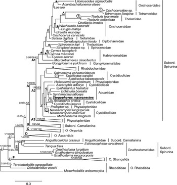
The final alignment for the 18S rDNA matrix consisted of 119 sequences of 1933 bp (718 bp were variable and informative characters). Using Gblocks on the complete matrix yielded a matrix consisting of 1523 characters (525 bp were variable and informative characters). Phylogenetic analyses of the complete and Gblocks matrices resulted in similar findings for the major groups. However, the complete matrix consisted of more informative characters, resulting in trees that showed greater resolution. Results of the phylogenetic analyses with the complete matrix using BI, ML and MP approaches are summarized in fig. 2.
Phylogenetic relationship between species of the Spirurida order. Tree topology was inferred by Bayesian analysis, based on 18S rDNA. A, B, C, D, E and F show the main clades. The numbers on the main branches show the Bayesian posterior probability and bootstrap support found under maximum parsimony and maximum likelihood criteria, respectively. 1 shows strongly very well-supported clades (pp = 1; bootstrap = 100). Stars mark other well-supported clades (pp ≥ 0.95; bootstrap ≥ 70). Circles mark pp ≥ 0.8; bootstrap ≥ 50. Triangles indicate pp ≥ 0.8 and bootstrap ≥ 50 for at least one method.

Stegophorus macronectes was situated within the cluster A, corresponding to the Spirurina suborder. Within this clade, S. macronectes appeared in a highly supported cluster (A2) that included other representatives of the family Acuariidae, and representatives from the Rhabdochonidae, Cystidicolidae and Physalopteridae families. However, most species from the Physalopteridae (collapsed in fig. 2) family grouped together (A3) outside of this monophyletic assemblage.
There was no clear structure among the different families in cluster A2 (fig. 2). The Acuariidae family cluster, which S. macronectes belongs to, was not highly supported, and was related to Ascarophis adioryx (Cystidicolidae) with high bootstrap and posterior probability. In fact, species considered as belonging to the Cystidicolidae family were distributed among different clusters.
The cluster A2 sister group was not clearly established due to a polytomy at this level. The relationships among clusters A2 and A3 (comprising the Physaloptera and Turgida (Physalopteridae) genera) and A1 (comprising representatives of the Onchocercidae, Tetrameridae, Thelaziidae, Setariidae, Diplotriaenidae, Spirocercidae, Habronematidae families, and Gongylonematidae) were not resolved.
The Philometridae, Dracunculidae, Skyrjabillanidae, Daniconematidae and Camallanidae families comprised a second large cluster in the Spirurida order (B), corresponding to the Camallanina suborder (this appears collapsed in fig. 2). Of the four different orders taken as outgroups (each one represented by three different species), two (Rhabditida and Strongylida) were at the base of the tree; the other two (Ascaridida and Oxyurida) were more closely related to the two main Spirurida clusters (A and B) than to cluster E, which consisted of two families also considered Spirurida (Anguillicolidae and Gnathostomatidae), thereby breaking the Spirurina and Camallanina suborder monophyly.
Discussion
The presence of a variable number of teeth on the posterior border of the collarette has been used as a diagnostic feature for delimiting species of the Stegophorus genera. However, our results in S. macronectes show that the number of teeth can exceed the range described in the literature (see table 2), suggesting that this feature increases asymmetrically during nematode development. Therefore, variations in the number of teeth on the collarette make this criterion a poor diagnostic feature, unless the difference in the number of teeth on each hemi-collarette is clearly delineated in the different species (e.g. ~10 in S. diomedeae vs. ~20 in S. macronectes). Moreover, deirid morphology and position are highly variable in this species. Deirids are commonly found at the level of the buccal capsule–oesophagus junction but can also be found at other levels, depending on specimen contraction and preservation.
The morphological deformities observed in some specimens are likely due to poor preservation, either because of the time elapsed from host death to collection or due to preservation by freezing. Parasites undergo internal and external changes, including internal decomposition and detachment of the cuticle, when frozen (Pritchard & Kruse, Reference Pritchard and Kruse1982). During this process, the cuticle tends to move frontally while the body of the parasite retracts backwards, hindering specific identification of some traits, such as deirid position. In addition, the oesophagus and buccal capsule can move back and, sometimes, the lateral lobes appear folded. The same problem was reported by Zdzitowiecki & Dróżdż (Reference Zdzitowiecki and Dróżdż1980), who provided morphometric data for two S. macronectes specimens, one straight and one contracted. Using altered traits (i.e. deirid position relative to the buccal capsule and oesophagus) could lead to erroneously describing different species. However, our molecular results show that, although relaxed and contracted specimens may appear different, they belong to the same species.
Primer pairs designed from the sequences of ITS regions were used to test for the molecular detection of S. macronectes. Four primer pairs were validated against different species of Stegophorus spp., with positive results, thus proving useful at the genus level. However, two of the primer pairs (Steg2-ITS1 and Steg4-ITS2) were also positive for another genus of the Acuariidae family. Therefore, we recommend that the other two primer pairs (Steg1-ITS1 and Steg3-ITS2) be used for greater specificity.
The phylogenetic relationships of spirurid nematodes have been studied for many years (Blaxter et al., Reference Blaxter, De Ley, Carey, Liu, Scheldeman, Vierstraete, Vanfleteren, Mackey, Dorris, Frisse, Vida and Thomas1998; Nadler et al., Reference Nadler, Carreno, Mejía-Madrid, Ullberg, Pagan, Houston and Hugot2007; Černotíková et al., Reference Černotíková, Horák and Moravec2011). Our results show that analyses of 18S rDNA sequences correctly classified S. macronectes within the Acuariidae family, though this was not strongly supported. The robustness of analyses were likely hampered by the scarcity of available sequences; for instance, for the 21 genera in this family (Skrjabin, Reference Skrjabin1949), there are only four 18S rDNA sequences available in the NCBI database. More sequences are therefore necessary to improve our knowledge of the relationships within the Acuariidae family.
The composition of the main Spirurida clades in our phylogenetic reconstructions is in agreement with previous studies (Černotíková et al., Reference Černotíková, Horák and Moravec2011): clade A corresponds to Spirurina (except for the Gnathostomatidae family), clade B to Camallanina (except for Anguillicoloides crassus), and clade E is comprised of A. crassus and Gnathostomatidae, supporting the non-monophyly of Dracunculoidea and Spirurina. However, the relationships within these clades differ slightly from those in other studies (Nadler et al., Reference Nadler, Carreno, Mejía-Madrid, Ullberg, Pagan, Houston and Hugot2007; Van Megen et al., Reference Van Megen, Van den Elsen, Holterman, Karssen, Mooyman, Bongers, Holovachov, Bakker and Helder2009; Černotíková et al., Reference Černotíková, Horák and Moravec2011). More Spirurida sequences were used in this study and may account for these differences. Results for species belonging to other orders, such as Strongylida, Oxyurida, Ascaridida and Rhabditida, were not always in agreement with previous phylogenetic studies. This is because only three species per order were chosen at random as outgroups for these analyses.
In any case, our analyses only consisted of a single gene (18S), which may account for the inconsistencies observed between our phylogenetic reconstructions and the classical taxonomy of these groups. Additional genes should be included to clarify whether these results are a consequence of homoplasy of some morphological characters or simply represent the phylogenetic relationships of a particular gene.
Supplementary material
To view supplementary material for this article, please visit http://dx.doi.org/10.1017/S0022149X15000218
Acknowledgements
We thank the Spanish Antarctic research base ‘Gabriel de Castilla’, the Spanish Polar ship Las Palmas, the Argentinean Antarctic Institute and the Maritime Technology Unit (UTM-CSIC) for logistical support and transport. Permission to work and collect samples in the study area was given by the Spanish Polar Committee. We thank Maria Cristina Estivariz (Centro de Estudios Parasitológicos y de Vectores (CEPAVE)), for help with the drawings. This is a contribution to International Polar Year Project 172 BirdHealth, the PINGUCLIM project and to the Unidad Asociada de Ecología e Inmunología parasitaria of the University of Murcia and the Spanish Council of Scientific Research (CSIC). Melinda Modrell corrected the English language usage. We greatly appreciate the reviewers’ comments, which have improved an early version of the manuscript.
Financial support
This study was funded by the Spanish Ministry of Economy and Competiveness and the European Regional Development Fund Projects CGL2004-01348, POL2006-05175, CGL2007-60369 and CTM2011-24427. V.V. was supported by a PhD grant from the Spanish Council of Scientific Research (CSIC) and the European Social Fund (JAEPre08-01053). M.J.P. was supported by a PhD grant from the Spanish Ministry of Science and Innovation (BES2005-8465). J.I.D. is a member of CONICET and is partially supported by PIP 698 (CONICET), N628 and N726 (UNLP).
Conflict of interest
None.