INTRODUCTION
‘Lithistid’ sponges are a polyphyletic group of Demospongiae (Phylum Porifera) with a rigid articulated skeleton of desma spicules of diverse morphology (Pisera & Lévi, Reference Pisera, Lévi, Hooper and Van Soest2002a, Reference Pisera, Lévi, Hooper and Van Soestb). Due to their articulated skeleton, lithistids preserve well as fossils, and most lithistid families have a long palaeontological record. The term ‘Lithistida’ is still used by palaeontologists to denote a morpho-functional group, not one evolutionary lineage (Pisera, Reference Pisera, Hooper and Van Soest2002).
Morphological data have long supported the inclusion of lithistids with astrose microscleres in the Order Astrophorida Sollas, Reference Sollas1888. Based on the integration of molecular and morphological data, Cárdenas et al. (Reference Cárdenas, Perez and Boury-Esnault2012) resurrected the order Tetractinellida Marshall, Reference Marshall1876, with suborders Astrophorina Sollas, Reference Sollas1888 and Spirophorina Bergquist & Hogg, Reference Bergquist and Hogg1969. Eight of the 13 extant lithistid families have affinities with the Astrophorina (Cárdenas et al., Reference Cárdenas, Xavier, Reveillaud, Schander and Rapp2011; Schuster et al., Reference Schuster, Erpenbeck, Pisera and Wörheide2012; Redmond et al., Reference Redmond, Morrow, Thacker, Diaz, Boury-Esnault, Cardenas, Hajdu, Lobo-Hajdu, Picton, Pomponi, Kayal and Collins2013): Corallistidae Sollas, Reference Sollas1888, Isoraphiniidae Schrammen, Reference Schrammen1924, Macandrewiidae Schrammen, Reference Schrammen1924, Neopeltidae Sollas, Reference Sollas1888, Phymaraphiniidae Schrammen, Reference Schrammen1924, Phymatellidae Schrammen, Reference Schrammen1910, Pleromidae Sollas, Reference Sollas1888 and Theonellidae von Lendenfeld, Reference von Lendenfeld1903. Lithistids with sigmaspire microscleres, as well as any with rhizoclone desmas and no microscleres, belong to the Suborder Spirophorina, while other groups are either incertae sedis or are attributable to other groups of demosponges.
Most lithistids occur worldwide in deep-water environments, but locally can be found in shallow water. They are not well known in the tropical western Atlantic, despite the fact that they were first described by Schmidt (Reference Schmidt1870, Reference Schmidt1879, Reference Schmidt1880). Van Soest & Stentoft (Reference Van Soest and Stentoft1988) described the lithistid fauna of Barbados as part of their survey of deep-water demosponges from that region. More recent biological and chemical interest in lithistid sponges is based on the fact that they are the source of more than 300 novel and diverse compounds, including polyketides, cyclic and linear peptides, alkaloids, pigments and sterols, many of which have potential human health applications (for review, see Wright, Reference Wright2010; Winder et al., Reference Winder, Pomponi and Wright2011). Systematic surveys of deep-water benthic habitats of the continental shelf and slope of the south-eastern USA, Bahamas, Caribbean and Gulf of Mexico have been conducted by the Harbor Branch Oceanographic Institute (HBOI) since 1984, as part of a marine natural products drug discovery programme. Pomponi et al. (Reference Pomponi, Kelly, Reed and Wright2001) described the diversity and bathymetric distribution of lithistids collected during these expeditions, and the chemotaxonomy of undescribed species of the genus Discodermia was discussed by Kelly-Borges et al. (Reference Kelly-Borges, Robinson, Gunasekera, Gunasekera, Gulavita and Pomponi1994), who reported at least seven chemotaxonomic groups, including D. dissoluta and D. verrucosa. Re-description of Setidium obtectum was presented by Pisera (Reference Pisera1999). Recently, Ruiz et al. (Reference Ruiz, Valderrama, Zea and Castellanos2013) described mariculture and production of the antitumour compounds of D. dissoluta from Colombia.
This paper describes one new and two known species of Theonellidae from deep waters off the Florida shelf, thus contributing to our knowledge of the biodiversity of lithistid sponges in the Caribbean.
MATERIALS AND METHODS
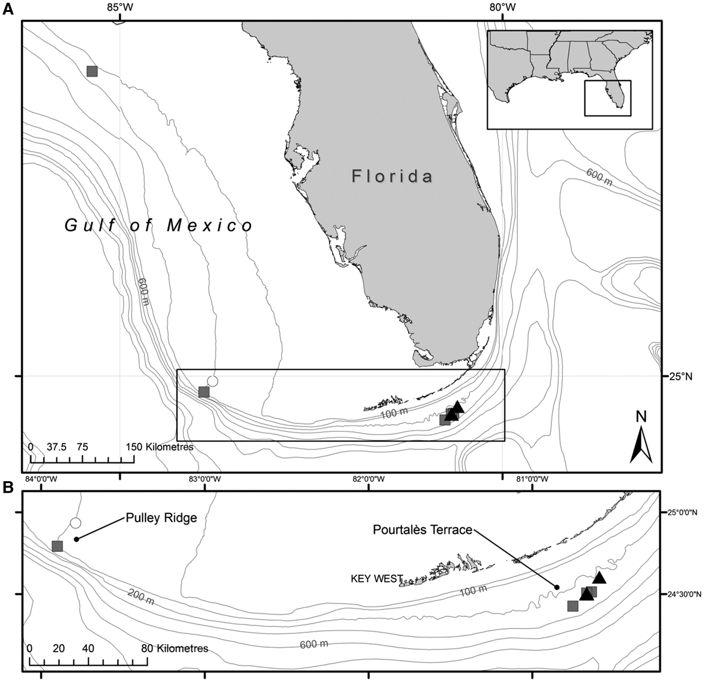
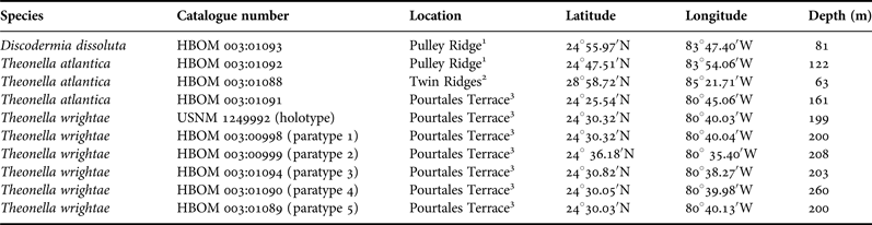
Samples were collected using the ‘Johnson Sea Link’ manned submersibles (HBOI) or remotely operated vehicles (‘Kraken II’, University of Connecticut; ‘Innovator’, Subsea International) during expeditions to the Florida shelf. Habitats and collection methods are described by Reed et al. (Reference Reed, Pomponi, Weaver, Paull and Wright2005). Collection site data (locations and depths) for each sample are detailed in Table 1 and Figure 1. Samples were preserved in ethanol (70 or 95%) immediately after collection. All samples are archived in the Harbor Branch Oceanographic Museum (HBOM) at Florida Atlantic University (FAU) in Fort Pierce, Florida. The holotype of Theonella wrightae is deposited in the US National Museum (USNM) in Washington, DC.
Collection locations of the lithistid sponges in this study. (A) Florida shelf areas (insert at upper right indicates location of box A within the USA; insert at bottom is enlarged in 1B). Open circle – Discodermia dissoluta; grey squares – Theonella atlantica; dark triangles – Theonella wrightae. (B) Enlarged area showing Pulley Ridge and Pourtalès Terrace.

Harbor Branch Oceanographic Museum (HBOM) catalogue numbers and collection data for studied material. All samples were collected off the coast of Florida (Figure 1).
1Gulf of Mexico, south-west Florida shelf.
2Gulf of Mexico, north-west Florida shelf.
3Straits of Florida, south Florida shelf.
Each specimen was examined microscopically to identify differences in spiculation due to functional (e.g. near ostia or oscules) and/or structural (e.g. upper surfaces, tips of branches) differences in the sponge body. In each such case, a separate sample was prepared to include part of the ectosome. Spicules were prepared by boiling fragments of each sample in a glass tube in 70% nitric acid to remove organic matter. After the acid cooled, the sample was washed several times with distilled water (until the pH was neutral), and twice more with pure propanol. The suspension of loose spicules was stored in propanol in a small plastic bottle. Remaining fragments of the choanosomal desma skeleton were washed again with propanol to remove loose spicules, air-dried, and attached to a scanning electron microscope (SEM) stub with LeitC carbon glue so that the upper, natural surface of the choanosomal skeleton could be observed under SEM. Because the architecture of the choanosomal skeleton can be diagnostic, it is important to never use a sectioned surface for basic observation of the desma architecture. The suspension of loose spicules in propanol was pipetted and dropped (one or more drops, depending on density of spicules in suspension) directly on the SEM stub (i.e. without the use of glue or adhesives), or on the clean surface of a round cover glass first attached with carbon glue to the SEM stub, then dried.
Another subsample (i.e. a fragment not treated with nitric acid) was dried and attached to an SEM stub with LeitC carbon glue, so that the upper ectosomal surface could be observed. Samples prepared for SEM analyses were then sputter-coated with platinum and observed at 25 kV using a Philips XL-20 scanning electron microscope (Institute of Paleobiology, Warsaw).
RESULTS
Ten specimens were analysed and assigned to two genera and three species within the Family Theonellidae. All are new records for Florida and one is a new species.
SYSTEMATICS
Class DEMOSPONGIAE Sollas, Reference Sollas1885
Order TETRACTINELLIDA Marshall, Reference Marshall1876
Suborder ASTROPHORINA Sollas, Reference Sollas1888
Family THEONELLIDAE von Lendenfeld, Reference von Lendenfeld1903
Genus Theonella Gray, Reference Gray1868
TYPE SPECIES
Theonella swinhoei Gray, Reference Gray1868, p. 566, figures 1–3.
REMARKS
Fourteen species of the genus Theonella Gray, Reference Gray1868 have been accepted as valid globally (Van Soest, Reference Van Soest, Van Soest, Boury-Esnault, Hooper, Rützler, de Voogd, Alvarez de Glasby, Hajdu, Pisera, Manconi, Schoenberg, Janussen, Tabachnick, Klautau, Picton, Kelly, Vacelet, Dohrmann and Díaz2012a), but most of them, with the exception of T. atlantica Van Soest & Stentoft, Reference Van Soest and Stentoft1988 known from Barbados, are from the Indo-West Pacific region. Most of these species have been described only once and usually without proper illustration of spiculation, including T. atlantica. On the other hand, numerous records of T. swinhoei, that has apparently very wide distribution, may actually represent a complex of similar species and may need revision.
Theonella atlantica Van Soest & Stentoft, Reference Van Soest and Stentoft1988
(Figures 2A–E, 4 & 5)
Theonella atlantica Van Soest & Stentoft, Reference Van Soest and Stentoft1988, p. 48, figure 21, pl. 6: 5–6.
Habitus of the studied lithistid sponges (A, C, underwater photos taken from the submersible). (A–E) Theonella atlantica Van Soest & Stentoft, Reference Van Soest and Stentoft1988: (A, B) HBOM 003:01091); (C, D) HBOM 003:01088; (E) HBOM 003:01092; (F) Discodermia dissoluta Schmidt, Reference Schmidt1880, HBOM 003:01093.
Habitus of Theonella wrightae sp. nov. (A, B, C, E, underwater photos taken from the submersible). (A) HBOM 003:00999; (B) HBOM 003:00998; (C, D) HBOM 003:01089; (E) holotype, USNM 1249992; (F) HBOM 003:01090.
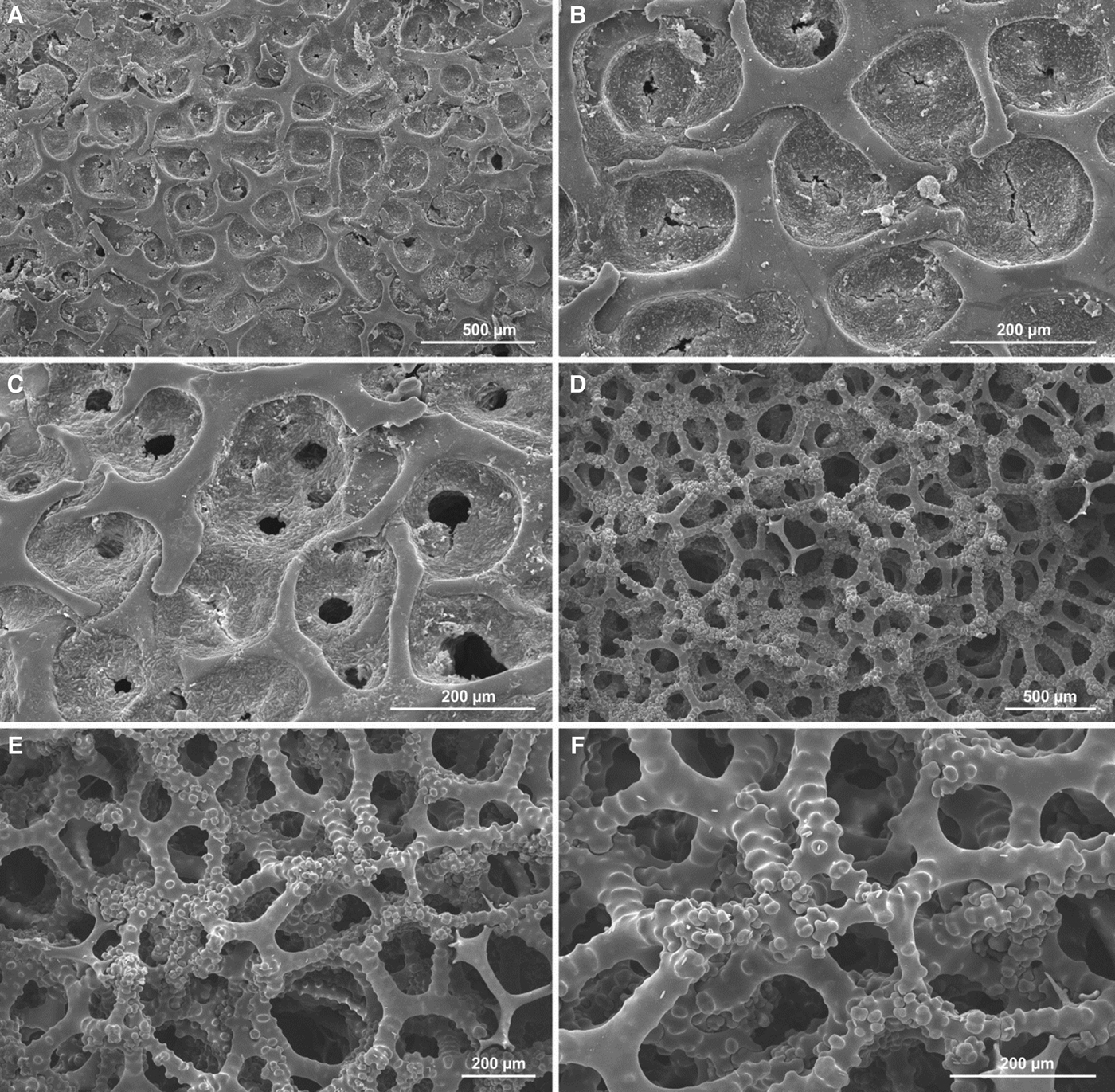
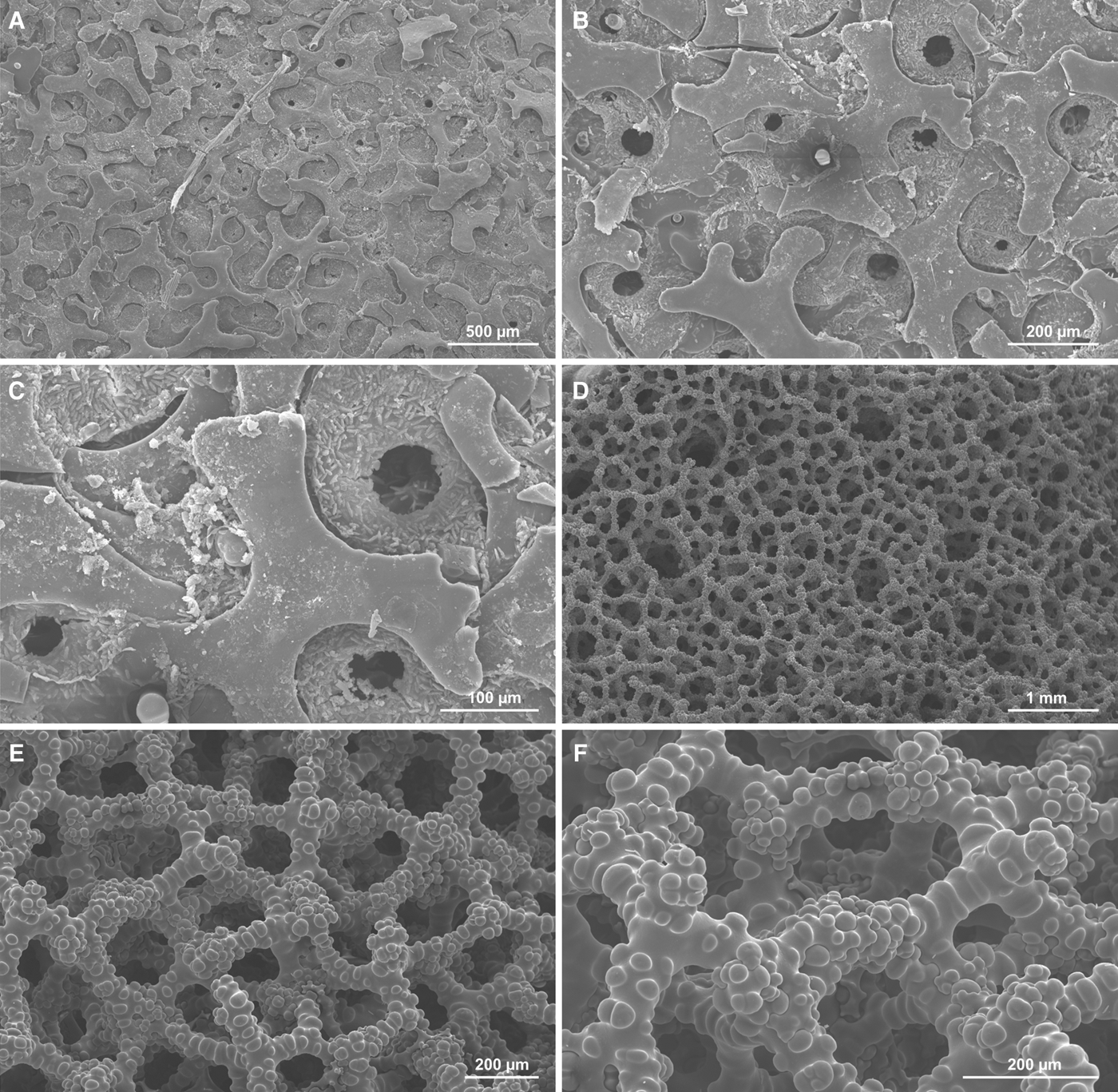
Theonella atlantica Van Soest & Stentoft, Reference Van Soest and Stentoft1988, HBOM 003:01092. (A–C) View of the natural ectosomal surface with smooth phyllotriaenes with narrow elongated cladomes; between is dense crust of microscleres; ostia openings visible in (C); (D–F) choanosmal skeleton of tetraclone desmas; note only partial tuberculation (E, F), young smooth desmas still not entirely articulated with the adult desmas (lower right on E) and strongly tuberculated zygomes of desmas (F).
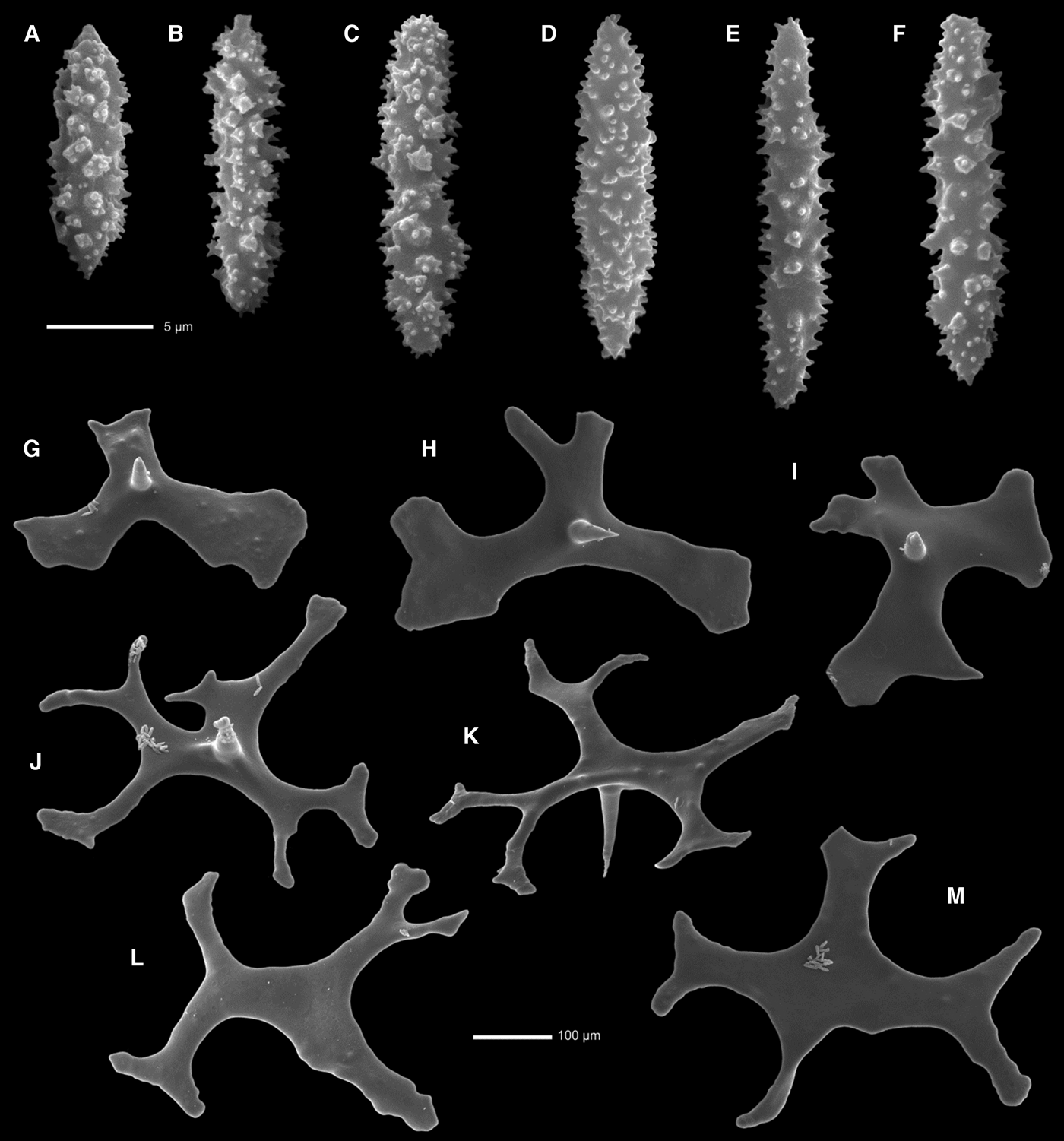
Theonella atlantica Van Soest & Stentoft, Reference Van Soest and Stentoft1988, HBOM 003:01092. (A–F) Fusiform acanthorhabd microscleres; (G–M) variously shaped ectosomal smooth phyllotriaenes, (G–J) in lower surface view, note conical short rhabd, and upper or oblique view (K–M).
MATERIAL
HBOM 003:01092, 122 m; HBOM 003:01088, 63 m; HBOM 003:01091, 161 m; widely distributed – northwest, southwest, and south Florida shelf (see Figure 1, Table 1).
DESCRIPTION
Variable in shape: amorphous encrusting to lobate, may be also pedunculate with cylindrical coalescing fingers (Figure 2A–E), reaching a size of 6 cm across. Colour: orange exterior (light orange interior) when alive. Consistency: firm/hard to stony and brittle. Oscules: apical, may be single or double, 1 mm in diameter, and also on lateral surface of branches, similar in size, also single, double, or triple, on slight elevations. Ostia: in vague fields, 43–57 µm in diameter (Figure 4C).
SPICULES
Ectosomal spicules as smooth phyllotriaenes with long narrow cladi that are sinuous and have rounded tips; in some areas of sponge surface, cladi are much wider or spicules resemble irregular discotriaenes (not shown), cladome clearly convex, rhabdome short conical and massive, 160–200 µm long, cladome diameter is 515–753 µm (Figures 4A–C & 5G–M). Space between cladomes with dense crust of microscleres (Figure 4B, C); desmas as massive tetraclones that are smooth near the centre and become progressively tuberculated toward tips, with strongly tuberculated zygomes (Figure 4E, F); they measure 380–490 µm in size; Microscleres are fusiform acanthorhabds (Figure 5A–F) that measure 13.2–17.9 × 3.0–5.15 µm.
REMARKS
This sponge is uncommon at the collecting sites and occurs at depths from 63–161 m. Theonella atlantica was earlier reported from Barbados by Van Soest & Stentoft (Reference Van Soest and Stentoft1988) from a depth of 144–153 m.
OCCURRENCE
This species is known only from Barbados and the Florida shelf (both Atlantic and Gulf of Mexico).
Theonella wrightae sp. nov.
(Figures 3 & 6–9)
Theonella wrightae sp. nov. HBOM 003:00998. (A–C) View of the natural ectosomal surface covered with phyllotriaenes with wide smooth cladome surface; between cladi, dense crust of microscleres; small openings are ostia; (D, E, F) details of upper surface of the choanosomal skeleton of tetraclone desmas to show sculpture and articulation; note openings of choanosomal canals in the desma skeleton (D).
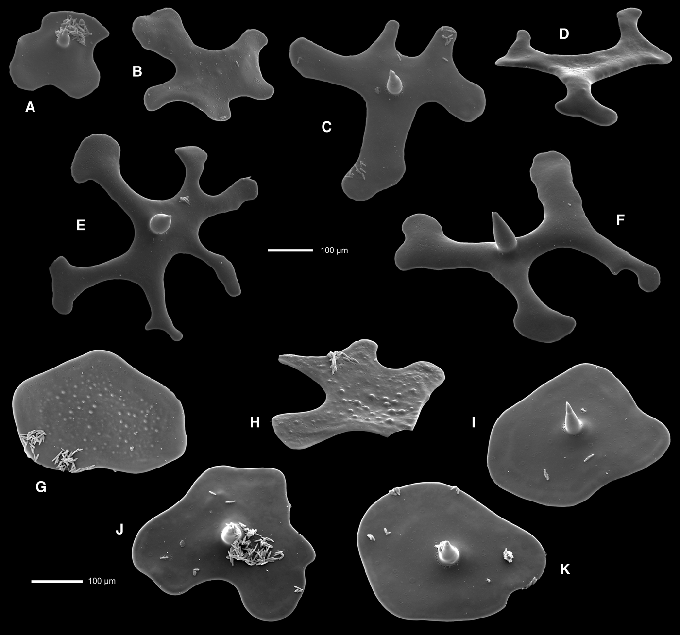
Theonella wrightae sp. nov. Ectosomal phyllo- to discotriaenes in various views to illustrate variability between and within specimens. (A–F) HBOM 003:00997; (G–K) HBOM 003:01094.
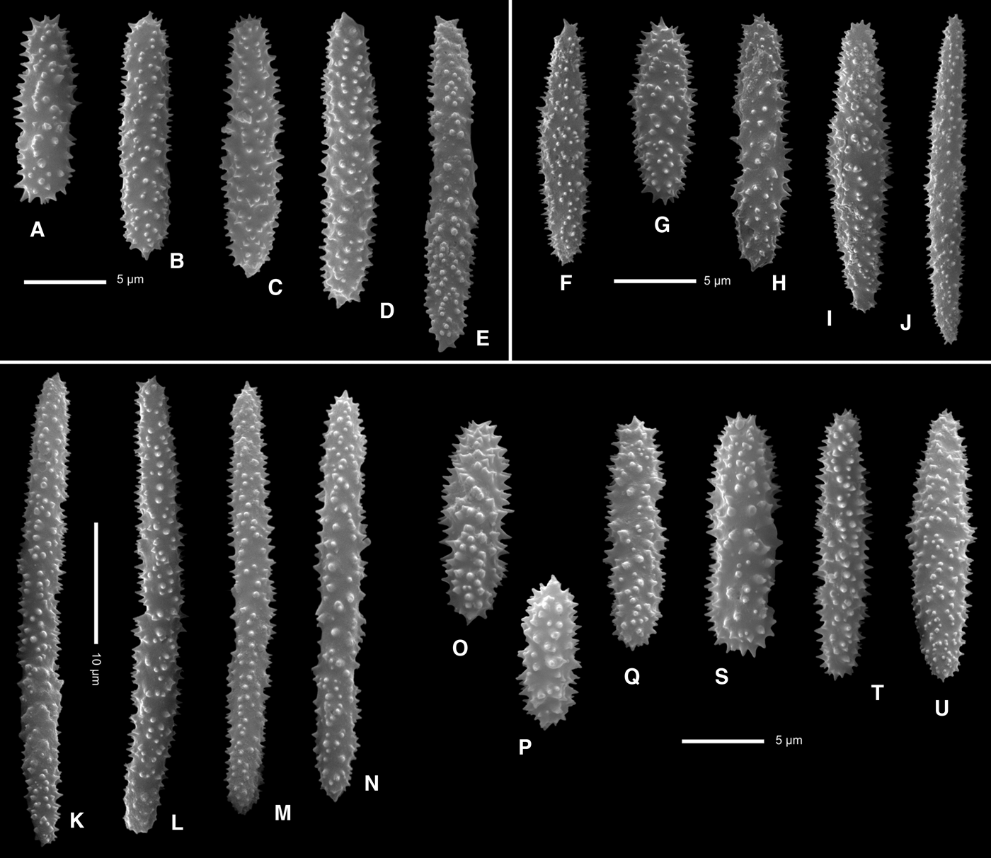
Theonella wrightae sp. nov., microscleres from three specimens to illustrate variability between and within specimens. (A–E) Holotype, USNM 1249992; (F–J) HBOM 003:00998; (K–U) HBOM 003:01094.
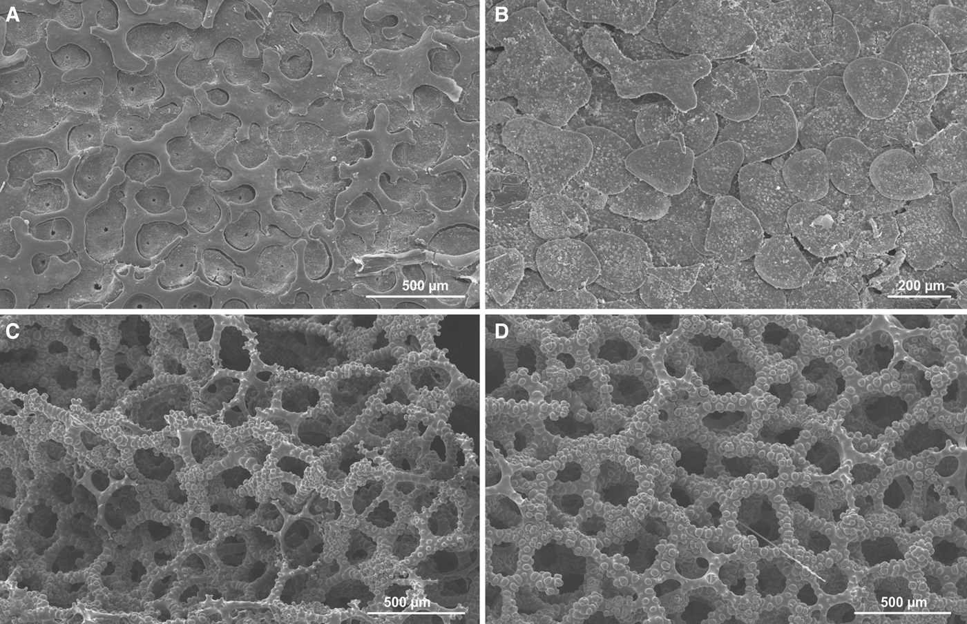
Theonella wrightae sp. nov., spiculation in various specimens to illustrate variability (similar variability may be observed within one specimen). (A) Ectosomal phyllotrianes, HBOM 003:01090; (B) tuberculated ectosomal discotriaenes, HBOM 003:01089; (C) choanosomal skeleton of tetraclone desmas, HBOM 003:00999; (D) choanosomal skeleton of tetraclone desmas, HBOM 003:01090.
TYPE MATERIAL
USNM 1249992 (holotype), 199 m; HBOM 003:00998 (paratype 1), 200 m; HBOM 003:00999 (paratype 2), 208 m; HBOM 003:01094 (paratype 3), 203 m; HBOM 003:01090 (paratype 4), 260 m; HBOM 003:01089 (paratype 5), 200 m; all from Pourtalès Terrace, south Florida shelf (see Figure 1, Table 1).
DIAGNOSIS
Theonella forming clusters of knobby fingers that are first round then flattened; oscules on elevations on one side of the branches; ectosomal phyllotriaenes to discotriaenes often tuberculated on the upper surface; desmas strongly tuberculated massive tetraclones; microscleres as very variable in size and shape (from cylindrical to fusiform) acanthorhabds.
ETYMOLOGY
The species is named in honour of Dr Amy E. Wright, HBOI-FAU, in recognition of her contributions to the discovery of new deep-water sponges, in general, and lithistid sponges, in particular, and her research on human health applications of natural products derived from lithistid sponges. Dr Wright selected and collected this sponge during a dive in the ‘Johnson-Sea-Link’ manned submersible.
DESCRIPTION
Sponge in the form of a cluster (10–20 cm across and 5 cm tall) of knobby fingers (sometimes anastomosing), fingers 0.7–1 cm in diameter, round when younger, later become flattened, sometimes coalescing to form a flat fan. Colour: purple brown, red to brick-red or dark red when alive (Figure 3), cream-coloured inside; cream-coloured in ethanol. Consistency: firm to hard, and crisp or brittle. Oscules: 1 mm or less, usually on lateral surface of branches but located only on one side, similar in size; located on slight elevations. There is a system of subectosomal canals (developed also on the surface of choanosomal skeleton) radiating from each oscule.
SPICULES
Ectosomal spicules are mostly phyllotriaenes (Figures 6A–C, 7 & 9A–B) with wide cladome and branches that are slightly convex; phyllotriaene cladomes are 348–717 µm in diameter; they may be substituted with discotriaenes (Figures 7G, I–K & 9B) that are 330–475 µm in diameter, and there is a complete set of transitional morphologies; upper surface, especially in discotriaenes, with numerous low tubercles; rhabd short massive and conical; dense crust of microscleres between cladomes of phyllotrianes (Figure 6C). Desmas: massive, strongly tuberculated tetraclones with very strong zygoses (Figures 6D–F & 9C, D). Microscleres: acanthorhabds, strongly variable in size and shape (Figure 8); they may be short and fusiform or more or less cylindrical (more common), 9.3.0–20.0 µm long and 3.17–4.27 thick in the middle, to long cylindrical (rare), 31.8–38.1 µm long and 3.7–4.4 µm thick in the middle.
REMARKS
The new species differs from other species of the genus Theonella in having oscules on slight elevations occurring on one side of the sponge, and in its ectosomal spicules that may be phyllotriaenes to discotriaenes, and that may be tuberculated on the upper surface. This sponge occurs on rock pavement, and is uncommon at the sampling sites. The spiculation of this species is very variable in the sense that in areas with various structures/functions (e.g. inhalant ostial vs non-inhalant areas, tips of the branch, areas near oscules, etc.), both ectosomal spicules and desmas vary considerably (Figures 7 & 9).
DISTRIBUTION
This sponge is known only from the south Florida shelf.
Genus Discodermia du Bocage, Reference du Bocage1870
TYPE SPECIES
Dactylocalyx polydiscus Bowerbank, Reference Bowerbank1869, p. 96, pl. 6: 10–13; non Discodermia polydiscus du Bocage, Reference du Bocage1870, p. 15, pl. 11:1
REMARKS
There are 29 species of the genus Discodermia accepted as valid globally, most from the Indo-Pacific region. Only four species are reported from the Caribbean region: D. inscripta (Schmidt, Reference Schmidt1879), D. dissoluta Schmidt, Reference Schmidt1880, D. polydiscus (Bowerbank, Reference Bowerbank1869) and D. verrucosa Topsent, 1928 (Pomponi et al., Reference Pomponi, Kelly, Reed and Wright2001, Van Soest, Reference Van Soest, Van Soest, Boury-Esnault, Hooper, Rützler, de Voogd, Alvarez de Glasby, Hajdu, Pisera, Manconi, Schoenberg, Janussen, Tabachnick, Klautau, Picton, Kelly, Vacelet, Dohrmann and Díaz2012b).
Discodermia dissoluta Schmidt, Reference Schmidt1880
(Figures 2F, 10 & 11)
Discodermia dissoluta Schmidt, Reference Schmidt1880, p. 87, pl. 5: 2; Sollas, Reference Sollas1888, p. 328. Discodermia dissoluta (Schmidt, Reference Schmidt1880): Lehnert & Van Soest, Reference Lehnert and Van Soest1996, p. 55, figures 2, 39–42; Zea, Reference Zea1987 p. 220, figure 80 (p. 221), pl. 5 (p. 265).
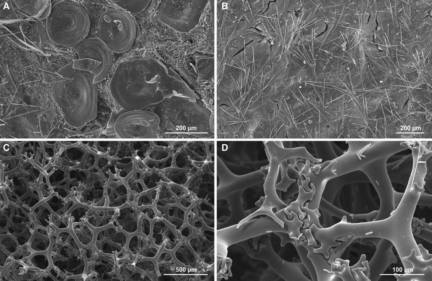
Discodermia dissoluta Schmidt, Reference Schmidt1880, HBOM 003:01093. (A) View of the natural ectosomal surface with loosely distributed discotriaenes and a dense crust of microscleres in between; (B) another region of the natural ectosomal surface where discotriaenes are entirely covered by soft tissue; note bunches of short oxeas protruding from the surface; (C) choanosomal skeleton of tetraclone desmas after nitric acid treatment; note smooth and delicate regular tetraclones; (D) articulation of tetraclone desmas, small objects on the desma surface are microscleres.
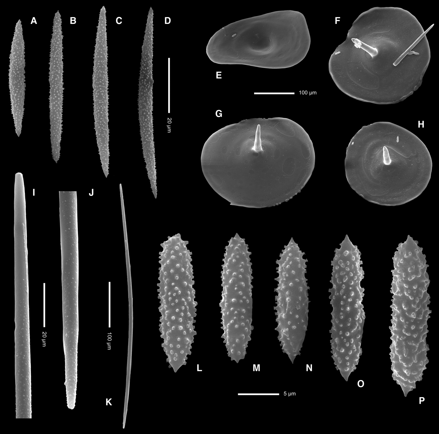
Discodermia dissoluta Schmidt, Reference Schmidt1880, HBOM 003:01093. (A–D) Fusiform acanthoxea microscleres; (E–H) ectosomal disctotrianes in various views (E –top view, F–H – underside views showing short conical rhabdome); (I–K) oxeas/styles (I, J enlargement to show rounded tips, K, general view); (L–P) acanthorhabd microscleres.
MATERIAL
HBOM 003:01093, Florida, 81 m (see Figure 1, Table 1).
Other material examined: D. dissoluta, Curaçao, 25 m depth. ZMA POR 4600.
DESCRIPTION
Shape: cluster of knobby fingers (Figure 2F), 20 cm diameter, 5 cm tall, fingers 1 cm diameter. Colour: purple brown exterior when alive, cream-coloured interior. Consistency: firm and brittle. Oscules: about 1 mm in diameter, scattered on the surface.
SPICULES
Ectosomal spicules are round, concave and smooth (except growth lines) discotriaenes, 203–294 µm in diameter, with a short delicate conical rhabd (Figures 10A & 11E–H). Discotriaenes usually loosely organized on the surface, with dense crust of microscleres between them (Figure 10A). In places, discotriaenes are completely covered by ectosome, and fan-like bunches of short oxeas protrude from the choanosome (Figure 10B). Desmas are smooth, regular tetraclones with weak zygosis (Figure 10C, D), 475–525 µm in size. Microscleres are of two types: the first are fusiform acanthoxeas (Figure 11A–D) that are variable in size, 41.6–68.0 µm in length and 5.5–6.1 µm thick in the middle; the second are fusiform acanthorhabds with pointed tips (Figure 11L–P); they measure 15.1–18.9 µm in length and 4.3–5.2 µm thick in the middle. There are also curved oxeas/styles (Figure 11I–K) that occur commonly and are approximately 500–530 µm long and about 9–10 µm thick.
REMARKS
Discodermia dissoluta has been reported from numerous sites in the tropical western Atlantic and Caribbean (Bahamas, Barbados, Cuba, Curacao, Colombia, Jamaica) (Zea, Reference Zea1987; Kelly-Borges & Pomponi, Reference Kelly-Borges and Pomponi1994; Kelly-Borges et al., Reference Kelly-Borges, Robinson, Gunasekera, Gunasekera, Gulavita and Pomponi1994; Lehnert & Van Soest, Reference Lehnert and Van Soest1996, Pomponi et al., Reference Pomponi, Kelly, Reed and Wright2001, Van Soest, Reference Van Soest, Van Soest, Boury-Esnault, Hooper, Rützler, de Voogd, Alvarez de Glasby, Hajdu, Pisera, Manconi, Schoenberg, Janussen, Tabachnick, Klautau, Picton, Kelly, Vacelet, Dohrmann and Díaz2012b), but not from Florida. Usually it is poorly illustrated and some of the species identifications may require verification, due to very different morphologies reported for the specimens described. We have observed large size variability of acanthoxea microscleres, even within one specimen (Figure 11). The examined specimen from Curaçao (ZMA POR 4600) is very similar to the material examined for this study, both in morphology and spicules. This sponge is uncommon at the sampling location and occurs on algal reef rock substrate at a depth of 81 m. Lehnert & Van Soest (Reference Lehnert and Van Soest1996) reported it from about 80–90 m depth in Jamaica.
DISCUSSION AND CONCLUSIONS
We report three species of theonellid demosponges, among them one new, from the north-west, south-west and south Florida shelf. This increases the number of lithistid sponges reported from the tropical western Atlantic region to 30, but our unpublished data suggest a much higher number of species present in this region. This report is also the second deep-water (81 m) occurrence of D. dissoluta that is common usually at 10–30 m depth in Colombia (Zea, Reference Zea1987).
There is considerable variability in spiculation in all lithistid species in this study, and especially in T. wrightae. If samples are taken randomly from different specimens and from functionally and structurally different locations, the specimens could be mistakenly identified or specimens of the same species could be described as different species. To a lesser extent, a similar situation applies to other lithistid species not included in this study (AP personal observations). To prevent misidentification of lithistid sponges, examination of spicules must be preceded by careful microscopic examination of the specimen. The sample must be oriented so that its upper and outer surfaces (and not a cut cross-section) are available to obtain spicules for examination.
ACKNOWLEDGEMENTS
The authors thank HBOI scientists John Reed, Amy Wright and Priscilla Winder, as well as the crews of the RV ‘Seward Johnson’, the ‘Johnson Sea Link’ submersibles, and the ‘Kraken II’ and ‘Innovator’ ROVs (University of Connecticut and Subsea International, respectively) for selection and collection of the samples. We also thank Stephanie Farrington (HBOI) for preparation of Figure 1 and John Reed (HBOI) for assistance with database searches. Part of the work here presented was conducted during the International Workshop on Taxonomy of Atlanto-Mediterranean Deep-Sea Fauna, held at CIBIO-Azores, University of the Azores in April 2012.
FINANCIAL SUPPORT
Funding for ship and submersible operations to collect sponges in this study was supported by grants (to SP) from the National Oceanic and Atmospheric Administration (NOAA).