Even for experienced operators, puncturing a surgically created intraatrial baffle can be a challenging procedure.Reference Hahn, Gal, Sarnoski, Kubota, Schmidt and Bajwa1–Reference Brockenbrough and Braunwald5 The magnetic navigation system, however, offers the advantage of using an extremely flexible, steerable, and non-traumatic catheter.Reference Thornton, Janse, Theuns, Scholten and Jordaens6, Reference Ernst, Ouyang and Linder7 In this presentation, we describe the utility of such a system in structurally and functionally modified hearts.
Case report
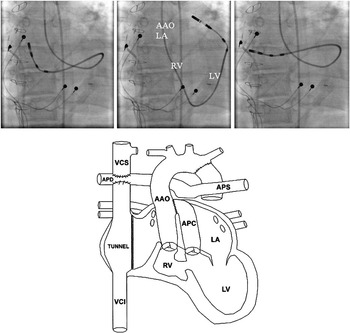
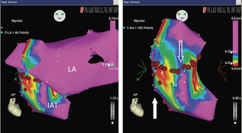
An 18-year-old female was referred because of multiple recurrences of atrial tachycardia 2 years after an apparently successful catheter ablation. At birth, she had been diagnosed with tricuspid atresia, discordant ventriculo-arterial connections, and aortic coarctation. At an initial surgical procedure, the coarctation was corrected and a band placed on the pulmonary trunk. After a period of 1 year, a bidirectional Glenn shunt was created, and the ventricular septal defect was enlarged. After another 3 years, the pulmonary trunk was closed proximally, and the systemic venous atrium was connected directly to the pulmonary arteries. She then developed severe mitral valvar insufficiency at the age of 10 years, requiring surgical repair. At the same intervention, a total cavo-pulmonary connection was made using an intraatrial tunnel, connecting the pulmonary trunk end-to-side to the aorta as the Damus-Kaye-Stansel procedure (Fig. 1). Subsequently, the patient developed multiple episodes of atrial tachycardia with a fast ventricular response, leading to near-syncope on many occasions, and necessitating frequent electrical cardioversions. Pharmacological treatment did not alleviate the symptoms, and caused unacceptable side-effects. An epicardial dual chamber pacemaker was implanted because of severe and symptomatic bradycardia. An electrophysiological study resulted in mapping and ablation of two scar-related tachycardias in the left atrium, using transoesophageal echocardiography to guide puncture of the intraatrial baffle. The symptoms recurred 2 years later, and a new electrophysiological study was undertaken. Under general anaesthesia, we punctured the right femoral artery and vein. A screw-in pacing lead (Medtronic, Minneapolis, MN, USA) was inserted into the right femoral vein and screwed into the lateral portion of the intraatrial tunnel. A magnetically steerable catheter (Navistar RMT DS 8 mm, Biosense-Webster, Diamond Bar, CA, USA) was also inserted into the right femoral vein, and the tip was positioned in the intraatrial tunnel. With programmed atrial stimulation, and atrial burstpacing, multiple episodes of non-sustained atrial tachycardias could be induced with slightly different cycle lengths, suggesting a complex substrate for the arrhythmia. All tachycardias, nonetheless, had a centrifugal sequence of activation, suggestive for an origin in the intraatrial tunnel. In the light of the history of arrhythmias in the left atrium, and using the CARTO™ navigation system (Biosense Webster, Diamond Bar, CA, USA) in combination with the magnetic navigation system (MNS) (Niobe, Stereotaxis, St. Louis, MO, USA), we were able to create safely a biatrial bipolar activation and voltage map (Fig. 2). We mapped the left atrium retrogradely using the femoral arterial access, a transseptal approach being deemed undesirable given the previous puncture of the baffle. The magnetically navigated catheter was piloted through the aortic arch, through the Damus-Kaye-Stansel shunt, into the incomplete morphologically right ventricle, through the ventricular septal defect into the morphologically left ventricle, and retrogradely through the surgically repaired mitral valve into the left atrium (Fig. 1). The voltage map showed high voltages in the intraatrial tunnel, albeit without any channels or scars. In the left atrium, however, we found multiple channels capable of conducting re-entry tachycardia. We modified the substrate by creating 2 lines. The first line bisected a channel between the septal and lateral parts of the intraatrial tunnel, while the second line joined the first line to the inferior caval vein (Fig. 2). Subsequent to creation of theses lines, we were no longer able to induce the tachycardia. The patient remained haemodynamically stable during the procedure. During the period of follow-up thus far, the patient has suffered only very short episodes of palpitations.
Top: Fluoroscopic images in the left anterior oblique projection of the magnetic navigation catheter retrogradely crossing the aortic valve, passing through the incomplete right ventricle, via the ventricular septal defect to the left ventricle, retrogradely through the mitral valve, and ending in the left atrium. The atrial and ventricular leads of an epicardial pacemaker are visible on all images. Bottom: Schematic reproduction of the heart after surgical repair. VCS superior caval vein, APD right pulmonary artery, APS left pulmonary artery, AAO ascending aorta, APC common pulmonary artery, TUNNEL intraatrial tunnel, LA left atrium, RV right ventricle, LV left ventricle, VCI inferior caval vein.

Left panel: the biatrial CARTO voltage map shows only large potentials in the left atrium (LA), and different channels in the intraatrial tunnel (IAT). Right panel: detail of CARTO voltage map in antero-posterior orientation (AP) of the intraatrial tunnel with ablation points (red dots), with the first ablation line (hollow arrow) bisecting a channel, and the second ablation line (solid arrow) joining the first line to the inferior caval vein.

Discussion
Conventional manually steerable electrophysiological catheters nowadays are designed to reach even remote areas in the heart. These catheters, nonetheless, have a relatively stiff body, making it very difficult to generate different consecutive bends, and at the same time to maintain a stable position for the tip of the catheter. It is virtually impossible to use a retrograde approach with a manually steered catheter to ablate an atrial tachycardia, and to our knowledge this has never been performed, even in structurally normal hearts. In our patient, we were confronted with several challenges to gain access to the left atrium to create activation and voltage CARTO™ maps. Transseptal mapping through the femoral vein was undesirable given the difficulties caused by the previous puncture of the atrial baffle. The likelihood of a possible cardiac perforation would put the patient at an unacceptable risk. A retrograde approach crossing the aortic valve, passing through the Damus-Kaye-Stansel shunt into the incomplete right ventricle, through the ventricular septal defect into the left ventricle, and retrogradely through the surgically repaired mitral valve into the left atrium was out of the question using a conventional manually steerable catheter. The magnetic navigation system, on the other hand, offers the opportunity to use extremely flexible and steerable catheters. The floppy tip of such catheters makes it possible to generate multiple consecutive bends. The soft and flexible nature of the catheter permits safe access to regions that are otherwise practically inaccessible. In addition, the chance of perforating the myocardial wall with the extremely floppy tip is very small. Another advantage is that as long as the magnetic field is applied, the tip remains stable at the desired position. Hence, we were able safely to generate a biatrial map, which provided valuable information about the origin of the postincisional macroreentrant tachycardia.
To our knowledge, this is the first report demonstrating successful retrograde mapping of the left atrium in a patient with complex congenital cardiac disease. Our experience, therefore, shows that such procedures can safely and effectively be performed, but we would suggest that they be attempted only in centres with extensive experience in magnetic navigation of congenitally malformed hearts.