Introduction
Timing and sites of protein synthesis are fundamentally important for promoting diverse biological processes including meiotic cell cycles, early development and neuronal plasticity. RNA-binding proteins have been shown to drive the localization and translational regulation of target mRNAs, by which protein synthesis is achieved at the right time and place (Kloc et al ., Reference Kloc, Zearfoss and Etkin2002; Martin and Ephrussi, Reference Martin and Ephrussi2009; Kotani et al., Reference Kotani, Maehata and Takei2017). Pumilio (Pum) is a sequence-specific RNA-binding protein that belongs to the Pumilio and Fem-3 mRNA-binding factor (PUF) family, which is highly conserved in eukaryotes from yeast to human (Wickens et al., Reference Wickens, Bernstein, Kimble and Parker2002; Spassov & Jurecic, Reference Spassov and Jurecic2003b). Pum was identified in Drosophila as a protein that is essential for posterior patterning of embryos and development of germline cells (Lehmann and Nussleinvolhard, Reference Lehmann and Nussleinvolhard1987; Lin and Spradling, Reference Lin and Spradling1997; Forbes and Lehmann, Reference Forbes and Lehmann1998; Asaoka-Taguchi et al., Reference Asaoka-Taguchi, Yamada, Nakamura, Hanyu and Kobayashi1999). Drosophila Pum targets hunchback mRNA in the posterior patterning and cyclin B mRNA in the germline development (Murata & Wharton, Reference Murata and Wharton1995; Asaoka-Taguchi et al., Reference Asaoka-Taguchi, Yamada, Nakamura, Hanyu and Kobayashi1999). In both cases, Pum represses the translation of target mRNAs in spatially and temporally regulated manners. FBF proteins, members of the PUF family in C. elegans, also function in germline development by regulating translation of their target mRNAs during embryogenesis (Zhang et al., Reference Zhang, Gallegos, Puoti, Durkin, Fields, Kimble and Wickens1997; Crittenden et al., Reference Crittenden, Bernstein, Bachorik, Thompson, Gallegos, Petcherski, Moulder, Barstead, Wickens and Kimble2002).
Two Pum proteins, Pumilio1 (Pum1) and Pumilio2 (Pum2), have been found in all vertebrates studied (Nakahata et al., Reference Nakahata, Katsu, Mita, Inoue, Nagahama and Yamashita2001; White et al., Reference White, Moore-Jarrett and Ruley2001; Spassov & Jurecic, Reference Spassov and Jurecic2002, Reference Spassov and Jurecic2003a; Padmanabhan & Richter, Reference Padmanabhan and Richter2006; Kurisaki et al., Reference Kurisaki, Iwai, Yamashita, Kobayashi, Ito and Matsuoka2007; Lee et al., Reference Lee, Lim, Kim, Zheng, Moon, Han, Song, Kim and Han2008). Recent studies have shown that Pum1-deficient mice are viable but have decreased fertility due to an extensive increase in apoptosis of spermatocytes in males (Chen et al., Reference Chen, Zheng, Lin, Uyhazi, Zhao and Lin2012) and due to a decrease in diplotene-stage oocytes in females (Mak et al., Reference Mak, Fang, Holden, Dratver and Lin2016). Pum1 deficiency also causes neuronal degeneration in the brain (Gennarino et al., Reference Gennarino, Singh, White, De Maio, Han, Kim, Jafar-Nejad, di Ronza, Kang, Sayegh, Cooper, Orr, Sillitoe and Zoghbi2015). Mouse Pum1 has been shown to target thousands of mRNAs in the testis and brain (Chen et al., Reference Chen, Zheng, Lin, Uyhazi, Zhao and Lin2012; Zhang et al., Reference Zhang, Chen, Xia, Han, Cui, Neuenkirchen, Hermes, Sestan and Lin2017). In addition, the amount of protein products, but not the amount of mRNAs, was increased in Pum1-deficient mice, indicating that Pum1 represses translation of target mRNAs. All findings from various species have shown the importance of Pum-mediated translational regulation in embryogenesis, spermatogenesis and neurogenesis. However, the mechanisms of Pum-mediated translational regulation remain to be elucidated.
Temporal regulation of mRNA translation has been most extensively studied in oocytes. In many vertebrates, oocytes are arrested at prophase I of the meiotic cell cycle and deposit tens of thousands of mRNAs for promoting oocyte maturation and development (Masui and Clarke, Reference Masui and Clarke1979; Harvey et al., Reference Harvey, Sealy, Kettleborough, Fenyes, White, Stemple and Smith2013). Fully grown oocytes in this stage are unable to be fertilized and they are called immature oocytes. It has been estimated that more than 1000 mRNAs are translationally repressed in immature oocytes and begin to be translated after resumption of the meiotic cell cycle at timings specific to distinct mRNAs (Chen et al., Reference Chen, Melton, Suh, Oh, Horner, Xie, Sette, Blelloch and Conti2011; Kotani et al., Reference Kotani, Maehata and Takei2017). Oocytes are arrested again at metaphase II until fertilization. This process is called oocyte maturation, and oocytes arrested at metaphase II are called mature oocytes.
Translation of cyclin B1 mRNA is repressed in immature oocytes and de-repressed after initiation of oocyte maturation. Synthesized Cyclin B1 protein from the stored mRNA functions as a regulatory subunit of maturation/M-phase-promoting factor (MPF), a master regulator in the progression of oocyte maturation. Extensive studies using Xenopus oocytes have shown that translational activation of dormant mRNAs including cyclin B1 mRNA is mainly directed by cytoplasmic polyadenylation elements (CPEs)-mediated polyadenylation of the mRNAs (McGrew et al., Reference McGrew, Dworkin-Rastl, Dworkin and Richter1989; Sheets et al., Reference Sheets, Fox, Hunt, Van de Woude and Wickens1994; de Moor and Richter, Reference de Moor and Richter1999). The CPE-binding protein CPEB has been shown to drive translational repression and activation of target mRNAs including cyclin B1 mRNA (Hake and Richter, Reference Hake and Richter1994; de Moor and Richter, Reference de Moor and Richter1999; Tay et al., Reference Tay, Hodgman and Richter2000). Pum1 targets cyclin B1 mRNA in zebrafish, Xenopus and mouse oocytes (Nakahata et al., Reference Nakahata, Katsu, Mita, Inoue, Nagahama and Yamashita2001, Reference Nakahata, Kotani, Mita, Kawasaki, Katsu, Nagahama and Yamashita2003; Pique et al., Reference Pique, Lopez, Foissac, Guigo and Mendez2008; Ota et al., Reference Ota, Kotani and Yamashita2011a; Kotani et al., Reference Kotani, Yasuda, Ota and Yamashita2013). We have previously shown that cyclin B1 mRNA forms RNA granules in immature oocytes of zebrafish and mouse (Kotani et al., Reference Kotani, Yasuda, Ota and Yamashita2013). These RNA granules disassemble at the timing of translational activation during oocyte maturation (Kotani et al., Reference Kotani, Yasuda, Ota and Yamashita2013; Nukada et al., Reference Nukada, Horie, Fukui, Kotani and Yamashita2015; Horie and Kotani, Reference Horie and Kotani2016). By generating transgenic zebrafish expressing reporter mRNAs in oocytes, we demonstrated that binding of Pum1 to cyclin B1 mRNA is essential for the formation of RNA granules. This Pum1-mediated granule formation seems to be important for regulating the timing of translational activation of cyclin B1 mRNA after initiation of oocyte maturation (Kotani et al., Reference Kotani, Yasuda, Ota and Yamashita2013). However, the mechanism by which translational timing of cyclin B1 mRNA is regulated by Pum1 remains unresolved.
In this study, we analyzed the phosphorylation of Pum1 in zebrafish oocytes. Pum1 was phosphorylated at multiple sites during oocyte maturation. This phosphorylation was initiated in an early period after induction of oocyte maturation, preceding the translational activation of cyclin B1 mRNA. In addition, Pum1 phosphorylation was found to be linked to disassembly of RNA granules and polyadenylation of cyclin B1 mRNA. Our findings provide the basis for understanding the mechanisms of translational regulation of mRNAs by Pum1.
Materials and methods
Preparation of oocytes
All animal experiments in this study were approved by the Committee on Animal Experimentation, Hokkaido University. Zebrafish ovaries were dissected from adult females in zebrafish Ringer’s solution (116 mM NaCl, 2.9 mM KCl, 1.8 mM CaCl2, and 5 mM HEPES; pH 7.2). Oocytes were manually isolated from ovaries with forceps under a dissecting microscope. Oocyte maturation was induced by stimulation with 1 µg/ml of 17α,20β-dihydroxy-4-pregnen-3-one, an maturation-inducing hormone (MIH) in fish. For in situ hybridization analysis, oocytes were fixed with 4% paraformaldehyde in PBS (4% PFA/PBS) overnight at 4°C. For poly(A) test (PAT) assay, oocytes were extracted with TRIzol reagent (Invitrogen) and total RNA was used for RNA ligation-coupled reverse transcriptase polymerase chain reaction (RT-PCR). For immunoblotting and immunoprecipitation analyses, oocytes were homogenized with an equal volume of ice-cold extraction buffer (EB: 100 mM β-glycerophosphate, 20 mM HEPES, 15 mM MgCl2, 5 mM EGTA, 1 mM dithiothreitol, 100 µM (p-amidinophenyl)methanesulfonyl fluoride, and 3 µg/ml leupeptin; pH 7.5). After centrifugation at 15,000 g for 10 min at 4°C, the supernatant was collected and used for SDS-PAGE analysis and immunoprecipitation.
Section in situ hybridization
Section in situ hybridization with the tyramide signal amplification (TSA) Plus DNP system (Perkin Elmer) was performed according to the procedure reported previously (Takei et al., Reference Takei, Nakamura, Kawamura, Takada, Satoh, Kimura and Kotani2018). Briefly, fixed oocytes were dehydrated, embedded in paraffin, and cut into 12-µm-thick sections. A digoxigenin (DIG)-labelled antisense RNA probe for the full length of cyclin B1 was used for detection of the cyclin B1 gene transcript. No signal was detected with sense probes. After hybridization and washing, samples were incubated with anti-DIG−horseradish peroxidase (HRP) antibody (Roche) (1:500 dilution) for 30 min. The reaction with tyramide–dinitrophenyl (DNP) was performed according to the manufacturer’s instructions. The samples were then incubated overnight with anti-DNP–Alexa 488 antibody (Molecular Probes) (1:500 dilution). To detect nuclei, the samples were incubated with 10 µg/ml Hoechst 33258 for 10 min. After being mounted with a Prolong Antifade Kit (Molecular Probes), the samples were observed under an LSM5LIVE confocal microscope (Carl Zeiss).
Poly(A) test (PAT) assay
RNA ligation-coupled RT-PCR was performed according to the procedure reported previously (Charlesworth et al., Reference Charlesworth, Cox and MacNicol2004; Yasuda et al., Reference Yasuda, Kotani, Ota and Yamashita2010). Two µg of total RNA extracted from pools of 15 zebrafish oocytes was ligated to 0.4 µg of P1 anchor primer (5′-P-GGTCACCTTGATCTGAAGC-NH2-3′) in a 10-µl reaction using T4 RNA ligase (New England BioLabs) for 30 min at 37°C. The ligase was inactivated for 5 min at 92°C. Half of the RNA ligation reaction was used in a 25-µl reverse transcription reaction using the Superscript III First Strand Synthesis System with a P1′ primer (5′-GCTTCAGATCAAGGTGACCTTTTT-3′). In total, 4 µl of the cDNA was used for the first PCR with the P1′ primer and a zcyclin B1−PAT-f1 primer (5′-GAGGGCCTTTCTAAGCATCTGGCTGTG-3′) for 17 cycles. Then, 2 µl of the first PCR reaction was used for the second PCR with the P1′ primer and a zcyclin B1–3′UTR-f primer (5′-TACGGATTTCTTCACTGCCATG-3′) for 35 cycles. The PCR product was resolved on a 2% agarose gel in TAE buffer. We confirmed that the increase in PCR product length was due to elongation of the poly(A) tails by cloning the second PCR products and sequencing them.
Immunoblotting and immunoprecipitation
Next, 10 µl of oocyte extracts was incubated with an anti-Xenopus Pum1 rabbit antibody, which was affinity-purified with zebrafish Pum1 (Kotani et al., Reference Kotani, Yasuda, Ota and Yamashita2013), for 1 h at 4°C. The extracts were then incubated with protein A-Sepharose beads (GE Healthcare) for 3 h at 4°C and washed three times with EB containing 1% Tween 20. The crude oocyte extracts and the immunoprecipitates of anti-Pum1 rabbit antibody were separated by SDS-PAGE, blotted onto an Immobilon membrane, and probed with an anti-Xenopus Pum1 monoclonal antibody (Pum2A5; Nakahata et al., Reference Nakahata, Katsu, Mita, Inoue, Nagahama and Yamashita2001). CPEB was detected by immunoblotting the crude extracts with anti-CPEB monoclonal antibody (9G3; Takahashi et al., Reference Takahashi, Kotani, Katsu and Yamashita2014). The Cyclin B1–Cdc2 complex was detected by immunoblotting Suc1 precipitates from crude oocyte extracts with anti-Cdc2 (MC2–21; Tanaka and Yamashita, Reference Tanaka and Yamashita1995) and anti-goldfish Cyclin B1 (B112; Katsu et al., Reference Katsu, Yamashita, Kajiura and Nagahama1993) monoclonal antibodies.
Phosphatase treatments
After immunoprecipitation of oocyte extracts with anti-Pum1 rabbit antibody, the immunoprecipitates were treated with calf intestinal alkaline phosphatase (CIAP; New England BioLabs) or λ-protein phosphatase (λ-PP; New England BioLabs) for 1 h at 30°C. The samples were then washed three times with EB and analyzed by immunoblotting.
Cytochalasin B treatment
To depolymerize actin filaments, oocytes were treated with 1 µg/ml cytochalasin B (Sigma) for 3 h. Cytochalasin B was dissolved in dimethyl sulfoxide (DMSO) as stocks and diluted in Ringer’s solution before use. As a control, oocytes were treated with 0.01% DMSO. After treatment with cytochalasin B or DMSO for 3 h, the oocytes were stimulated with MIH for induction of oocyte maturation.
RT-PCR analysis after IP (IP/RT-PCR)
Here, 100 µl oocyte extracts was incubated with affinity-purified anti-Pum1 rabbit antibody or control rabbit IgG for 1 h at 4°C. The extracts were then incubated with protein A-Sepharose beads for 3 h at 4°C and washed five times with EB containing 1% Tween 20. After extraction of mRNAs from the beads with TRIzol reagent, RT-PCR was performed using a zcyclin B1-f2 primer (5′-GAGGGCCTTTCTAAGCATCTGGCTGTG-3′) and a zcyclin B1-r2 primer (5′-TTATTTGAATTCAAATGTACAAACTTGC-3′).
Overexpression of Pum1 N-terminus and immunostaining
mRNAs encoding GFP–Pum1N (Pum1N; 1–790 amino acids) (Kotani et al., Reference Kotani, Yasuda, Ota and Yamashita2013) and GFP were synthesized with an mMESSAGE mMACHINE SP6 kit (Life Technologies). In total, 2 ng of the mRNAs were injected into fully grown oocytes using a microinjector (CellTram vario). The oocytes were incubated for 3 h and stimulated with MIH. Before or at 90 min after MIH stimulation, the oocytes were fixed with 4% PFA/PBS for in situ hybridization analysis or extracted with ice-cold EB for detection of Pum1. To detect GFP–Pum1N and GFP, the oocytes were immunostained with anti-GFP mouse antibody (1:200 dilution; Roche) followed by anti-mouse IgG–Alexa Fluor 488 antibody (1:200 dilution; Molecular Probes), after hybridization and washing of the cyclin B1 RNA probe in in situ hybridization analysis.
Results and Discussion
Phosphorylation of Pum1 during zebrafish oocyte maturation
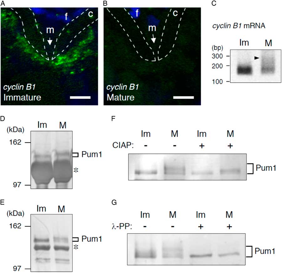
As reported previously, in situ hybridization of zebrafish ovaries showed the distribution of cyclin B1 mRNA as granular structures in the animal polar cytoplasm of fully grown immature oocytes beneath the micropyle (Fig. 1A) (Kotani et al., Reference Kotani, Yasuda, Ota and Yamashita2013; Horie & Kotani, Reference Horie and Kotani2016). The micropyle is a structure located at the animal pole of the fish chorion, through which a sperm enters into the oocyte cytoplasm. The cyclin B1 RNA granules disassembled during oocyte maturation (Fig. 1B) (Kotani et al., Reference Kotani, Yasuda, Ota and Yamashita2013; Horie & Kotani, Reference Horie and Kotani2016). The PAT assay showed that poly(A) tails of cyclin B1 mRNA were short in immature oocytes and became elongated during oocyte maturation (Fig. 1C) (Yasuda et al., Reference Yasuda, Kotani, Ota and Yamashita2010), accompanied by translational activation of mRNA (Kotani et al., Reference Kotani, Yasuda, Ota and Yamashita2013). Pum1 has been shown to bind to Pumilio-binding element (PBE) located in the 3′ untranslated region (UTR) of cyclin B1 mRNA (Kotani et al., Reference Kotani, Yasuda, Ota and Yamashita2013). As binding of Pum1 to cyclin B1 mRNA persists during oocyte maturation (Ota et al., Reference Ota, Kotani and Yamashita2011a; Takahashi et al., Reference Takahashi, Kotani, Katsu and Yamashita2014) even after the mRNA is translated, we predicted that modification of Pum1 may be involved in the change of translational regulation of target cyclin B1 mRNA from repression to activation states. One candidate is phosphorylation as observed in Xenopus Pum1 (Ota et al., Reference Ota, Kotani and Yamashita2011a). To assess this possibility, we examined whether zebrafish Pum1 is phosphorylated during oocyte maturation.
Phosphorylation of Pum1 during oocyte maturation in zebrafish. (A, B) In situ hybridization analysis of cyclin B1 mRNA (green) in immature (A) and mature (B) oocytes. DNA is shown in blue. The chorion is outlined by broken lines. Dormant cyclin B1 mRNA is stored as RNA granules localized in the animal polar cytoplasm of immature oocytes. These granules disappear during oocyte maturation. m, micropyle; f, follicle cells; c, chorion. Bars: 10 µm. (C) PAT assay of cyclin B1 mRNA in immature (Im) and mature (M) oocytes. The poly(A) tails of cyclin B1 mRNA are short in immature oocytes and become elongated during oocyte maturation (arrowhead). (D, E) SDS-PAGE analyses of crude extracts (D) and immunoprecipitations with anti-Pum1 antibody (E) from immature (Im) and mature (M) oocytes followed by immunostaining with anti-Pum1 antibody. Asterisk shows background signals of yolk protein. The electrophoretic mobility of Pum1 is reduced in mature oocytes. (F, G) SDS-PAGE analyses of immunoprecipitations with anti-Pum1 antibody from immature (Im) and mature (M) oocytes stimulated with (+) or not stimulated with (−) CIAP (F) and λ-PP (G). The reduction in electrophoretic mobility of Pum1 in mature oocytes is recovered partially by CIAP and almost completely by λ-PP. Similar results were obtained from two independent experiments.
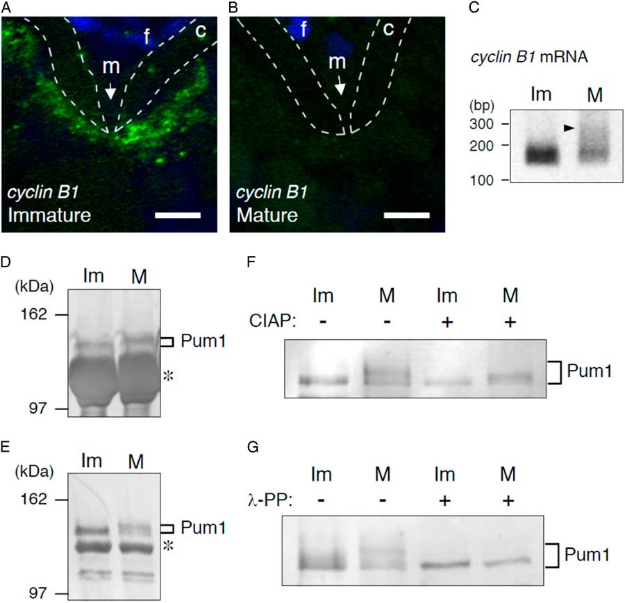
Pum1 was detected as an approximately 120 kDa protein in crude extracts of zebrafish immature oocytes by SDS-PAGE analysis followed by immunostaining with anti-Pum1 antibody (Fig. 1D). The electrophoretic mobility of Pum1 appeared to be reduced in mature oocytes, resulting in an apparent increase in the molecular weight of Pum1 (Fig. 1D). This reduction in Pum1 mobility in mature oocytes was confirmed by immunoprecipitation of Pum1 (Fig. 1E). The electrophoretic mobility of Pum1 in mature oocytes was partially but not completely recovered by treatment of the precipitations with CIAP (Fig. 1F). Treatment of the precipitations with λ-protein phosphatase (λ-PP) resulted in almost complete recovery of the mobility of Pum1 as a single band (Fig. 1G). These results indicate that Pum1 is phosphorylated at multiple sites and suggest that serine and threonine residues of Pum1 are mainly phosphorylated because λ-PP specifically dephosphorylates these residues (Barik, Reference Barik1993). Notably, partial reduction in Pum1 mobility was observed in immature oocytes of some, but not all, batches (4 out of 18 batches) (Fig. 1G, compare with Fig. 1D−F ), suggesting that a kinase responsible for Pum1 phosphorylation is at least partially activated and that the state of phosphorylation is equilibrated with that of dephosphorylation in immature oocytes.
Timing of Pum1 phosphorylation after induction of oocyte maturation
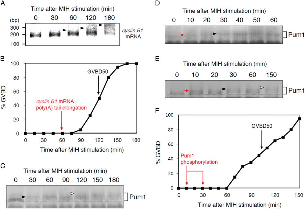
To assess whether Pum1 phosphorylation is involved in the translational regulation of target cyclin B1 mRNA, we examined the timing of Pum1 phosphorylation after induction of oocyte maturation. Polyadenylation of cyclin B1 mRNA began 60 min after induction of oocyte maturation (Fig. 2A), at which no oocyte underwent GVBD (Fig. 2B). As reported previously, the timing of cyclin B1 mRNA poly(A) tail elongation (60 min) was approximately half of the time point at which 50% of the oocytes have undergone GVBD (120 min in this batch; Fig. 2B) (Kotani et al., Reference Kotani, Yasuda, Ota and Yamashita2013; Horie & Kotani, Reference Horie and Kotani2016). Time course analysis of Pum1 phosphorylation showed that partial but apparent phosphorylation had occurred at 30 min and then significant phosphorylation had occurred at 120 min after induction of oocyte maturation (Fig. 2C). We then focused on the initiation of Pum1 phosphorylation. Interestingly, slight phosphorylation of Pum1 was detected at 10 min and then apparent phosphorylation was detected at 30 min after induction of oocyte maturation (Fig. 2D). As crude extracts were unable to clearly show the changes in Pum1 electrophoretic mobility, we performed immunoprecipitation of Pum1 and confirmed that Pum1 was slightly phosphorylated at 10 min and apparently phosphorylated at 30 min after induction of oocyte maturation (Fig. 2E). Significant phosphorylation of Pum1 was also observed 150 min after induction of oocyte maturation in this assay (Fig. 2E). The results showing a gradual increase in the molecular weight of Pum1 support our conclusion that Pum1 is phosphorylated at multiple sites during oocyte maturation (Fig. 1). Taken together, these results indicate that Pum1 phosphorylation precedes the polyadenylation of target cyclin B1 mRNA during normal oocyte maturation (Fig. 2F, compared with Fig. 2B).
Time course analyses of Pum1 phosphorylation after initiation of oocyte maturation. (A, B) Time course of PAT assay for cyclin B1 mRNA (A) and GVBD (B) after MIH stimulation. Arrowheads indicate signals of poly(A) tail elongation. (C, D) Time course of Pum1 phosphorylation after MIH stimulation using crude oocyte extracts. A red arrow indicates an initial signal of phosphorylation. Black arrowheads indicate apparent phosphorylation. White arrowheads indicate significant phosphorylation. Dotted lines indicate the basal size of Pum1. Similar results were obtained from two independent experiments. (E, F) Time course of Pum1 phosphorylation using immunoprecipitations with anti-Pum1 antibody (E) and GVBD (F) after MIH stimulation. A red arrow indicates an initial signal of phosphorylation. A black arrowhead indicates apparent phosphorylation. A white arrowhead indicates significant phosphorylation. Pum1 phosphorylation began from 10 to 30 min after MIH stimulation. Similar results were obtained from five independent experiments.
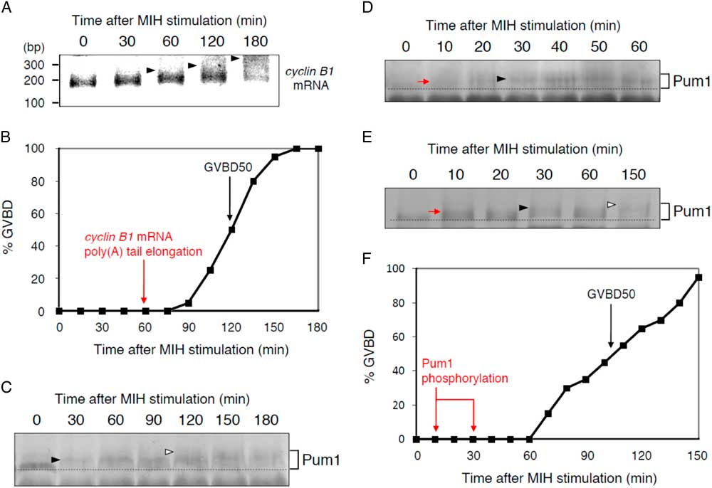
Phosphorylation of Pum1 was previously reported in Xenopus oocytes (Ota et al., Reference Ota, Kotani and Yamashita2011a). However, a shift up of the Pum1 protein band, which was caused by phosphorylation as in the case of zebrafish Pum1, was shown to begin at a timing similar to that of Cyclin B1 protein synthesis. Therefore, whether Pum1 phosphorylation was the cause or consequence of translational activation of cyclin B1 mRNA remains unclear. Our results show for the first time that Pum1 is phosphorylated before translational activation of its target mRNA, implying that this Pum1 modification may be a cause of translational activation of target mRNAs.
Effect of actin filament depolymerization on Pum1 phosphorylation
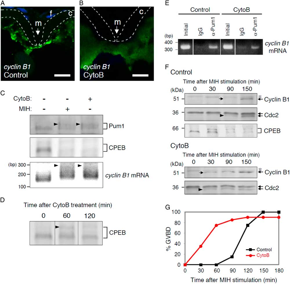
The granule formation of cyclin B1 mRNA has been shown to be dependent on the binding of Pum1 and on the actin filaments assembled in the oocyte cytoplasm (Kotani et al., Reference Kotani, Yasuda, Ota and Yamashita2013; Nukada et al., Reference Nukada, Horie, Fukui, Kotani and Yamashita2015). However, the relationship between Pum1 modification and actin filament depolymerization has been completely unknown. We then examined the effects of depolymerization of actin filaments on Pum1 phosphorylation. Treatment of immature oocytes with cytochalasin B caused depolymerization of actin filaments (Nukada et al., Reference Nukada, Horie, Fukui, Kotani and Yamashita2015) and disassembly of cyclin B1 RNA granules (Fig. 3A, B) (Kotani et al., Reference Kotani, Yasuda, Ota and Yamashita2013). In the oocytes treated with cytochalasin B for 3 h, Pum1 was phosphorylated at a level similar to that in oocytes stimulated with MIH for 3 h (Fig. 3C). RT-PCR analysis after immunoprecipitation with anti-Pum1 antibody of the oocyte extracts showed that the binding of Pum1 to cyclin B1 mRNA was not affected by actin filament depolymerization (Fig. 3E).
Effects of actin filament depolymerization on Pum1 phosphorylation and translational regulation of cyclin B1 mRNA. (A, B) In situ hybridization analysis of cyclin B1 mRNA (green) in oocytes treated with DMSO (Control) and cytochalasin B (CytoB). DNA is shown in blue. The chorion is outlined by broken lines. cyclin B1 RNA granules are maintained in control oocytes (A), while they are dispersed throughout the cytoplasm in CytoB-treated oocytes (B). m, micropyle; f, follicle cells; c, chorion. Bars: 10 µm. (C) Immunoblotting of Pum1 (upper) and CPEB (middle) and PAT assay for cyclin B1 mRNA (lower) in oocytes treated with (+) or not treated with (−) CytoB or MIH. Arrowheads indicate Pum1 phosphorylation (upper) and poly(A) tail elongation of cyclin B1 mRNA (lower). Similar results were obtained from four independent experiments. (D) Time course of CPEB phosphorylation after CytoB treatment. An arrowhead indicates a phosphorylated form of CPEB. (E) RT-PCR analysis of immunoprecipitations with control IgG (IgG) or anti-Pum1 (α-Pum1) antibody in control and CytoB-treated oocytes. (F) Time course of Cyclin B1 synthesis after stimulation with MIH in control and CytoB-treated oocytes. Arrows indicate initial signals of Cyclin B1 synthesis. Arrowheads indicate active forms of the Cdc2 kinase. Asterisks show nonspecific bands. Time course of CPEB phosphorylation and degradation is shown in control oocytes. (G) Time course of GVBD after stimulation with MIH in control and CytoB-treated oocytes. Similar results were obtained from three independent experiments.
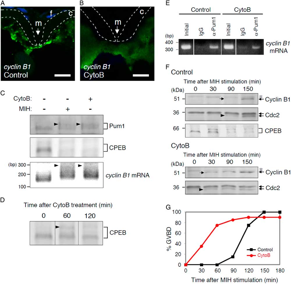
As reported previously, Cyclin B1 protein was not detected in oocytes treated with cytochalasin B for 3 h (Fig. 3F, see the lane 0 min after stimulation with MIH) (Kotani et al., Reference Kotani, Yasuda, Ota and Yamashita2013), indicating that translation of cyclin B1 mRNA is still repressed. However, interestingly, the PAT assay showed that poly(A) tails of cyclin B1 mRNAs were elongated in oocytes treated with cytochalasin B (Fig. 3C). We then examined modifications of CPEB in the cytochalasin B-treated oocytes. CPEB was shown to be phosphorylated and degraded after induction of oocyte maturation in zebrafish (Takahashi et al., Reference Takahashi, Kotani, Katsu and Yamashita2014) as in the cases of Xenopus (Mendez et al., Reference Mendez, Barnard and Richter2002), mouse (Tay et al., Reference Tay, Hodgman and Richter2000; Chen et al., Reference Chen, Melton, Suh, Oh, Horner, Xie, Sette, Blelloch and Conti2011) and porcine (Komrskova et al., Reference Komrskova, Susor, Malik, Prochazkova, Liskova, Supolikova, Hladky and Kubelka2014) (see Fig. 3F control oocytes stimulated with MIH after DMSO treatment). In Xenopus oocytes, CPEB degradation has been shown to control the translational activation of cyclin B1 mRNA (Mendez et al., Reference Mendez, Barnard and Richter2002). In the cytochalasin B-treated oocytes, CPEB was degraded at a level similar to that in the MIH-stimulated oocytes (Fig. 3C). We confirmed that CPEB was phosphorylated shortly after cytochalasin B treatment, followed by gradual degradation (Fig. 3D). Collectively, our results indicate that neither Pum1 phosphorylation, CPEB degradation, nor polyadenylation of cyclin B1 mRNA is sufficient for translational activation of the mRNA in oocytes. Instead, these events seemed to be linked to rapid activation of cyclin B1 mRNA translation after the response to MIH, because synthesis of Cyclin B1 protein was detected immediately (30 min) after MIH stimulation in these oocytes (Fig. 3F), compared with the synthesis of Cyclin B1 in a later period (90 min) in control oocytes. Consistent with the rapid synthesis of Cyclin B1, appearance of active form of Cdc2 kinase (Fig. 3F, arrowheads) and occurrence of GVBD (Fig. 3G) were significantly accelerated in oocytes treated with cytochalasin B. Our results also indicated the existence of an unknown event(s) or factor(s) downstream of the MIH receptor that is required for the final trigger of translational activation of cyclin B1 mRNA after Pum1 phosphorylation, CPEB degradation, and polyadenylation of the mRNA.
Effect of overexpression of GFP–Pum1N on Pum1 phosphorylation
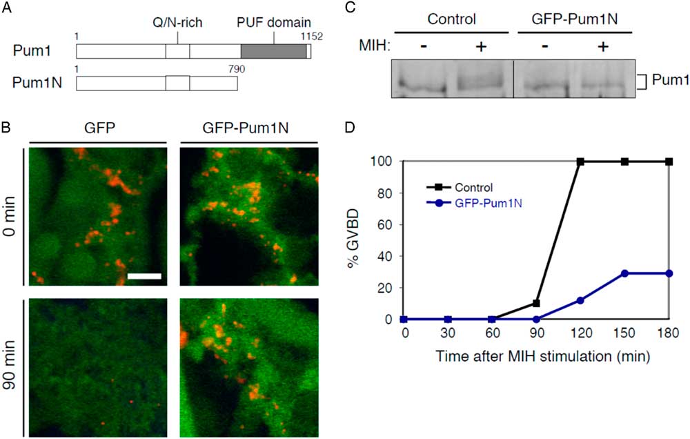
Pum1 contains the highly conserved PUF domain at the C-terminal region that is responsible for binding to mRNAs (Fig. 4A) (Zamore et al., Reference Zamore, Williamson and Lehmann1997). In the central region, Pum1 contains a Q/N-rich region that is thought to promote ordered aggregation of the proteins (Salazar et al., Reference Salazar, Silverman, Menon and Zinn2010). Overexpression of Pum1N, which lacks the PUF domain, affected the assembly of cyclin B1 RNA granules and the translational activation of mRNA (Kotani et al., Reference Kotani, Yasuda, Ota and Yamashita2013). This result suggested that overexpression of Pum1N affects Pum1 phosphorylation. We therefore examined the effects of overexpression of GFP–Pum1N on Pum1 phosphorylation.
Effects of overexpression of Pum1N on endogenous Pum1 phosphorylation and translational regulation of cyclin B1 mRNA. (A) Schematic diagrams of zebrafish Pum1 and Pum1N. The PUF domain is responsible for binding to PBE in target mRNAs. The Q/N-rich region is thought to promote protein aggregation. (B) In situ hybridization of cyclin B1 mRNA (red) and immunostaining of GFP (green) in oocytes expressing GFP or GFP–Pum1N before (0 min) or at 90 min after stimulation with MIH. Bars: 5 µm. (C) Pum1 phosphorylation in control or GFP–Pum1N-expressing oocytes stimulated with (+) or not stimulated with (−) MIH. Pum1 is phosphorylated in control oocytes but not in GFP–Pum1N-expressing oocytes after stimulation with MIH. Similar results were obtained from three independent experiments. (D) Time course of GVBD after stimulation with MIH in control and GFP–Pum1N-expressing oocytes. Similar results were obtained from three independent experiments.
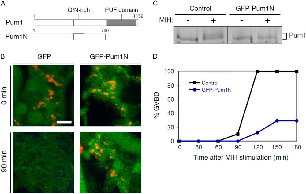
As reported previously, GFP–Pum1N formed large aggregates surrounding cyclin B1 RNA granules in the animal polar cytoplasm of immature oocytes, while GFP was diffused throughout the cytoplasm (Fig. 4B) (Kotani et al., Reference Kotani, Yasuda, Ota and Yamashita2013). These GFP–Pum1N aggregates prevented the disassembly of cyclin B1 RNA granules. The cyclin B1 RNA granules had almost completely disassembled in control GFP-expressing oocytes 90 min after induction of oocyte maturation, while the granules were maintained in GFP–Pum1N-expressing oocytes (Fig. 4B) (Kotani et al., Reference Kotani, Yasuda, Ota and Yamashita2013). In response to MIH, endogenous Pum1 was phosphorylated in the control oocytes, while endogenous Pum1 phosphorylation was prevented in the oocytes expressing GFP–Pum1N (Fig. 4C). Consistent with our previous study, oocytes expressing GFP–Pum1N showed delay in the occurrence of GVBD and 70% of the oocytes did not undergo GVBD (Fig. 4D), accompanied by failure in Cyclin B1 synthesis (Kotani et al., Reference Kotani, Yasuda, Ota and Yamashita2013). An explanation for the effects of overexpression of GFP–Pum1N is that Pum1N proteins surrounding cyclin B1 RNA granules prevented endogenous Pum1 phosphorylation by sequestering phosphorylation sites of Pum1 and therefore attenuated granule disassembly and translational activation of the mRNA. Alternatively, GFP–Pum1N affected endogenous Pum1 phosphorylation independently of the effects on granule disassembly and translational activation of the mRNA. Nevertheless, these results suggest a relation between Pum1 phosphorylation and disassembly of cyclin B1 RNA granules, which may promote translational activation of the mRNA.
Pum1 phosphorylation and translational activation of cyclin B1 mRNA
Pum1 has been shown to play fundamental roles in diverse biological processes by regulating translation of target mRNAs. However, the mechanisms of Pum1-mediated translation remain to be elucidated. In this study, we demonstrated that:
(a) Pum1 was phosphorylated at multiple sites during oocyte maturation (Fig. 1).
(b) This phosphorylation preceded the polyadenylation and translational activation of its target mRNA, cyclin B1 mRNA (Fig. 2).
(c) Depolymerization of actin filaments resulted in Pum1 phosphorylation, cyclin B1 RNA granule disassembly and polyadenylation of the mRNA without induction of oocyte maturation (Fig. 3).
(d) Overexpression of Pum1N prevented Pum1 phosphorylation, cyclin B1 RNA granule disassembly and translational activation of the mRNA even after induction of oocyte maturation (Fig. 4).
As binding of Pum1 to cyclin B1 mRNA is required for RNA granule formation (Kotani et al., Reference Kotani, Yasuda, Ota and Yamashita2013) and persists during oocyte maturation (Ota et al., Reference Ota, Kotani and Yamashita2011a; Takahashi et al., Reference Takahashi, Kotani, Katsu and Yamashita2014), our findings suggest that Pum1 phosphorylation is an initial key step to promote the disassembly of cyclin B1 RNA granules and polyadenylation of mRNA, which accelerates the sequential activation of mRNA translation.
The signalling pathway upstream of Pum1 phosphorylation in zebrafish oocytes remains unknown. One candidates is Nemo-like kinase (NLK), a typical mitogen-activated protein kinase. NLK is present in Xenopus immature oocytes and is activated in an early phase of oocyte maturation (Ota et al., Reference Ota, Kotani and Yamashita2011b). This kinase can directly phosphorylate Pum1 as well as CPEB and Pum2 in vitro. Whether NLK is expressed in zebrafish oocytes and is involved in Pum1 phosphorylation will be an interesting issue. Another possibility is that a kinase responsible for Pum1 phosphorylation is at least partially active in zebrafish immature oocytes, because our immunoprecipitation analysis showed that Pum1 was partially phosphorylated in immature oocytes of some batches (Fig. 1G and data not shown) and that Pum1 was immediately phosphorylated in response to MIH in most batches (Fig. 2). If this is the case, a phosphatase responsible for dephosphorylation of Pum1 would be present in immature oocytes, which would maintain the non-phosphorylated state of Pum1. In this scenario, MIH stimulation immediately inhibits the phosphatase in addition to fully activating the kinase responsible for Pum1 phosphorylation. As a next step to understand the mechanism of Pum1-mediated translation, identification of the kinase responsible for Pum1 phosphorylation will be necessary. Our time course analysis also showed significant phosphorylation of Pum1 after GVBD (Fig. 2). This phosphorylation would be caused by activated MPF as for CPEB hyperphosphorylation in Xenopus, porcine and mouse oocytes (Mendez et al., Reference Mendez, Barnard and Richter2002; Komrskova et al., Reference Komrskova, Susor, Malik, Prochazkova, Liskova, Supolikova, Hladky and Kubelka2014; Han et al., Reference Han, Martins, Yang, Kang, Daldello and Conti2017). The kinase responsible for significant phosphorylation of Pum1 and the importance of this second phase of phosphorylation remain to be elucidated.
An intriguing finding in this study is that cytoplasmic polyadenylation is not sufficient for translational activation of cyclin B1 mRNA (Fig. 3). Extensive studies using Xenopus and mouse oocytes have demonstrated that cytoplasmic polyadenylation of dormant mRNAs stored in oocytes direct translational activation of the mRNAs (McGrew et al., Reference McGrew, Dworkin-Rastl, Dworkin and Richter1989; Vassalli et al., Reference Vassalli, Huarte, Belin, Gubler, Vassalli, O’Connell, Parton, Rickles and Strickland1989; Sheets et al., Reference Sheets, Fox, Hunt, Van de Woude and Wickens1994; Stutz et al., Reference Stutz, Conne, Huarte, Gubler, Volkel, Flandin and Vassalli1998). Polyadenylation of cyclin B1 mRNA has been shown using Xenopus oocytes (Radford et al., Reference Radford, Meijer and de Moor2008; Weill et al., Reference Weill, Belloc, Bava and Mendez2012). In immature oocytes, both deadenylase PARN and the poly(A) polymerase GLD2 associate with cyclin B1 mRNA. The high activity of PARN sequesters GLD2 activity, shortening mRNA poly(A) tails. Multiple CPEBs bind to the CPEs of cyclin B1 mRNA (de Moor and Richter, Reference de Moor and Richter1999). Pum1 binds CPEB, PARN and GLD2 in immature oocytes (Nakahata et al., Reference Nakahata, Kotani, Mita, Kawasaki, Katsu, Nagahama and Yamashita2003; Ota et al., Reference Ota, Kotani and Yamashita2011a). After initiation of oocyte maturation, several CPEBs were degraded (Mendez et al., Reference Mendez, Barnard and Richter2002), and the remaining CPEB recruits cleavage and polyadenylation specificity factor CPSF on the poly(A) signal AAUAAA in the terminal end of mRNA and excludes PARN from the mRNA. This remodelling promotes the mRNA polyadenylation by GLD2. The subsequent binding of the poly(A)-binding protein PABP on the elongated poly(A) tail circularizes mRNA by interaction between PABP and cap-binding factor eIF4E, which is mediated by a scaffold protein eIF4G, resulting in induction of the translational activation of mRNA (Cao & Richter, Reference Cao and Richter2002). Although we could not rule out the possibility that polyadenylation caused by actin filament depolymerization is partially different from that caused by MIH stimulation, our findings suggested that the induction of translational activation of cyclin B1 mRNA requires another factor(s) or additional remodelling of the complex, even after the polyadenylation of mRNA.
In Xenopus oocytes, Pum2 has been shown to function in oocyte maturation by regulating translation of mRNAs other than cyclin B1 mRNA (Padmanabhan & Richter, Reference Padmanabhan and Richter2006; Ota et al., Reference Ota, Kotani and Yamashita2011a). In addition, Xenopus Pum2 was shown to be phosphorylated during oocyte maturation (Ota et al., Reference Ota, Kotani and Yamashita2011a, Reference Ota, Kotani and Yamashita2011b), implying that phosphorylation is one of the important processes in Pum2-mediated translational regulation. The expression and involvement of Pum2 in zebrafish oocyte maturation remain to be elucidated. Our results in this study and future studies on the phosphorylation of Pum1 including the identification of phosphorylation sites will contribute to an understanding of the molecular mechanisms of Pum-mediated translational control in diverse systems and organisms.
Acknowledgements
We thank Dr Yamashita for providing anti-Pum1 antibodies and Dr Katsu for providing anti-CPEB antibody.
Financial support
This work was supported by a Grant-in-Aid for Scientific Research (16K07242 to T.K.) from the Ministry of Education, Culture, Sports, Science and Technology, Japan.
Conflicts of interest
The authors have no conflicts of interest to declare.
Ethical standards
All animal experiments in this study were approved by the Committee on Animal Experimentation, Hokkaido University, Japan.