Introduction
Dementia with Lewy bodies (DLB) accounts for 15–20% of late onset dementia (Hansen et al., Reference Hansen1990; McKeith et al., Reference McKeith2004), whilst recent longitudinal studies indicate that the majority of older patients with Parkinson's disease (PD) eventually develop dementia (Aarsland et al., Reference Aarsland, Andersen, Larsen, Lolk and Kragh-Sorensen2003; Reid et al., Reference Reid1996). DLB and Parkinson's Disease Dementia (PDD) are distressing conditions characterized by parkinsonism, visual hallucinations, fluctuating cognition, REM sleep behavioral disorder, marked sensitivity to neuroleptic drugs and other psychotic symptoms (Boeve et al., Reference Boeve, Silber and Ferman2004; McKeith et al., Reference McKeith2005; Aarsland et al., Reference Aarsland2005), resulting in major difficulties for clinical management. The defining neuropathological and neurochemical characteristics include widespread cortical Lewy bodies, α-synuclein and ubiquitin inclusions (McKeith et al., Reference McKeith2004), a major loss of cholinergic neurons within the nucleus basalis magnocellularis (nBM) and extensive cortical cholinergic deficits (Lippa et al., Reference Lippa, Smith and Perry1999; Ballard et al., Reference Ballard2000).
Accurate diagnosis is essential to avoid harm and optimize clinical management. Although the development of operationalized clinical criteria has been an important step, their sensitivity is only 50% and accurate diagnosis of DLB remains challenging (McKeith et al., Reference McKeith2004; Reference McKeith2005). There is therefore an urgent imperative to identify reliable biomarkers, in particular for early-stage disease, where the clinical differentiation between Alzheimer's disease (AD) and DLB is particularly difficult, since only a minority of DLB cases with early-stage dementia exhibit key diagnostic features such as parkinsonism and visual hallucinations (Tiraboschi et al., Reference Tiraboschi, Salmon, Hansen, Hofstetter, Thal and Corey-Bloom2006). Such biomarkers are also essential to underpin the development of new therapies targeting disease mechanisms. Without biomarkers of pathology, it is hard to determine if a therapy is only targeting symptoms or whether it is also reducing pathological burden. However, with the exception of functional imaging of the striatal dopamine transporter, which is expensive and not generally available, there are no established biomarkers for DLB (McKeith et al., Reference McKeith2007).
Although α-synuclein, the key protein in the pathogenesis of DLB and PDD, is a non-secreted pre-synaptic protein, a secreted form of this protein has been identified and detected in both cerebrospinal fluid (CSF) and plasma (El-Agnaf et al., Reference El-Agnaf2006). However, reports on CSF have been inconsistent. Some studies have not found the monomeric form of soluble α-synuclein with molecular size in the range 14–19 kDa in CSF (Jakowec et al., Reference Jakowec, Petzinger, Sastry, Donaldson, McCormack and Langston1998). Other studies using enzyme-linked immunosorbent assays (ELISAs) have reported reduced levels of α-synuclein and an inverse relationship with the severity of parkinsonism in PD patients (Tokuda et al., Reference Tokuda2006) and in patients with DLB compared to AD patients or controls (Mollenhauer et al., Reference Mollenhauer2008). In contrast, higher concentrations have also been reported in post-mortem CSF (Mukaetova-Ladinska et al., Reference Mukaetova-Ladinska2008). Of interest, CSF of DLB patients enhances the fibrillization of α-synuclein, and insoluble oligomers of high molecular weights have been reported in several of the studies using Western blotting (Jakowec et al., Reference Jakowec, Petzinger, Sastry, Donaldson, McCormack and Langston1998; Borghi et al., Reference Borghi2000).
The aim of this study was to establish a valid method for detecting α-synuclein monomers in the CSF of patients with DLB, and to compare α-synuclein in the CSF of DLB patients and aged matched controls. In addition, we wanted to undertake a preliminary investigation to examine whether there was a relationship between any of these measurements and cognition.
Methods
Participants
Dementia patients attending a memory clinic in Malmö, Sweden were prospectively evaluated. CSF samples were collected from 12 patients with an operationalized clinical diagnosis of DLB, made according to the consensus clinical criteria (McKeith et al., Reference McKeith1996; Reference McKeith2005). CSF samples were also obtained from nine control subjects in Malmö. The first portion of CSF (1 ml) was discarded and the following 10 ml was collected and centrifuged at 2,000g at 4°C for 10 min to eliminate cells and other insoluble materials. The CSF samples were immediately frozen and stored at −80°C. Cognition was evaluated with the Mini-mental State Examination (MMSE). The mean age of DLB patients was 74.8 years, and their mean MMSE was 23.0. Controls had a mean age of 73.4 years, and a mean MMSE score of 29.7; 58.3% of the DLB patients and 44.4 % of controls were male.
The study was approved by the Lund University Hospital Ethics Committee and informed consent was obtained in each instance.
Western blotting to detect α-synuclein in CSF
Six hundred microlitres of each CSF sample were concentrated sixfold using 3 KDa molecular weight cut-off filters (Amicon Millipore) at 4°C, 2,000g. Total protein concentrations were determined using the Bradford Assay (Sigma) according to the manufacturer's instructions. All samples were mixed with Laemmli buffer, with or without reducing agent, and boiled for 2 minutes at 100°C. A positive control containing 0.05–0.1μg pure recombinant α-synuclein (BIOMOL, U.S.A.) was run with the CSF samples. Equivalent amounts (10–15μg) of total protein of CSF samples were separated by SDS-PAGE and transferred to polyvinylidene difluoride (PVDF) membranes (Amersham Biosciences). Membranes were then blocked for 1 hr in phosphate buffered saline (PBS) with 5% milk powder and 0.05% tween-20 (PBST + milk). Incubation in PBST + milk with primary antibody, α-synuclein (211) (Santa Cruz) was carried out for 16 hrs at a dilution of 1:250, followed by incubation with anti-mouse HRP secondary antibody (GE Healthcare) at a dilution of 1:3,000 in PBST + milk. α-synuclein (211) is a monoclonal antibody recognizing amino acids 121–125 of human α-synuclein. Detection used ECL plus (GE Healthcare) and hyperfilm MP (GE Healthcare) exposure. Bands were quantified using Image J. This process resulted in detection of several bands, which could correspond to α-synuclein aggregates; bands varied in size between 52 and 150 kDa in the reduced samples, whilst non-reduced samples showed bands greater than 200 kDa. The positive control showed a band of approximately 15 kDa.
Mass spectrometry (MS) analysis of aggregated bands detected by Western blotting
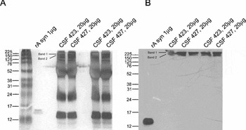
Further parallel SDS-PAGE gels were run using samples prepared in non-reducing buffer. Gels were stained using MS-compatible silver staining or α-synuclein (211) Western blotting was carried out (Figure 1). Bands on the silver-stained gel corresponding to the bands detected by the antibody, were excised and analyzed by liquid chromatography-mass spectrometry (LC/MS) using the following protocol. Gel pieces were reduced and alkylated prior to digestion with trypsin. Digestion was carried out overnight at room temperature following two hours’ incubation at 37°C. Cysteine residues were reduced with dithiothreitol and derivatized by treatment with iodoacetamide to form stable carbamidomethyl derivatives. Acetonitrile and aqueous washes were used to extract the pepetide fragments from the gel pieces, and the extract was combined with the initial supernatant and lyophilized. Samples were resuspended in 23 μl ammonium bicarbonate (50mM). Chromatographic separations were performed using an Ultimate LC system (Dionex, U.K.) and peptides were resolved by reversed phase chromatography on a 75 μm C18 PepMap column. Peptides were eluted at a flow rate of 200nl/min using a gradient of acetonitrile in 0.05% formic acid. Peptides were ionized using a z-spray source fitted to a QTof-micro (Waters Corp.). The instrument was set to run in automated switching mode, selecting precursor ions based on their intensity, for sequencing by collision-induced fragmentation. The tandem mass spectrometry (MS/MS) analyses were conducted using collision energy profiles that were chosen based on the m/z and the charge state of the peptide.
Confirmation of the presence of α-synuclein in CSF using mass spectrometry and Western blotting. Parallel gels were run: one stained with α-synuclein (211), and the second with mass spectrometry compatible silver staining. On both gels, the recombinant form of α-synuclein is detected at around 15 kDa. (A) 1D PAGE gel of recombinant α-synuclein and CSF samples. Bands 1 and 2 represent the excised areas for LC/MS/MS analysis. (B) Corresponding Western blot using primary antibody α-synuclein (211) showing possible aggregation of α-synuclein in CSF.

The MS data was processed into peak lists using ProteinLynx Global Server v2.2.5. The list of peaks was searched against the Swiss Prot and NCBI non-redundant databases using Mascot software v2.2 (http://www.matrixscience.com). Sequence information confirmed the presence of α-synuclein in the detected band.
Western blotting analysis of CSF samples using α-synuclein antibodies
Three further antibodies were tested using Western blotting: α-synuclein (BD, Transduction Laboratories, U.S.A.), and α synuclein antibody [4B12] and α synuclein antibody [syn204] (Abcam) in 1:500 dilution. α-synuclein was raised against amino acids 15–123 of rat synuclein-1, but also recognizes human α-synuclein. 4B12 is a monoclonal antibody which is manufactured using E.coli-produced human α-synuclein. Syn204 is also a monoclonal antibody produced using recombinant human α-synuclein. These antibodies detected the same bands as the α-synuclein (211) antibody. The protocol was further optimized, and using reducing conditions and a primary antibody incubation time of 36hrs, we detected an approximately 24 kDa band. All 12 DLB and nine control CSF samples were then analyzed using these three antibodies. Samples were prepared in Laemmli buffer with reducing agents.
Statistics
As data were non-parametric, α-synuclein concentrations were compared between DLB patients and controls using the Mann-Whitney U test. Preliminary correlations with MMSE were evaluated using Spearman's rank correlation. All evaluations were undertaken with the SPSS computerized statistics package, version 15.0.
Results
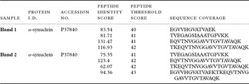
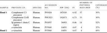
In the first experiment, soluble monomeric α-synuclein was not detected in CSF using α-synuclein (211), although we were able to detect highly aggregated bands > 150 kDa. In an attempt to verify if these highly aggregated bands contain α-synuclein, parallel Western blot and silver stained gels were run followed by analysis of gel extracts by mass spectrometry. Four peptides originating from α-synuclein were identified and showed no identity with other proteins using database searches (Table 1A). These amino acids covered 47% of full-length 140 amino acid α-synuclein (amino acids 32–97). This process confirmed that the silver stained band corresponding to the Western blot band representing the aggregated form of α-synuclein, contained a peptide originating from human α-synuclein (Figure 1 and Table 1A). Further proteins detected in the aggregated bands with sequence coverage are shown in Table 1B. As a control for the monomeric form of α-synuclein, recombinant α-synuclein was also run on these parallel gels and α-synuclein (211) detected a band of 15 kDa. Thus, the antibody recognized both the aggregated form of α-synuclein in CSF and the monomeric soluble form of the recombinant protein (Figures 1A and 1B).
Results of mass spectrometry analyses Four peptides from α-synuclein were present in each of bands 1 and 2. The table shows peptide identity scores and the sequences for each fragment. The aggregated bands detected using Western blotting contain α-synuclein: results of LC/MS/MS analysis of 1D gel bands for proteins with >30% sequence coverage (see Figure 1).

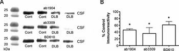
Three α-synuclein antibodies with similar detection results to α-synuclein (211) were used to measure α-synuclein in 12 DLB samples and nine control samples with a modified method. Each of the three α-synuclein antibodies identified a band of approximately 24 kDa, which was semi-quantified. There was a significant reduction in the levels of this band in the DLB patients compared to controls (Figure 2 and Table 2) (Mann-Whitney U test, α-synuclein P = 0.05, α synuclein antibody [4B12] P = 0.001 and α synuclein antibody [syn204] P = 0.03). This difference is still significant when comparing only mild DLB patients (MMSE >24) with controls using α synuclein antibody [4B12] (Mann-Whitney U, p = 0.007).
Analysis of three α-synuclein antibodies in CSF: α synuclein antibody [4B12], α synuclein antibody [syn204], α-synuclein. (A) Western blots derived from two age-matched controls and two DLB cases. (B) Mean integral of band density of 24 kDa band in DLB patients for each antibody expressed as a percentage of control.
* P < 0.05, Mann-Whitney U test.

Lower density of the 24 kDa band in DLB patients than in controls: semi-quantitative levels of approximately 24 kDa band measured using antibodies α-synuclein, α synuclein antibody [4B12] and α synuclein antibody [syn204].

Note: Values are band density measurements in arbitrary units, and show means ± SEM.
DLB = dementia with Lewy bodies; MMSE = Mini-mental State Examination
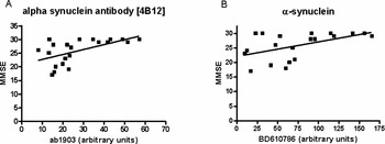
A preliminary analysis of the correlation between MMSE score and the 24 kDa band densities was undertaken. When using antibody α synuclein antibody [4B12], there is a positive correlation between band density and MMSE score (Figure 3) (Spearman's correlation, r = 0.59, P = 0.005) and when using α-synuclein (r = 0.479, P = 0.032) (Figure 3).
MMSE score and density of the 24 kDa band are correlated. Associations between MMSE and α-synuclein measurements in DLB patients using (A) α synuclein antibody [4B12] and (B) α-synuclein. Arbitrary units were measured using Image J to quantify Western blotting bands.

Discussion
Our findings demonstrate that aggregated α-synuclein can be measured in CSF in controls and people with DLB. By optimizing incubation conditions we were able to detect a band of approximately 24 kDa using α-synuclein-detecting antibodies, and semi-quantitative techniques identified significant differences between controls and DLB patients. This 24 kDa band was significantly reduced in DLB patients compared to controls, with a preliminary suggestion of a relationship with the severity of cognitive impairment meriting further investigation. When using α synuclein antibody [4B12], this 24 kDa band was still found to be reduced when comparing controls with patients with mild DLB, indicating that it may be useful as a biomarker in the early disease stages. This result in particular warrants further investigation as many preliminary biomarker studies have examined patients in advanced stages of dementia. This further verifies the potential value of this 24 kDa band as a CSF biomarker of cortical Lewy body dementias by demonstrating a relationship with the severity of cognitive impairment and emphasizes the importance of further methodological developments to enable robust quantitative analysis. Future work must include the identification of the protein in this approximately 24 kDa band using mass spectrometry, as used for the verification of the presence of α-synuclein in the aggregated bands.
Other studies using Western blotting techniques and CSF have detected monomeric α-synuclein (Borghi et al., Reference Borghi2000; El-Agnaf et al., Reference El-Agnaf2003). One notable difference between these studies and our current work is that we have used molecular weight cut-off filters to concentrate CSF samples, whilst other groups have used immunoprecipitation with α-synuclein antibodies prior to Western blotting. It is possible that our chosen technique is a less effective method of sample preparation when measuring monomeric α-synuclein. A further study (Jakowec et al., Reference Jakowec, Petzinger, Sastry, Donaldson, McCormack and Langston1998) also was not able to detect monomeric α-synuclein, but it did detect the presence of a 42 kDa band in CSF. This group used ethanol precipitation rather than immunoprecipitation, further suggesting that immunoprecipitation may be the key to successful α-synuclein detection.
The molecular form of α-synuclein determined in many CSF studies is unknown, although some of the studies claim that they measured oligomeric forms of α-synuclein using ELISAs (El-Agnaf et al., Reference El-Agnaf2006; Tokuda et al., Reference Tokuda2006). Further refinement of our method is required before we can routinely measure different oligomeric forms and aggregates in CSF.
The measurement of this approximately 24 kDa band required incubation with a high concentration of primary antibody and a long exposure of membrane, and it should be acknowledged that the techniques utilized to enable measurement have the potential to introduce bias.
Our study, is consistent with emerging data (Mollenhauer, Reference Mollenhauer2006), but uses longer incubation with primary antibody to quantify protein levels. Furthermore, we performed a structural validation using MS to establish that the antibodies did in fact identify α-synuclein.
The current study, although including a smaller number of patients than others (Mollenhauer, Reference Mollenhauer2006), is the first to carry out a preliminary evaluation of a relationship between a CSF protein and cognition. Although the numbers are too small for robust statistical analysis and we have assessed cognition using MMSE only, there is a preliminary suggestion of a possible relationship between the concentration of this protein in CSF and cognition, emphasizing the need for further studies to clarify this relationship.
In conclusion, the current study confirms a CSF protein, which is potentially important as a biomarker of DLB, extending our previous knowledge by suggesting a possible relationship with the severity of cognitive impairment.
Conflict of interest
None.
Description of authors' roles
Professor Clive Ballard carried out the statistical analysis and writing of the paper, and was responsible for study design. Elisabet Londos, Lennart Minthon and Emma Jones assisted with writing the paper and Paul Francis assisted with study design. Dag Aarsland assisted with the statistical analysis and writing the paper.
Acknowledgments
We gratefully acknowledge Dr. Ezra Mulugeta and Marina Vietri of the Clinical Neuroscience Research, Stavanger University Hospital, Norway, for carrying out the laboratory experiments. This work was funded by the Trustees of Guy's and St Thomas’ Hospital, London.