Background
The Fontan procedure and its modifications have been used as a palliative strategy for patients with single-ventricle physiology for over 45 years. Advances in patient selection, as well as refinements in surgical and postoperative care, have led to low perioperative morbidity and mortality. Consequently, the growing population of patients who undergo this procedure in childhood has been increasingly reported to develop multi-organ dysfunction over time, particularly at the hepatic level. Reference Johnson, Cetta and Graham1,Reference Tsuchihashi, Cho and Tokuhara2
Fontan-associated liver disease encompasses a broad spectrum of structural, functional, and clinical abnormalities secondary to the haemodynamic alterations induced by the Fontan circulation. Reference Tellez, Payance and Tjwa3 Passive pulmonary blood flow, which depends on elevated venous pressure, leads to congestive liver disease and histological changes. These, combined with ischaemic events, trigger the release of profibrotic factors, ultimately resulting in sinusoidal fibrosis and the formation of regenerative nodules, which are associated with the progression to liver fibrosis, cirrhosis, and hepatocellular carcinoma. Reference Goldberg, Surrey and Glatz4,Reference Gordon-Walker, Bove and Veldtman5
The optimal screening modality for Fontan-associated liver disease and its progression remains undefined. The gold standard for diagnosis is liver biopsy, which also allows for assessing the degree of hepatic involvement. Key histopathological findings include architectural distortion, massive sinusoidal dilation, perisinusoidal fibrosis, and an absence of significant parenchymal inflammation. However, the invasive nature of this procedure limits its routine use, reserving it for patients undergoing heart transplantation evaluation or when an alternative aetiology of liver disease is suspected. Noninvasive methods, such as abdominal ultrasound, elastography, and laboratory tests, have been explored with variable results, often demonstrating limited sensitivity. Reference Tsuchihashi, Cho and Tokuhara2
The natural history of Fontan-associated liver disease remains unclear, particularly regarding the timing of onset and the rate of progression in the paediatric population. Therefore, we present our institutional experience in the clinical, laboratory, imaging, and histological evaluation of paediatric patients with Fontan-associated liver disease at a paediatric cardiovascular referral centre in Colombia.
Materials and methods
Study design
This was a retrospective study including all patients diagnosed with FALD during a 23-year follow-up period at Clínica CardioVID, a referral centre for paediatric cardiology and cardiovascular surgery in Medellín, Colombia. The study was approved by the institutional ethics committee.
Patients
All patients diagnosed with Fontan-associated liver disease who met the inclusion criteria between January 2001 and October 2024 were included. At the cardioVID clinic, we have had a functional univentricular heart program for over 20 years, with guidelines and strategies for strict follow-up for each patient, based on national and international consensus and guidelines. Reference Rychik, Atz and Celermajer6 If patients during follow-up in the post-Fontan period met one or more of the following criteria, they were diagnosed with Fontan-associated liver disease:
-
Blood tests: elevated γ-glutamyl transpeptidase, total bilirubinemia, aspartate aminotransferase, and alanine amino-transferase; thrombocytopenia; elevated alpha fetoprotein; and other findings suggestive of progressive liver disease (elevated liver fibrosis markers).
-
Imaging: hepatomegaly or hepatic atrophy, hepatic vein dilatation, enlargement of the caudate lobe, heterogeneous parenchymal echotexture, hyperechoic spots, liver surface nodularity, splenomegaly, and other findings suggestive of progressive liver disease (ascites, portosystemic shunts, or oesophagogastric varices)
-
Presence of liver tumours: benign hyperplastic nodules and focal nodular hyperplasia-like nodules to malignant lesions including HCC and cholangiocarcinoma
-
Liver biopsy: sinusoidal dilatation, liver fibrosis
If a patient had suspected Fontan-associated liver disease, during the functional univentricular heart follow-up program, additional tests were requested for the appropriate approach and the patient was referred for evaluation by paediatric hepatology. Data were obtained from medical records. Per institutional protocol, all included patients underwent extracardiac fenestrated Fontan surgery (5 mm) between 4 and 6 years of age and all patients were under treatment with aspirin thromboprophylaxis. Data collection was conducted using Microsoft Excel, with records maintained from the time of admission until the most recent consultation or hospitalisation.
Variables
The study variables included age, sex, weight, height, cardiac anatomy, and ventricular morphology. Additionally, a history of previous surgical procedures—such as systemic-to-pulmonary shunts, pulmonary artery banding, or bidirectional cavopulmonary shunt—was recorded. Variables related to the Fontan procedure itself were also analyzed, including Fontan type (extracardiac and fenestrated), time of failure diagnosis, laboratory parameters, hemodynamic variables, and outcomes (death, transplantation listing, and survival). The variables of Fontan failure and concomitant presence of protein-losing enteropathy and plastic bronchitis were related to the diagnostic criteria proposed by Rychik et al. Reference Rychik, Atz and Celermajer6 and the institutional protocol. This variable was considered a concomitant diagnosis at the time of Fontan-associated liver disease diagnosis.
Echocardiographic variables at diagnosis were extracted from institutional aediatric echocardiography reports. Atrioventricular valve regurgitation was defined qualitatively by the evaluator at the time of diagnosis. By convention, systolic dysfunction was defined qualitatively and, in some cases, quantitatively, based on ventricular ejection fraction. By convention, a cut-off value of < 50% was used, regardless of the method used.
Haemodynamic variables included mean pulmonary artery pressure, pulmonary vascular resistance index, end-diastolic pressure, and valvular regurgitation. Liver function parameters, imaging findings, elastography results, and histological features were also evaluated. Follow-up during the study period included outcomes of interest such as cirrhosis and hepatocellular carcinoma.
Statistical analysis
A descriptive analysis was performed for quantitative variables. Normality was assessed using the Shapiro–Wilk test, which determined the measures of central tendency (median and mean), positional measures (quartiles, deciles, and percentiles), and dispersion (standard deviation and interquartile range). For qualitative variables, absolute and relative frequencies were calculated. Statistical analyses were performed using Jamovi, a free statistical software program.
Results
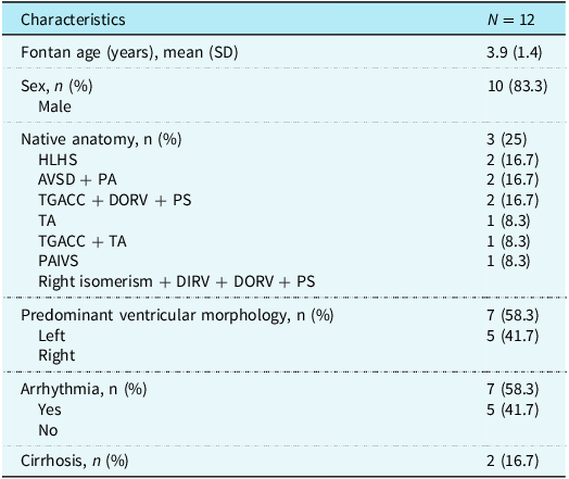
During the study period, 108 patients were at the Fontan stage at the time of data collection. Among them, 12 patients (11.1%) met the criteria for Fontan-associated liver disease. Of these, 10 were male (83.3%), and the most common native cardiac anatomy was hypoplastic left heart syndrome. The mean age at which extracardiac Fontan surgery was performed was 3.9 years (SD±1.4). See Table 1.
Socio-demographic and clinical characteristics

*HLHS: Hypoplastic left heart syndrome. AV: Atrioventricular. TGACC: Congenitally corrected transposition of the great arteries. DORV: Double outlet right ventricle. PS: Pulmonary stenosis. TA: Tricuspid atresia. PAIVS: Pulmonary atresia with intact ventricular septum. DIRV: Double inlet right ventricle. IQR: Interquartile range. SD: Standard deviation.
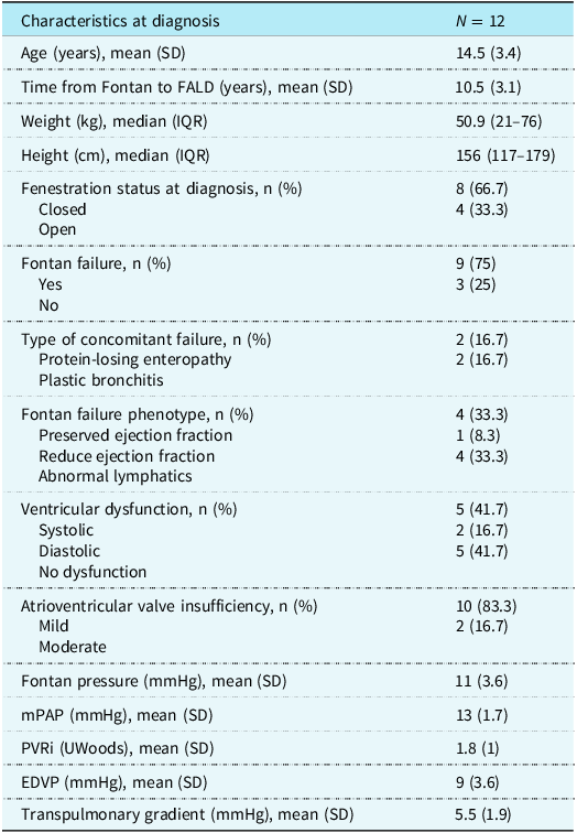
The mean age at Fontan-associated liver disease diagnosis was 14.5 years (SD±3.4). At the time of diagnosis, 75% of patients had concomitant Fontan failure, and 66% had a closed fenestration. Additionally, 33% presented with concomitant protein-losing enteropathy and/or plastic bronchitis.
Echocardiographic follow-up revealed systolic dysfunction in 41% of patients and diastolic dysfunction in 16%. All patients exhibited some degree of atrioventricular valve insufficiency, which was mild in 83.3% and moderate in 16%. Only one patient had liver elastography findings consistent with severe liver fibrosis. Haemodynamic parameters at diagnosis were within normal ranges, as detailed in Table 2.
Fontan characteristics at diagnosis and evidence of concomitant failure

EF: Ejection fraction. mPAP: Mean pulmonary artery pressure. PVRi: Pulmonary vascular resistance index. EDVP:End-diastolic ventricular pressure.
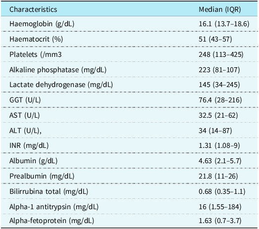
Table 3 presents the laboratory values for liver function enzymes, tumour markers, and metabolic markers at the time of diagnosis. Gamma-glutamyl transferase levels were elevated beyond the normal range for age.
Laboratory values at diagnosis of FALD

GGT: Gamma-glutamyl transferase. AST: Aspartate aminotransferase. ALT: Alanine aminotransferase. INR: International normalised ratio. IQR: Interquartile range.
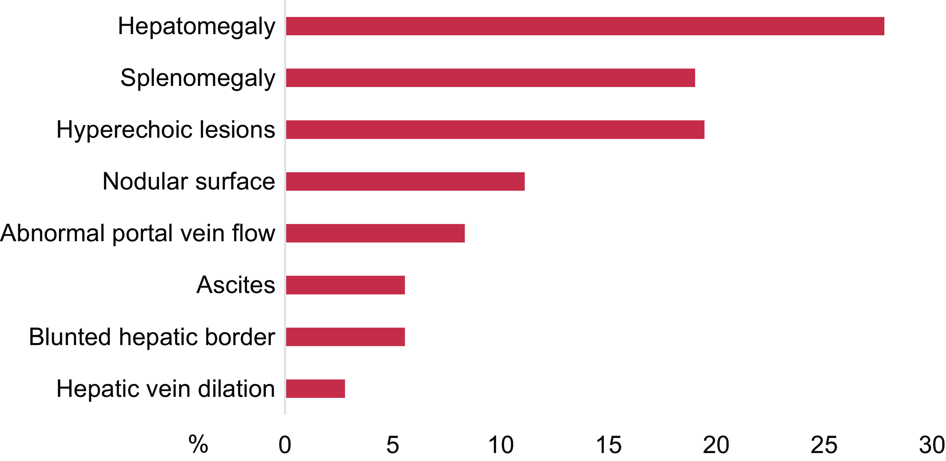
In the liver and biliary tract ultrasound, the main findings were hepatomegaly (28%), followed by splenomegaly and hyperechoic lesions (20%) (see Figure 1).
Findings in liver and biliary tract ultrasound.

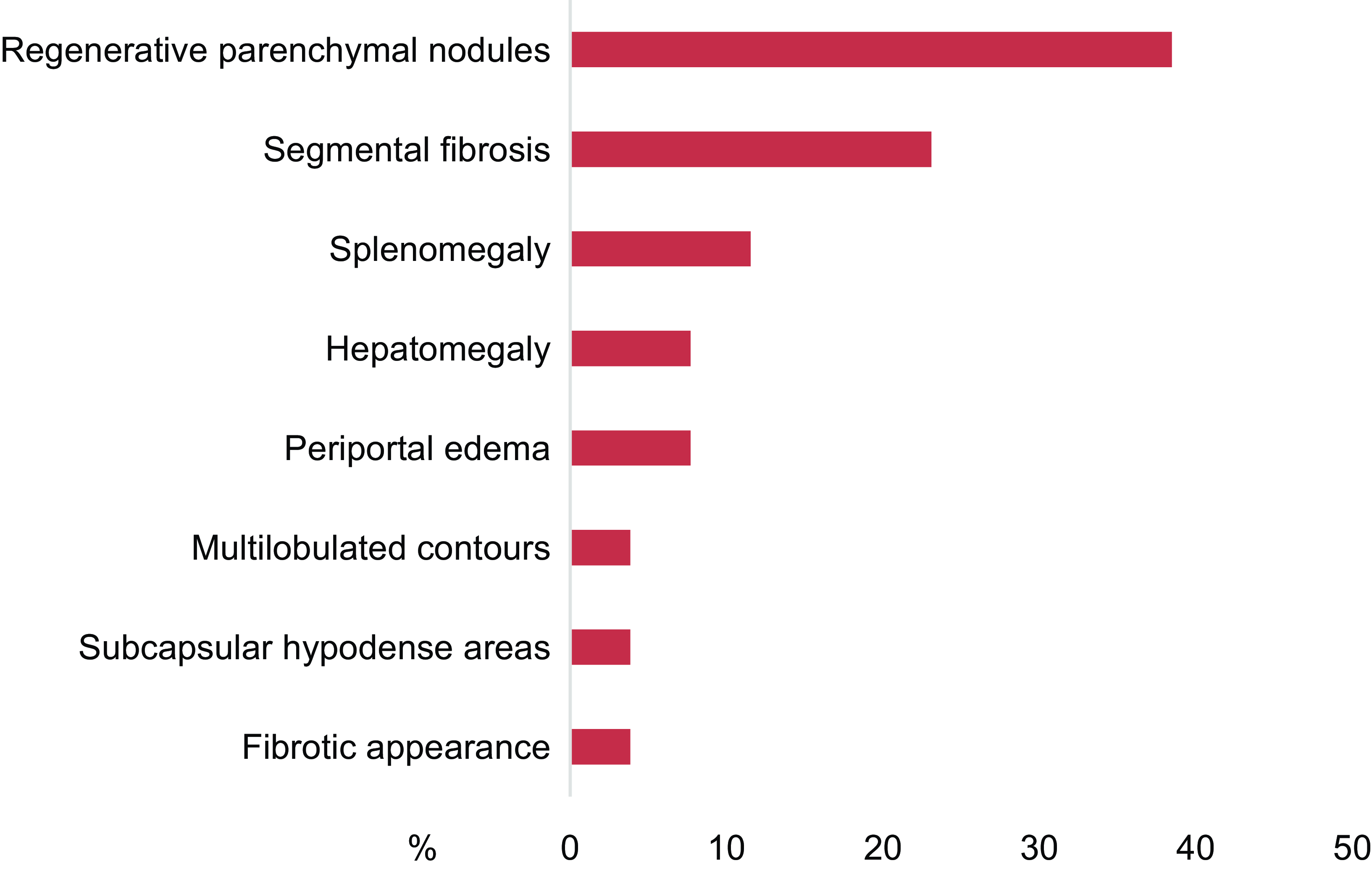
Nine patients underwent liver MRI during follow-up and the most frequent findings were segmental fibrosis and regenerative parenchymal nodules (38%), followed by segmental fibrosis (23%) and splenomegaly (12%) (see Figure 2).
MRI findings of the liver and bile ducts.

A liver biopsy was performed in six patients, four of whom underwent the procedure using the transjugular approach. The most frequent finding was fibrosis of the portal and sinusoidal spaces in three patients, followed by oedema and polymorphic inflammatory infiltrate in the portal triad, focal nodular hyperplasia, and micronodular cirrhosis, each observed in two patients. During follow-up, two patients developed clinical cirrhosis, and none were diagnosed with hepatocellular carcinoma.
Discussion
FADL is a well-recognized consequence in this patient population, potentially leading to liver fibrosis, cirrhosis, and hepatocellular carcinoma. Reference Rychik, Atz and Celermajer6 There is limited evidence on the progression of this condition in paediatric patients, as many of its manifestations depend on the time elapsed between Fontan surgery and the onset of Fontan-associated liver disease.
This study describes the clinical, laboratory, imaging, and invasive characteristics of a series of paediatric patients diagnosed with Fontan-associated liver disease. The disease was predominantly identified in adolescents, with a mean age of presentation of 14.5 years and a median interval of 10.5 years from Fontan surgery to diagnosis. These findings align with other paediatric series, such as those reported by DeBruyne et al. Reference De Bruyne, Vandekerckhove and Van Overschelde7 and in the meta-analysis by Seol et al. Reference Seol, Song and Kim8 In contrast, studies in the adult population, such as those by Hitawala et al. Reference Hitawala, Gopalakrishna and Mironova9 and Pundi et al. Reference Pundi, Pundi and Kamath10 , describe cirrhosis onset occurring approximately 20 to 30 years after Fontan surgery.
Although the exact cause of death in patients with Fontan circulation is sometimes unclear, Fontan-associated liver disease has been identified as a significant contributor to morbidity and mortality. Various studies estimate a mortality rate up to three times higher compared to controls, with reports from Japan indicating rates as high as 17.8%. These findings highlight the importance of early prediction, timely diagnosis, and appropriate management to mitigate mortality associated with liver dysfunction.
Noninvasive evaluation for the diagnosis of Fontan-associated liver disease through protocolised follow-up has certain limitations, which may lead to delays in diagnosis. Reference Tsuchihashi, Cho and Tokuhara2 Laboratory markers have limited diagnostic value, with gamma glutamyl transferase being the most consistently reproducible across different studies. In the present study, gamma glutamyl transferase levels were elevated above the normal range for age, but they did not correlate with other cholestasis markers or parameters of hepatocellular injury, similar to the findings reported by Bruyne et al. Reference Tellez, Payance and Tjwa3
Imaging studies can provide important clues regarding structural alterations secondary to physiological and histological changes. Ultrasound and magnetic resonance imaging MRI have an established role in Fontan-associated liver disease evaluation, demonstrating acceptable sensitivity and specificity. Reference Tsuchihashi, Cho and Tokuhara2 In recent years, magnetic resonance elastography and shear wave elastography have shown good sensitivity for early fibrosis detection, correlating well with liver biopsy findings. Reference Sugimoto, Oka and Kajihama11 However, in our setting, access to these imaging modalities is limited, and elastography was only performed in one patient, with good correlation to biopsy results.
Liver biopsy remains the gold standard for diagnosing Fontan-associated liver disease. In our study, 50% of the patients underwent biopsy, with four of them undergoing the procedure via the transjugular approach. This technique not only allows for safer tissue sampling with a reduced risk of bleeding but also enables the concurrent acquisition of haemodynamic data, including central venous pressure, hepatic venous pressure gradient, and Fontan system pressures, through simultaneous catheteris ation. Reference Borquez, Silva-Sepulveda and Lee12– Reference Butikofer, Greutmann-Yantiri and Gubler14
Systolic and diastolic ventricular dysfunction are well-established risk factors for liver dysfunction and late mortality. Reference Anderson, Sleeper and Mahony15 –Reference Grattan and Mertens16 However, objective assessment of ventricular function in patients with univentricular circulation remains challenging due to the lack of consistent interobserver correlation. Advanced imaging techniques such as strain analysis and 3D echocardiography have improved the evaluation of ventricular function in Fontan patients.
The correlation between haemodynamic variables and the development of Fontan-associated liver disease does not follow a strictly linear pattern regarding diagnosis and severity assessment. In the present study, Fontan pressure, pulmonary vascular resistance index, and ventricular end-diastolic pressure showed no significant alterations at diagnosis. This finding aligns with previous studies and meta-analyses, such as that of Seol et al. Reference Seol, Song and Kim8 , which identified a direct correlation between a Fontan pressure greater than 15 mmHg and advanced liver fibrosis. In our study, Fontan system pressure measured during hemodynamic evaluation averaged ≤ 15 mmHg at the time of diagnosis, with only two patients exceeding this threshold. These findings suggest that assessing the trend of pressure values over time may be more informative than relying on a single measurement. However, this type of longitudinal analysis was not feasible given the retrospective nature of our study. Furthermore, it is important to note that haemodynamic evaluations at our institution are routinely performed under general anaesthesia, which may influence pressure measurements in paediatric patients. This factor should be taken into consideration when interpreting the data.
In our study, no patients underwent liver, heart, or combined transplantation as a management strategy. In addition to interventional approaches, Kigoso et al. Reference Kogiso, Ogasawara and Taniai17 , in a series of 220 patients, demonstrated that the use of ursodeoxycholic acid significantly reduced liver enzyme levels and mitigated the development of hepatocellular carcinoma, suggesting a potential benefit for patients with Fontan-associated liver disease.
This study has some limitations. The retrospective design and the relatively small number of patients with this complication limited our ability to compare specific risk factors for Fontan-associated liver disease53. Additionally, elastography data were scarce, as this diagnostic tool is relatively new in our setting. Standardised paediatric values for elastography analysis are still being validated, which prevented us from obtaining data from most patients.
Conclusions
Liver disease is a persistent concern in paediatric patients with Fontan physiology, posing diagnostic and therapeutic challenges. Healthcare professionals managing these patients should be aware of its early identification and appropriate treatment.
Financial support
This research received no specific grant from any funding agency, commercial, or not-for-profit sectors
Competing interests
None
Ethical standard
The CardioVID Clinic ethics committee approved this study.