INTRODUCTION
Human lymphatic filariasis (LF) is a disease caused by the nematode parasites Wuchereria bancrofti, Brugia malayi and Brugia timori. At present, 120 million people in 81 countries are infected by this parasite, and millions more are at risk of acquiring the infection. Disease pathogenesis is linked to host inflammation invoked by the death of the parasite, causing hydrocoele, lymphoedema and elephantiasis (Taylor et al. Reference Taylor, Hoerauf and Bockarie2010). The current control efforts that rely on the repeated annual mass administration of antifilarial combination are unsustainable and the threat of drug resistance to mainstay drugs is increasing. Therefore, there is an urgent need to develop an effective drug or a vaccine candidate. Understanding the structure and the functions of the essential filarial proteins and determination of their immunoprophylactic potential seems to be imperative.
RNA helicases are some of the most abundant essential proteins in B. malayi (Elodie et al. 2007) that participate in all the biological processes involving RNA such as transcription, splicing, translation and, in addition, they utilize the energy derived from ATP hydrolysis to modulate the structure of RNA. Considering the merits of RNA helicase in B. malayi, we attempted to construct the 3D structure of this protein. In our earlier study, the cDNA clone of B. malayi helicase was picked up via immunoscreening of the infective larval (L3) cDNA library and the recombinant protein demonstrated high reactivity with the endemic normal W. bancrofti area (Singh et al. Reference Singh, Srivastava and Bhattacharya2009b). These findings impelled us to explore the immunoprophylactic potential of BmL3-Helicase in the experimental rodent model. It is also well documented that the immune response in LF is most likely to be operative in the host during early larval development and therefore the antigens derived from L3 represent the key targets of protective immunity because they are important in the establishment of infection (Devaney and Osborne, Reference Devaney and Osborne2000; Gregory et al. Reference Gregory, Atmadja, Allen and Maizels2000). The parallel observations were also made to divulge the immune response generated by this protein. To the best of our knowledge, this is the first report on the 3D structure and immunoprophylactic efficacy of B. malayi RNA helicase.
MATERIALS AND METHODS
Molecular modelling of BmL3-Helicase
The 3D structure of BmL3-Helicase was constructed by homology modelling using the Modeler program (Sali and Blundell, Reference Sali and Blundell1993) interfaced with Insight II 2000.1 (Accelrys Inc., San Diego, CA, USA). All the computational studies were performed on a SGI Origin300 server equipped with four 600-MH3-R-12000 processors. Stereochemical evaluations were performed with the program PROCHECKv3.4.4 (Laskowski et al. Reference Laskowski, MacArthur, Moss and Thornton1993). Amino acid sequence of BmL3-Helicase was submitted to BLAST 2.0 (Altschul et al. Reference Altschul, Thomas, Alejandro, Jinghui, Zheng, Webb and David1997) to identify any structural homologue. Out of the various BLAST hits, the crystal structure of human RNA Helicase DDX48 (c) with 25% sequence identity was chosen as a structural template for homology modelling.
In silico analysis of immunogenicity of BmL3-Helicase
The antigenicity of BmL3-Helicase was determined using Kolaskar's method; available at http://imed.med.ucm.es/Tools/antigenic.pl (Kolaskar and Tongaonkar, Reference Kolaskar and Tongaonkar1990). The Propred algorithm (www.imtech.res.in/raghava/propred/) was used to predict the presence of promiscuous human major histocompatibility complex (MHC) class II-restricted epitopes (Singh and Raghava, Reference Singh and Raghava2001). Promiscuous epitopes are defined as peptides that are predicted to bind to at least 80% of the MHC class II molecules expressed by the 51 human leukocyte antigen (HLA) subregion DR alleles available for analysis in the ProPred algorithm. The threshold for the ProPred analyses was set at a relatively high stringency of 3%.
Parasite host
The experimental animals, Mastomys coucha were maintained under appropriate housing conditions at the Laboratory Animal Facility of our Institute. All of the animals and the experimental protocols involving animal handling were duly approved by the Institutional Animal Ethics Committee (IAEC). Brugia malayi L3 for challenge experiments were recovered from the laboratory-bred vector mosquitoes (Aedes aegypti) that had been fed on donor mastomys 9 days previously (Singh et al. Reference Singh, Misra, Muthy, Katiyar, Agrawal and Sircar1997).
Cloning, expression and purification of recombinant BmL3-Helicase
The 1·16 kb immunodominant cDNA clone of BmL3-Helicase was picked up by immunoscreening of the larval stage cDNA library of B. malayi using resistant sera raised against irradiated L3. The recombinant construct was subcloned into pET28a, transformed into Escherichia coli BL21 (DE3) cells and the protein was over-expressed after induction with 1·0 mm isopropyl-β-D-thiogalactopyranoside (IPTG, Sigma). The histidine-tagged recombinant protein was purified through a nickel nitrilotriacetic acid agarose affinity column (Ni-NTA Column, Qiagen) (Singh et al. Reference Singh, Srivastava and Bhattacharya2009b).
Immunization and challenge
Mastomys coucha, the susceptible host for B. malayi infection, were divided into 3 groups each consisting of 5 mastomys: (i) control group, (ii) adjuvant group and (iii) BmL3-Helicase immunized group. Then 25 μg protein/animal was administered along with Freund's complete adjuvant (FCA, Sigma) subcutaneously (s.c.), followed by 2 booster doses of an equivalent quantity of protein in Freund's incomplete adjuvant (FIA) on day 14 and day 21 of the first immunization. The adjuvant group received FCA/FIA in PBS and the normal control group received phosphate-buffered saline (PBS, pH 7·2). One week later, all the groups of animals were challenged with 100 L3 of B. malayi by the s.c. route. The experiment was repeated using the same number of mastomys and groups.
Effect of immunization on microfilaraemia, adult parasite recovery and female worm fecundity
The observation of microfilaraemia was initiated in 10 μL of tail blood on day 90 post-challenge (p.c.) and followed until day 270 by preparing thick blood smears at fortnightly intervals (Vedi et al. Reference Vedi, Dangi, Hajela and Misra-Bhattacharya2008). Mastomys were euthanized on day 270 p.c. to assess the recovery of adult worms. Various tissues, namely heart, lungs, testes and lymph nodes were gently teased in 0·85% saline to isolate and count adult worms. Female worms were teased and observed microscopically to assess the effect of immunization on worm fecundity. Arithmetic means were calculated for the total worm burden and the percentage protection was calculated. Female worm sterilization was calculated as the ‘number of sterile worms/total worm teased × 100’.
Measurement of specific IgG antibody and antibody isotypes by enzyme-linked immunosorbent assay (ELISA)
Blood for sera was collected from the retro-orbital plexus of animals at various times to measure the serum IgG antibody titre (Singh et al. Reference Singh, Misra, Muthy, Katiyar, Agrawal and Sircar1997). The ELISA plates were coated with 1 μg mL−1 of BmL3-Helicase and blocked with 1% gelatine in PBS-T (phosphate-buffered saline with 0·05% Tween-20). Sera of immunized and control mastomys were used as primary antibodies and rabbit anti-mouse-IgG-HRP served as the secondary antibody (Sigma, USA). The absorbance was read at 492 nm using an ELISA reader (Infinite M200, Tecan, Switzerland) after adding the substrate, orthophenylenediamine (OPD), and stopping the reaction with 20 μL of H2SO4.
IgG isotypes and IgA levels were determined using an antibody isotyping kit (Sigma, USA) as mentioned earlier (Vedi et al. Reference Vedi, Dangi, Hajela and Misra-Bhattacharya2008). ELISA plates were coated with 0·1 μg mL−1 of protein followed by blocking with 1% gelatin in PBS-T. After antigen coating and blocking, 1 : 100 dilutions of sera were added followed by a 1 : 1000 dilution of monoclonal antibodies to each IgG isotype (goat anti-mouse IgG1, IgG2a, IgG2b, IgG3) and IgA and a 1 : 5000 dilution of HRP-rabbit anti-goat IgG. The absorbance was read at 492 nm using an ELISA reader after adding OPD as substrate.
In vitro antibody-dependent cellular adhesion and cytotoxicity (ADCC)
Cellular adhesion to microfilariae, L3 and adult parasites was carried out as described previously (Vedi et al. Reference Vedi, Dangi, Hajela and Misra-Bhattacharya2008). Microfilariae and adult worms were isolated from the peritoneal cavity of infected gerbils while L3 were isolated from an experimentally infected mosquito, A. aegypti. Then 100 live microfilariae, 4 adult females and 10 L3 were individually co-cultured with 1 × 10⁶ peritoneal exudate cells (PECs) isolated from normal mastomys in the presence of a polyclonal serum raised against BmL3-Helicase. The adherence of cells on the surface of parasites and worm motility were observed microscopically at 1, 3, 6 and 24 h of incubation (Vedi et al. Reference Vedi, Dangi, Hajela and Misra-Bhattacharya2008).
Cell surface staining of B and T cells by flow cytometry
Immunophenotyping was carried out to assess lymphocyte proliferation on FACS Calibur (BD Biosciences) using fluorescein isothiocyanate (FITC)-labelled CD3, CD45, CD80, CD19, CD16/32 or phycoerythrin (PE)-labelled CD4, CD8 fluorochrome-conjugated monoclonal antibodies directed against specific receptors following the manufacturer's protocol (BD Biosciences). The splenocyte suspension was prepared as mentioned earlier (Singh et al. Reference Singh, Shakya, Soni, Dangi, Kumar and Bhattacharya2009a) and 1 × 10⁶ cells were incubated with CD3/CD4 or CD3/CD8 or CD4/CD45, CD80, CD16/32 antibodies for 10 min at room temperature. Aliquots of cells were also labelled with B-cell surface-specific CD19. After washing, cells were suspended in PBS and analysed on FACS.
Measurement of Th1/Th2 cytokine response
Intracellular cytokines in the spleen cells were estimated as described earlier (Singh et al. Reference Singh, Shakya, Soni, Dangi, Kumar and Bhattacharya2009a). Briefly, splenocytes from each animal (4 × 10⁶ mL−1) were first incubated with Brefeldin A (10 μg mL−1) in the dark for 4 h in a CO2 incubator at 37 °C followed by re-incubation in the dark at 4 °C in the presence of FITC-labeled CD4+ antibody. The cells were fixed with Leucoperm A, permeabilized with the Leucoperm B and dispensed in 4 tubes each containing 1 × 10⁶ cells 100 μL−1. PE-labelled rat anti-mouse monoclonal antibodies to the cytokines, IL-2, IL-4, IL-10 and IFN-γ were added and cells were incubated for 15 min in the dark. Cells were washed and suspended in 250 μL of 0·5% paraformaldehyde for FACS readings.
Content of nitric oxide (NO) and reactive oxygen species (ROS) in peritoneal macrophages
PECs were isolated from all the groups of mastomys after introducing cold RPMI medium (Gibco) in the peritoneal cavity via a sterile syringe as described previously (Singh et al. Reference Singh, Shakya, Soni, Dangi, Kumar and Bhattacharya2009a). Then 3 × 10⁶ cells mL−1 of cells (100 μL/well) were plated in 96-well plates in triplicate and stimulated with 2·5 μg mL−1 of BmL3-Helicase or 1·0 μg mL−1 of LPS for 24 h at 37 °C in a CO2 incubator. The production of NO in the cell supernatant was measured by Griess reagent (Sigma) by taking the optical density at 540 nm. Real-time monitoring of ROS was carried out by fluorometric assay using 2′–7′-dichlorofluorescein diacetate (DCF-DA) as described previously (Vedi et al. Reference Vedi, Dangi, Hajela and Misra-Bhattacharya2008).
Statistical analysis
Data were analysed using Student's t-test and one-way analysis of variance (ANOVA) as appropriate. Individual comparisons following ANOVA were made using the Dunnet's t-test method with the help of statistical software PRISM 3.0. A value of P < 0·05 was considered significant, and P < 0·01 as highly significant.
Nucleotide sequence data reported in this paper are available in the GenBank™, EMBL and DDBJ databases under the accession number EF409381.
RESULTS
3D homology model of BmL3-Helicase has DEAD and SAT domains in close vicinity
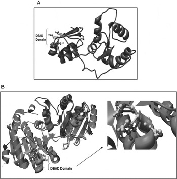
The alignment of the amino acid sequence of the functional domains of DEAD box RNA Helicase from B. malayi with the sequence from RNA Helicase DDX48 protein from Homo sapiens (PDB Id: 2J0U_B; 3·0 Å resolution) obtained from NCBI Blast is shown in Fig. S1 (Online version only). The 3D structure model of BmL3-Helicase is shown in Fig. 1A. Refinement of the homology model removed the steric constraints and a stable conformation was obtained by energy minimization of 1000 iterations. After the refinement process, the model was validated using the PROCHECK program. The Ramachandran plot showed normal distribution of points with phi (ϕ) values and psi (ψ) values clustered in a few distinct regions with 88·3% and 10·6% of residues occupying core and allowed regions, respectively. Only a few residues (0·6%) were in the disallowed regions. Superimposition of the BmL3-Helicase homology model on the template also revealed a close structural resemblance along with non-variant similarities in DEAD and SAT domains of both the proteins, indicating high conservation (Fig. 1B).
Three-dimensional homology model of Brugia malayi RNA Helicase using Modeler and PROCHECK. (A) Homology model structure of BmL3-Helicase showing conserved DEAD domain. (B) Superposition of BmL3-Helicase (model, light grey shade) with the Homo sapiens RNA helicase crystal structure (template, dark grey shade). The DEAD box domain of BmL3-Helicase (light grey shade) and template (dark grey shade) is depicted by the ball and stick model.

BmL3-Helicase possesses antigenic property
The results obtained after analysis of the amino acid sequence of BmL3-Helicase revealed a high antigenic propensity for this protein (1.0263) with a total of 15 antigenic determinants (Table S1 – Online version only). Six antigenic determinants were located in the N-terminal region and the remaining 9 were in the rest of the region including the conserved DEAD and SAT domains. In addition, 12 promiscuous T-cell epitopes (Table S2 – Online version only) were identified by ProPred.
Subcutaneous administration of BmL3-Helicase notably impairs establishment of the challenged infective larvae
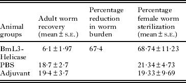
There was a marked reduction in adult B. malayi recovery in the BmL3-Helicase group (6·1±2·0) as compared with worms recovered from the PBS (18·7±2·7) or adjuvant (19·4±3·7) control groups (Table 1). Thus, the immunization with BmL3-Helicase signifies a noticeable reduction in the larval development resulting in a reduced (67·4%) adult worm burden compared with the PBS control group (Table 1).
Summarized table for adult worm recovery, reduction in worm burden and female worm sterilization in immunized and control groups
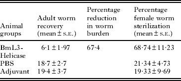
Immunization also weakened (68·74±11·2%; P < 0·01) the reproductive potential of adult female B. malayi since a significant proportion of established females carried degenerated intrauterine stages that were unable to transform into microfilariae in contrast to normal embryos in the uteri of females recovered from control groups (Table 1).
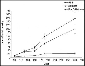
The microfilariae started to appear in the tail blood of mastomys after day 90 post-L3 challenge. However, the counts were low in all the experimental groups during initial monitoring but increased subsequently in the PBS or adjuvant control groups in contrast to the BmL3-Helicase immunized group that demonstrated an 86·7% decrease (P < 0·01) compared with that of the PBS control group (Fig. 2).
Microfilarial kinetics in Mastomys coucha immunized with BmL3-Helicase, adjuvant or PBS. Blood microfilaraemia was determined after 12–28 weeks post challenge. In all the experimental groups microfilarial density was low during the initial monitoring period and increased subsequently in the following weeks, but remained low in the BmL3-Helicase immunized group throughout the observation period. Each point represents the mean±s.e.

Serum antibody isotypes demonstrate the generation of a polarized Th2 immune response
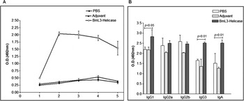
The BmL3-Helicase immunized group developed high titres of antigen-specific IgG antibody incongruent to non-immunized mastomys (Fig. 3A). The IgG level continuously rose following each immunizing dose, reaching a maximum on day 7 post final protein booster and did not augment further post L3 challenge. However, the level was comparatively much higher in the BmL3-Helicase immunized group. The pattern of BmL3-Helicase specific antibody isotypes revealed a predominant elevation of IgG1 (P < 0·05), IgG3 (P < 0·01) and IgA (P < 0·01) while IgG2a and IgG2b remained unaffected (Fig. 3B).
Humoral antibody response in various experimental groups. (A) Kinetics of BmL3-Helicase specific IgG antibody response in the sera of mastomys. All animals were free of BmL3-Helicase specific antibodies on the day of initiation of vaccination (1). The recombinant protein generated a gradually increasing IgG antibody level following each immunizing dose, reaching a maximum on day 7 post-final protein booster (2) and did not increase further post-L3 challenge (3, 4) but remained high even up to the day of autopsy (5) in the BmL3-Helicase immunized group. The PBS and adjuvant groups did not develop a BmL3-Helicase specific IgG response. (B) BmL3-Helicase specific IgG isotypes levels in sera of mastomys of different groups. The IgG1, IgG2a, IgG2b, IgG3 and IgA antibodies were checked by use of ELISA for specific antibodies against BmL3-Helicase. The BmL3-Helicase immunized group (dark shaded bar), showed a significant increase in IgG1 (P < 0·05), IgG3 isotypes and IgA (P < 0·01) antibody as compared with the PBS control (open bars), adjuvant control group (light shaded bar). OD values (at 492 nm) are shown as the mean±s.e.

Antibodies generated to BmL3-Helicase induced sizeable adherence of cells to the surface of L3 and microfilariae
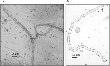
The BmL3-Helicase immunized animal serum led to marked cellular adherence on the surface of microfilariae and L3 at 1 : 16 dilution within 24 h of parasite co-culture with peritoneal exudate cells (Fig. 4) that resulted in complete immobility and subsequent death of the parasite. In contrast, cell adhesion to the adult worm surface was almost negligible.
Antibody dependent cellular adhesion to the surface of (A) microfilariae, (B) infective larvae. Photographs were captured on a phase-contrast fluorescent microscope (Nikon, Japan) at 40 × magnification. Arrows indicate adhered cells. Scale bar=100 μm.

BmL3-Helicase induces CD4+ T-helper cell proliferation
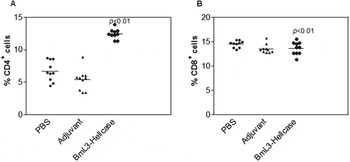
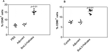
The function and state of activation of different cell types can be determined by the expression or presence of CD antigens on the cell surface. The flow cytometric analysis illustrated an increased population of CD3+/CD4+ T cells (P < 0·01) accompanied by a marginally decreased CD3+/CD8+ cell population (Fig. 5), clearly displaying induction of a T-helper cell population in response to the immunogenic enzyme as a result of larval invasion. Further, expression of CD4/CD45 markers and CD80 marker on antigen-presenting cells which provide the necessary co-stimulatory signals for Th cell activation and proliferation was also elevated (P < 0·01) in the BmL3-Helicase immunized group (Fig. 6).
Immunophenotyping of CD4+, CD8+ cell surface marker. (A) The flow-cytometric analysis of CD4+ T cells in BmL3-Helicase immunized group (open circular dots), PBS (closed circular dots) or adjuvant control group (triangular dots). (B) The flow-cytometric analysis of CD8+ T cells in BmL3-Helicase immunized group (open circular dots), PBS (closed circular dots) or adjuvant control group (triangular dots). Data are represented as scatter plots where each dot represents the value for an individual animal. The lines drawn parallel to the X-axis represent the mean values. P < 0·01 was considered as highly significant.

Immunophenotyping of CD45+, CD80+ cell surface marker. (A) The flow-cytometric analysis of CD45+ markers in the BmL3-Helicase immunized group (open circular dots), PBS (closed circular dots) or adjuvant control group (triangular dots). (B) The flow-cytometric analysis of CD80+ marker in the BmL3-Helicase immunized group (open circular dots), PBS (closed circular dots) or adjuvant control group (triangular dots). Data are represented as scatter plots where each dot represents the value for an individual animal. The lines drawn parallel to the X-axis represent the mean values. P < 0·01 was considered as highly significant.

BmL3-Helicase immunization also leads to an increase in the population of CD19+ B cells and CD16/32
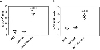
FACS analysis showed a significantly higher (P < 0·01) density of CD19+ cells suggesting the generation of an antibody-mediated protective immune response in immunized animals. Increased levels of CD16/32 (Fcϒ receptor) (P < 0·01) indicated the possible involvement of innate effector cells in the protective mechanism (Fig. 7).
Immunophenotyping of CD19 and CD16-32 cell surface marker. (A) The flow-cytometric analysis of CD19 cell surface marker in BmL3-Helicase immunized group (open circular dots), PBS (closed circular dots) or adjuvant control group (triangular dots). (B) The flow-cytometric analysis of CD16-32 cell surface marker in BmL3-Helicase immunized group (open circular dots), PBS (closed circular dots) or adjuvant control group (triangular dots). Data are represented as scatter plots where each dot represents the value for an individual animal. The lines drawn parallel to the X-axis represent the mean value. P < 0·01 was considered as highly significant.

An early T-cell response to BmL3-Helicase is dominated by a mixed Th1/Th2 cytokine response
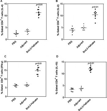
The levels of both pro-inflammatory and anti-inflammatory cytokines were determined intracellularly in the splenic cell population of immunized-challenged mastomys by FACS analysis. There was an increase in the level of pro-inflammatory as well as anti-inflammatory cytokines namely IL-2, IFN-γ and IL-4, IL-10 (Fig. 8).
Measurement of pro-inflammatory and anti-inflammatory cytokines. The flow-cytometric analysis of (A) IL-4, (B) IL-2, (C) IFN-γ and (D) IL-10 cytokines in BmL3-Helicase immunized group (open circular dots), PBS (closed circular dots) or adjuvant control group (triangular dots). Data are represented as scatter plots where each dot represents the value for an individual animal. The lines drawn parallel to the X-axis represent the mean value. P < 0·01 was considered as highly significant.

BmL3-Helicase immunization causes profound activation of macrophages
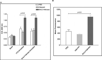
Peritoneal macrophages of BmL3-Helicase immunized animals generated a significantly higher level of NO as compared with the 2 control groups (P < 0·01) following L3 challenge (Fig. 9A). Real-time monitoring of the oxidative burst generated from peritoneal macrophages to determine the in vivo milieu of macrophage activity (Fig. 9B) demonstrated a considerably significant oxidative burst (P < 0·01) by the BmL3-Helicase immunized group.
Nitric oxide and reactive oxygen species determination by macrophages of immunized and control groups. (A) NO production in the BmL3-Helicase immunized group (dark shaded bar), PBS control (open bar), and adjuvant control (light shaded bar). (B) ROS levels in the BmL3-Helicase immunized group (black bar), PBS control (open bar), and adjuvant control (light shaded bar). Each bar represents the mean±s.e.P < 0·01 was considered as highly significant.

DISCUSSION
The structural elucidation of BmL3-Helicase can be exploited for drug or vaccine development based on the differences from its human homologue. This could not have been possible by sequence analysis only. Thus, by comparing the 3D structure of BmL3-Helicase with Human Helicase we can get better insights into the major and minor differences between the two, in order to (i) avoid any cross-reactivity with the human homologue during vaccine development and (ii) develop novel inhibitors which specifically target B. malayi. In addition, a study on the 3D structure of BmL3-Helicase provided more insight into the specificity of the binding mechanism of conserved domains, namely the DEAD domain and SAT domain, to exhibit an ATP-dependent RNA unwinding property. The DEAD domain is an ATP-binding domain and is indispensable for ATPase and helicase activity, while any mutation in the SAT domain leads to impaired helicase activity (Pause et al. Reference Pause, Méthot and Sonenberg1993). The position of both domains in close vicinity in the 3D structure suggests strict coordination with respect to the cascade of events after ATP-binding followed by unwinding of dsRNA.
Although, in an earlier study we established that the protein showed strong reactivity with human sera residing in the Wuchereria bancrofti endemic area, the correct prediction of the immunogenicity of the protein to be used as vaccine candidate, before in vivo analysis in an animal model, forms a great challenge. In the present study, the in silico analysis of BmL3-Helicase further confirmed that this enzyme is highly immunogenic and carries 15 antigenic determinants. Subsequent in vivo immunization of BmL3-Helicase into M. coucha followed by L3 challenge resulted in a considerable level of protection against B. malayi infective larval challenge. Notwithstanding, the majority of the female parasites recovered from the immunized group were also found to be sterilized. To explore the immunological characteristics of the recombinant protein, the immune effector mechanism, both humoral and cellular immune arms, were investigated in the immunized animals. The recombinant protein promoted generation of upregulated IgG and IgA antibodies with elevated levels of IgG1, IgG3 antibody isotypes; however, IgG2a and IgG2b remained unaffected. This pattern of antibody response shows a predominant Th2 immune response. The raised antibody production in immunized animals demonstrates the high antigenicity of BmL3-Helicase as reflected in the in silico analysis, and an inverse correlation between elevated IgG1, IgG3, IgA and the establishment of infection suggests a possible involvement of antibodies in imparting partial protection against infective larval development. Since IgG has been reported to be the major antibody for ADCC against microfilariae and infective larvae in vitro (Chandrashekar et al. Reference Chandrashekar, Rao, Parab and Subrahmanyam1985), these two life-stages when incubated with PECs in the presence of polyclonal serum raised against BmL3-Helicase showed considerable adherence of cells to the surface of parasites, followed by their immobility and death. This correlated well with the raised expression of CD16/32 (FcϒRIII/FcϒRII) on the spleen cells in the immunized group. CD16/32 is the Fc gamma receptor on natural killer cells, macrophages and neutrophils that binds to the Fc region of IgG in the ADCC reaction.
Antigens present on the surface of T and B cells and cytokine production by CD4+ T cells provided further insight into the possible immune effector mechanisms operating in BmL3-Helicase immunized animals. The administration of recombinant BmL3-Helicase stimulated the proliferation of CD4+ MHC class II restricted T-helper cells, along with CD80 which is a surface marker on APC (antigen presenting cell) that provide the necessary co-stimulatory signal for full activation and clonal expansion of the Th cells. In addition, increased expression of CD45, a common leukocyte surface antigen on activated B and T cells and CD19, a B cell specific marker also demonstrated active involvement of both T-helper and B cells. However, the cytokine profiles revealed a mixed Th1/Th2 response since the levels of both pro-inflammatory (Th1) and anti-inflammatory (Th2) type cytokines were augmented. As expected, there was no effect on the CD3+/CD8+ cytotoxic T cells, as these cells are basically involved in the killing of intracellular pathogens. The increased release of NO and oxidative burst in BmL3-Helicase immunized animals might have contributed to filarial larval killing. The Th1 cytokine, IFN-γ, activates the macrophages which are involved in parasite killing, either indirectly via ADCC through the release of ROS, or directly by generation of reactive nitrogen intermediates. NO is widely recognized as an antimicrobial effector molecule capable of killing microfilariae in vitro and L3 in vivo (Rajan et al. Reference Rajan, Porte, Yates, Keefer and Shultz1996; Thomas et al. Reference Thomas, Crossan and Selkirk1997).
Thus the present study highlights the immunological and structural characterization of the BmL3-Helicase. It was inferred from the current findings that ATP-dependent DEAD box RNA Helicase of B. malayi is an important Th1/Th2 stimulatory enzyme that provides long-term protection against microfilariae, L3 and adult parasites. In addition, 3D structure determination has given more insight into the structure and function relationship of the important domains of the BmL3-Helicase. The superimposition of the 3D structure of BmL3-Helicase with Human Helicase indicated the N-terminal region which is immunogenic as well as being dissimilar from Human RNA Helicase. This region of the protein is of interest in avoiding the problem of cross-reactivity with the human host. Our earlier work on BmL3-Helicase gene silencing with small-size siRNA validated its antifilarial drug target potential. Further studies on inhibitor design through docking with the 3D structure are underway to search for an adulticidal antifilarial agent.
ACKNOWLEDGEMENTS
We thank Mr A. K. Roy and Mr R. N. Lal for their excellent technical assistance in maintaining the experimental B. malayi infection. We gratefully acknowledge the help extended by Mr A. L. Vishwakarma, of the Sophisticated and Analytical Instrumentation Facility (SAIF) for FACS analysis. This article bears CSIR-CDRI communication number 8423.
FINANCIAL SUPPORT
The authors are thankful to University Grant Commission and Council of Scientific and Industrial Research, New Delhi, India for providing the Research Fellowships to M.S. and N.S. respectively.