Introduction
Sperm delivers the paternal genome with its highly condensed chromatin structure to the oocyte (D’Occhio et al., Reference D’Occhio, Hengstberger and Johnston2007). For many years, this paternal gamete had been considered to just pass on its own haploid genome. However, for the past few decades, new insights regarding sperm have focused on their mRNA and protein levels in many animal studies (Boerke et al., Reference Boerke, Dieleman and Gadella2007; Rodriguez-Martinez, Reference Rodriguez-Martinez2007). Various sperm proteins have been reported to participate in the fertility process, such as sperm binding to the zona pellucida of the oocyte (fertilin beta, also known as ADAM2; Primakoff et al., Reference Primakoff, Hyatt and Tredick-Kline1987) and cytoplasm activation induced by calcium oscillation (phospholipase C zeta, PLCz; Saunders et al., Reference Saunders, Larman, Parrington, Cox, Royse, Blayney, Swann and Lai2002). In addition to these functional studies of the sperm protein, comparative studies of sperm proteins or mRNA levels have also been carried out to identify sperm fertility markers. For example, the amount of protamine 1 and 2 (PRM1 and PRM2, respectively), which is known to manage the condensation of the sperm nucleus during spermiogenesis, showed different amounts in fertile and infertile men (Aoki et al., Reference Aoki, Liu and Carrell2005). Also, bulls with low fertility had low quantities of CD36 transcript compared with high-fertile bulls (Feugang et al., Reference Feugang, Rodriguez-Osorio, Kaya, Wang, Page, Ostermeier, Topper and Memili2010). These findings led to the application of sperm transcripts and proteins for evaluating sperm qualities in domestic animals and diagnosing sperm abnormalities in humans (Nolan et al., Reference Nolan, Babcock, Wennemuth, Brown, Burton and McKnight2004; Bissonnette et al., Reference Bissonnette, Levesque-Sergerie, Thibault and Boissonneault2009; Liao et al., Reference Liao, Xiang, Zhu and Fan2009; Park et al., Reference Park, Kwon, Oh and Pang2012b). However, only a few studies investigating protein markers for evaluating sperm quality have been performed in pigs.
The mRNA in sperm had been thought to be remnant RNA, which is produced during spermatogenesis (Kleene et al., Reference Kleene, Distel and Hecht1984; Kashiwabara et al., Reference Kashiwabara, Arai, Kodaira and Baba1990). Not long after this theory was proposed, the presence of mRNA in human sperm was confirmed (Kumar et al., Reference Kumar, Patel and Naz1993), and various studies for analyzing the relationship between the abundance of mRNA and sperm quality were performed in mammals (Jodar et al., Reference Jodar, Selvaraju, Sendler, Diamond and Krawetz2013). These investigations demonstrated that mRNA levels could be related with the normality and quality of sperm. In our previous study, some types of mRNA showed different levels among the sperm samples, which resulted in different embryo development rates (Hwang et al., Reference Hwang, Mulligan, Kim, Yang and Lee2013). Although the reports suggested the possibility of various genes as candidate markers for evaluating sperm quality at the mRNA level, the influence of the suggested marker gene on sperm quality was not clear, and the relevant relationship with sperm quality still needed to be confirmed.
Our previous report suggested that the transcript level of CYP19A1, which is also known as aromatase, may be a quantitative marker of porcine sperm fertility. This enzyme has been known to convert testosterone to estrogen by hydroxylation of the 19-methyl group of testosterone (Akhtar et al., Reference Akhtar, Calder, Corina and Wright1982); deletion of the gene resulted in reduced fertility in mice (Robertson et al., Reference Robertson, Simpson, Lacham-Kaplan and Jones2001). Carreau and colleagues suggested that Cyp19A1 may have some role in spermatogenesis, even after ejaculation, in mice (Carreau et al., Reference Carreau, Delalande, Silandre, Bourguiba and Lambard2006, Reference Carreau, Delalande and Galeraud-Denis2009). An abundance of CYP19A1 protein and mRNA has been suggested to be related to sperm motility and fertility in various species. In humans, the quantity of CYP19A1 transcript was lower in immotile than in motile sperm (Lambard et al., Reference Lambard, Silandre, Delalande, Denis-Galeraud, Bourguiba and Carreau2005). Also, the expression of CYP19A1 was higher in bull sperm with a relatively higher motility than it was in bull sperm with low motility (Tiwari et al., Reference Tiwari, Singh, Kumar and Sharma2008). An abundance of CYP19A1 transcript may possibly be an applicable marker for evaluating sperm quality in mammals. However, the effect of CYP19A1 on porcine sperm fertility and whether the protein could also be applied as a fertility marker in pigs has not been thoroughly studied.
In this study, we examined the relationship between the amount of CYP19A1 and sperm fertility to confirm the effect on sperm fertility and determine whether CYP19A1 may have a possible application as a fertility marker protein in pigs.
Materials and methods
The care and experimental use of pigs and mice was approved by the Institute of Laboratory Animal Resources, Seoul National University (SNU-140328–2).
Unless otherwise stated, we obtained all chemicals from Sigma-Aldrich Corp. (St. Louis, MO, USA).
Exemestane treatment on porcine sperm
To suppress CYP19A1 activity, we used the CYP19A1 inhibitor, exemestane. Sperm were incubated in Dulbecco's phosphate-buffered saline (DPBS, Welgene, Daegu, Korea) containing 0, 10, 20, 50, 100, 200 or 500 μM of exemestane for 0.5, 1, 2, 4 or 8 h, respectively. Exemestane-treated sperm was incubated at 17ºC, the general temperature for liquid sperm storage (Johnson et al., Reference Johnson, Weitze, Fiser and Maxwell2000). After the treatment, the entire medium was centrifuged at 2600 rpm for 5 min, and the pellet was used to evaluate sperm viability.
Sperm viability evaluation
The viability of sperm was evaluated with the hypo-osmotic swelling test (HOST) following the process described in our previous report (Park et al., Reference Park, Lee, Choi and Lee2009). Briefly, 1 × 10⁶ cells of sperm were incubated in 1 ml of hypo-osmotic solution (16.6 mM sodium citrate dehydrate and 50 mM fructose; 100 mOsmol/kg) at 37.8ºC for 1 h. At least 200 incubated sperm were counted under an inverted microscope; sperm having either coiled or bent tails were considered viable.
In vitro production of fertilized embryo
The ovaries of pre-pubertal gilts were obtained from a local slaughterhouse and transferred to the laboratory within warmed saline. Cumulus–oocyte complexes (COCs) were collected by aspirating 3–7-mm follicles of pre-pubertal gilts with a 10-ml syringe and an 18-gauge needle. COCs with compact multiple layers of cumulus cells and fine cytoplasm were collected from the aspirated porcine follicular fluid (pFF) and allowed to maturate for 44 h in tissue culture medium 199 (TCM 199) (Gibco, Grand Island, NY, USA) supplemented with 10% pFF, l-cysteine (0.1 mg/ml), sodium pyruvate (44 ng/ml), epidermal growth factor (10 ng/ml), and insulin (1 mg/ml) and kanamycin (75 μg/ml) at 39°C. The COCs were matured with 10 IU/ml of gonadotropin hormones, equine chorionic gonadotropin (eCG) (Intervet, Millsboro, DE, USA) and human chorionic gonadotropin (hCG) (Intervet) only for the first 22 h. After the maturation, cumulus cells were removed from the oocytes with hyaluronidase. Sperm that had separated by density or had been treated with exemestane were washed twice with 0.1% bovine serum albumin (BSA) supplemented with DPBS at 1400 rpm for 3 min. Washed sperm were co-incubated with the matured oocytes in modified Tris-buffered medium (mTBM) drops and covered with mineral oil for 6 h (Abeydeera and Day, Reference Abeydeera and Day1997). Each drop contained 50 μl of total medium, 20–25 oocytes and 2 × 10⁵/ml of sperm. mTBM consisted 113.1 mM sodium chloride, 3 mM potassium chloride, 7.5 mM calcium chloride, 20 mM Trizma® base, 11 mM glucose, 5 mM pyruvate, 1 mM caffeine, and 0.8% BSA. After this process, 20–25 fertilized eggs were incubated in 5% CO2 and 5% O2 at 39°C in 20 μl of porcine zygote medium 3 (PZM3) (Park et al., Reference Park, Jeong, Jeong, Lee, Jeong, Shin, Kim, Jeung, Hyun, Lee, Lee and Hwang2012a). The cleavage rate was measured at day 2 after the insemination.
Examination of fertility
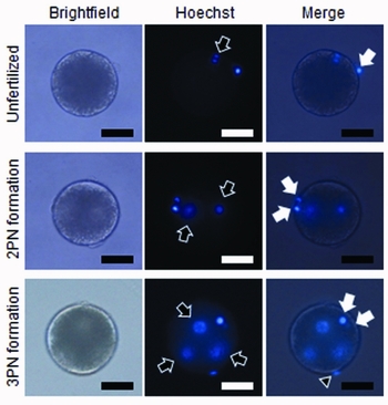
The fertility of each assessed sperm group was examined by counting the number of sperm that penetrated an oocyte and by the cleavage rate of in vitro fertilized (IVF) embryos. To determine the penetration rate of each sperm population, 15–20 of the IVF-assessed oocytes (or embryos) were examined for each of the sperm samples in each trial. The zona pellucida of the fertilized embryos were lysed with acid Tyrode's solution 12 h after the insemination. The embryos without their zona pellucida were stained with 0.2% Hoechst 33342 (Molecular Probes, Eugene, OR, USA) in 0.1% BSA-supplemented DPBS for 5 min. Penetrated sperm and formed pronuclei were counted under the microscope (Fig. 1). The cleaved embryos were enumerated 2 days after insemination.
Evaluation of penetrated sperm and pronuclear formation. Zona pellucida denuded unfertilized oocytes and zygotes were stained with Hoechst 33342 and observed for 12 h after in vitro fertilization. The polar body and the pronucleus are represented by filled and empty arrows, respectively. The arrowhead indicates penetrating sperm without decondensed nuclei. The scale bar represents 100 μm.

Sperm preparation by Percoll discontinuous gradient centrifugation
The layers for Percoll gradient discontinuous centrifugation contained 90, 60.5 or 45% of Percoll™ (GE Healthcare, Buckinghamshire, UK) in DPBS supplemented with 0.1% BSA, and 0.3% phenol red was added to the 90% and 45% Percoll solutions. Each Percoll solution (5 ml of 90% and 10 ml of both 60.5 and 45%) was added to a 50 ml tube with serial layers. For the control condition, 5 ml of the 90% and 20 ml of the 45% solution were used. Sperm were washed with 0.1% BSA-supplemented DPBS by centrifugation at 1500 rpm for 5 min and diluted in 5 ml of 0.1% BSA-supplemented DPBS prior to their separation by Percoll gradient centrifugation. The washed sperm were placed on top of the Percoll gradient layers. The whole discontinuous gradient with sperm was centrifuged at 1500 rpm for 20 min. Sperm populations were taken from the layer between 90 and 60.5% (high-density population), 60.5 and 45% (low-density population) and 90 and 45% (control population). The grouped sperm populations were stored at 17°C before use.
RNA extraction, reverse transcription and polymerase chain reaction
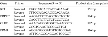
RNA extraction was carried out following the procedure described in our previous report (Hwang et al., Reference Hwang, Mulligan, Kim, Yang and Lee2013). Briefly, before RNA extraction, sperm samples were separated by Percoll and somatic cell contamination and then were examined with a microscope. In succession, RNA were extracted using Trizol® reagent (Invitrogen, Carlsbad, CA, USA) following the manufacturer's guide. The extracted RNA were reverse transcribed using a High Capacity RNA-to cDNA Kit™ (Applied Biosystems, Foster City, CA, USA) in a final volume of 20 μl. Synthetized sperm cDNA were subjected to polymerase chain reaction (PCR) to examine the somatic cell contamination. The cDNA samples were amplified using the primer pairs listed in Table 1. The reaction was conducted with a 2× master mix solution (iNtRON, Seongnam, Korea) with 0.5 μM of each primer and 0.5 μl of the cDNA sample in a final volume of 10 μl. For the somatic cell contamination examination, KIT, PRPTC and CDH1 were used, which are immature germ cell, leukocyte and epithelial cell markers, respectively. PRM1 was selected as a positive marker for RNA in sperm. The amplification was performed at the following conditions: 95°C for 5 min followed by 35 cycles of 95°C for 15 s, 62°C for 20 s and 72°C for 25 s, with a final step at 72°C for 7 min.
Primer sequences used for reverse transcriptase polymerase chain reaction

Western blot assay
Protein was extracted with a Laemmli sample buffer (Kwon et al., Reference Kwon, Park, El Mohamed and Pang2013). Sperm populations separated by Percoll were washed with DPBS by centrifugation at 1500 rpm for 10 min. The sperm pellets were diluted in a Laemmli buffer and incubated for 20 min at room temperature. The lysates were centrifuged at 13,000 rpm for 20 min, and the supernatant was boiled for 5 min at 95°C and then chilled on ice. Extracted sperm protein samples were examined using sodium dodecyl sulfate-polyacrylamide gel electrophoresis (SDS-PAGE) at 150 V and transferred to a nitrocellulose transfer membrane (Schleicher & Schuell BioScience, Inc., Dassel, Germany) at 200 V for 90 min. The membranes were incubated with 5% skimmed milk in Tris-buffered saline containing 0.1% TWEEN®-20 (TBST) for 1 h. The rabbit anti-human CYP19 (#sc-30086, Santa Cruz Biotechnology, Inc., CA, USA), and mouse anti-pig α-tubulin (#ab7750, Abcam, Cambridge, UK) were used for the western blot. Each antibody was diluted to 1:200 for use. Primary antibodies were agitated on the membranes at 4°C overnight, and the membranes were washed three times with TBST for 10 min. Goat anti-rabbit secondary antibody with horseradish peroxidase (HRP) (1:2000, #sc-2004, Santa Cruz) and goat anti-mouse secondary antibody with HRP (1:2000, #sc-2005, Santa Cruz) were agitated with the membranes for 1 h at room temperature, and the membranes were washed three times. HRP on the secondary antibody was activated with enhanced chemiluminescence (ECL) (Amersham Bioscience Corp., Little Chalfont Bucks, UK) for 2 min, and the membrane was imaged with ChemiDoc (ChemiDoc™ XRS+ System, Bio-Rad, Hercules, CA, USA). Quantification of protein expression using images from ChemiDoc was conducted with the Image J program.
Statistical analysis
Statistical significance was determined using GraphPad Prism Software for Windows, version 5.01 (GraphPad Software, San Diego, CA, USA). All data were analyzed using a one-way analysis of variance (ANOVA), and Tukey's multiple comparison test with 95% confidence intervals was employed to determine differences among the experimental groups. When the P-value was less than 0.05, the difference between the compared groups was considered significant. The data are represented as mean ± standard error of the mean (SEM).
Results
Purification of sperm with differential densities
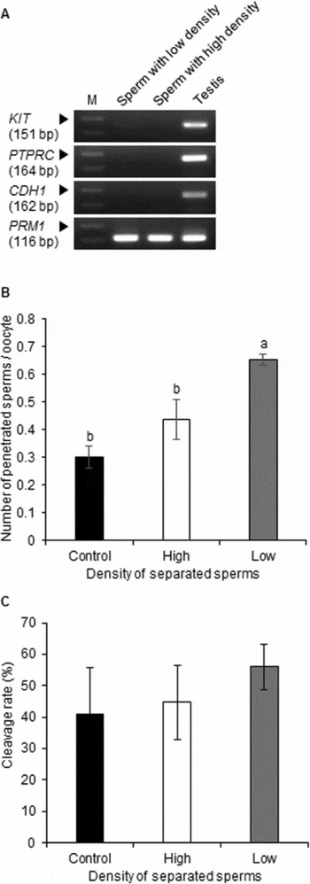
The sperm population from a single ejaculate of each boar was classified by density using a Percoll gradient because sperm with different densities have shown varying motility and viability in other species (Parrish et al., Reference Parrish, Krogenaes and Susko-Parrish1995). Before analyzing the CYP19A1 level of the sperm with differential densities, contamination of the sample with non-sperm cells was examined using RT-PCR to confirm the purity of the sperm population showing differential densities. Non-sperm cell markers KIT (immature germ cell), PCPRT (leukocyte) and CDH1 (epithelial cell) were not detected. However, PRM1, which is a male germ cell-specific gene, was present in both prepared sperm groups (Fig. 2 A). The samples with pure sperm were applied to western blot.
Fertility of Percoll separated sperm. (A) Purity evaluation of Percoll separated sperm. Primers of immature germ cell (KIT), leukocyte (PTPRC) and epithelial cells (CDH1) were used to evaluate non-sperm cell contamination, and primers of sperm cell (PRM1) were employed as a positive marker. Testis tissue was used as a positive control for each gene expression. M and the arrowhead indicate the DNA size ladder and the 200 bp size marker, respectively. Penetration rate of CYP19A1 separated sperm. Total pronucleus minus one and any additionally penetrating sperm were considered to be penetrated sperm. a,bValues with different letters in the graph are significantly different (P < 0.05). This experiment was replicated three times, and 20–30 oocytes were used in each condition. (C) Cleavage rate of embryos fertilized with separated sperm. The cleavage rate was measured on day 2 of culture. This experiment was replicated three times, and 90–175 oocytes were used in each condition.

Fertility of porcine sperm grouped by density
The prepared sperm populations, which were divided by density using a Percoll gradient (high and low), showed a significantly different penetration rate. The sperm fraction with a higher penetration rate into the oocytes had a lower density than the control and the high-density groups (Fig. 2 B). However, there was no significant difference between the prepared sperm groups for the cleavage rate of fertilized embryos (Fig. 2 C). This result indicated that the two sperm populations from a single ejaculate that had been divided by Percoll showed differential fertility levels.
Quantitative comparison of CYP19A1 levels in sperm
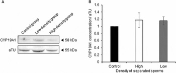
To investigate the CYP19A1 levels in porcine sperm samples with different densities, which showed different fertility levels, CYP19A1 expression was compared quantitatively between these sperm populations. Both groups contained the CYP19A1 protein (Fig. 3 A), and the amount of protein had no significant difference among these separated sperm populations (Fig. 3 B).
Western blot analysis of separated sperm. (A) Western blot image of separated sperm. CYP19A1 in sperm is detected on 58 kDa, and alpha-tubulin protein is detected on 55 kDa. Molecular weights are indicated on the right side of the blot image. (B) Relative concentrations of CYP19A1 in Percoll separated sperm. The values were normalized with the value of the control group. This experiment was replicated four times.

Determination of the CYP19A1 inhibitor treatment condition
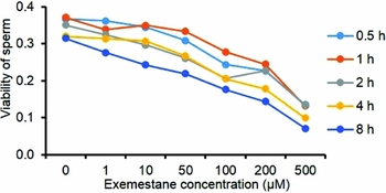
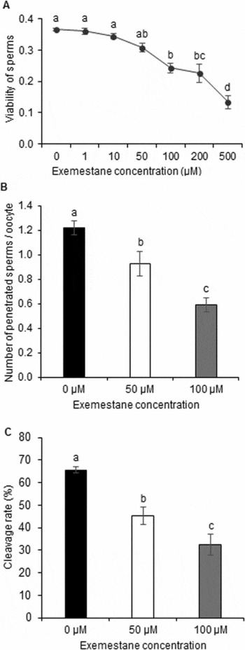
To examine the effect of CYP19A1 on sperm fertility, the CYP19A1 inhibitor exemestane was applied to the sperm. Before analyzing the effect of the CYP19A1 inhibitor on porcine sperm fertility, proper treatment conditions that would have no effect on viability had to be determined. The viability of sperm showed a tendency to decrease as the exemestane treatment time and concentration increased (Fig. 4). Because the viability of sperm was affected by the inhibitor concentration during the short period of treatment, the inhibitor was treated with a diverse range of inhibitor concentrations for 0.5 h. In this treatment, the viability of sperm subjected to more than 100 μM of inhibitor treatment was significantly different compared to 0 μM (Fig. 5 A). Therefore, the inhibitor was treated with 50 μM and 100 μM for 0.5 h in the following experiment.
Effect of exemestane on porcine sperm viability. Decision of inhibitor treatment condition. Exemestane was treated for 0.5–8 h with concentrations ranging from 0–500 μM. Viability of the sperm was measured six times in each condition.

Effect of exemestane on porcine sperm. (A) Decision of inhibitor treatment condition. Exemestane was treated for 0.5 h with concentrations ranging from 0–500 μM. Viability of the sperm was measured six times in each condition. (B) Penetration rate of CYP19A1 inhibited sperm. Total pronucleus minus one and additionally penetrated sperm were considered to be penetrated sperm. Experiments were replicated four times, and 20–30 oocytes were used in each condition. (C) Cleavage rate of embryos fertilized with inhibitor-treated sperm. The cleavage rate was measured on day 2 of culture. Twenty-two to 30 oocytes were used in each condition. The experiment was repeated five times. a,b,cDifferent characters indicate significant differences.

Effect of CYP19A1 inhibition on porcine sperm fertility
To confirm the effect of CYP19A1 on porcine sperm fertility, inhibitor-treated sperm received IVF. The number of penetrating sperm decreased significantly as the concentration of exemestane rose (Fig. 5 B). After a 2-day culture, the inhibitor-treated groups exhibited a significantly low cleavage rate of embryos compared to the control sperm (Fig. 5 C). Therefore, inhibition of CYP19A1 activity affected sperm fertility in the pig.
Discussion
Sperm quality evaluation is an important assessment for both human infertility and livestock animal fertilization studies. To complement physiological markers like morphology and motility, the requirement of molecular markers to delicately evaluate sperm quality has been suggested (Petrunkina et al., Reference Petrunkina, Waberski, Gunzel-Apel and Topfer-Petersen2007). Recent studies on mammalian sperm have discovered several molecular markers for sperm fertility. Human PRM1 and PRM2 (Aoki et al., Reference Aoki, Liu and Carrell2005), bovine ENO1 (Park et al., Reference Park, Kwon, Oh and Pang2012b) and CD36 transcript levels have been suggested as fertility markers of sperm (Feugang et al., Reference Feugang, Rodriguez-Osorio, Kaya, Wang, Page, Ostermeier, Topper and Memili2010). In porcine animals, our previous study showed that the quantity of some transcripts, including ADAM2 and CYP19, indicates sperm fertility (Hwang et al., Reference Hwang, Mulligan, Kim, Yang and Lee2013). These data of CYP19 were parallel to the results of reports that dealt with human and bovine sperm (Lambard et al., Reference Lambard, Silandre, Delalande, Denis-Galeraud, Bourguiba and Carreau2005; Tiwari et al., Reference Tiwari, Singh, Kumar and Sharma2008). The effect of the CYP19A1 protein and its activity have also been investigated in rodents (Carreau et al., Reference Carreau, Delalande and Galeraud-Denis2009; Janulis et al., Reference Janulis, Bahr, Hess, Janssen, Osawa and Bunick1998), but not in pigs. In the present study, we focused on the relationship between the CYP19A1 protein and sperm fertility.
CYP19A1 is an enzyme that converts steroid hormones from testosterone to estrogen (Simpson et al., Reference Simpson, Zhao, Agarwal, Michael, Bulun, Hinshelwood, Graham-Lorence, Sun, Fisher, Qin and Mendelson1997). This enzyme is thought to play an important role in male reproduction as levels of estrogen and testosterone are related to sperm fertility. Previous studies have shown that the balance of steroid hormones in seminal plasma is important for sperm fertility (Carani et al., Reference Carani, Qin, Simoni, Faustini-Fustini, Serpente, Boyd, Korach and Simpson1997; Luboshitzky et al., Reference Luboshitzky, Kaplan-Zverling, Shen-Orr, Nave and Herer2002); estrogen, which is a product of CYP19A1, is known to induce a downstream reaction of the estrogen receptor pathway in sperm, but excessive estrogen also reduces sperm fertility (Bujan et al., Reference Bujan, Mieusset, Audran, Lumbroso and Sultan1993; Aquila et al., Reference Aquila, Sisci, Gentile, Carpino, Middea, Catalano, Rago and Ando2003; Lambard et al., Reference Lambard, Silandre, Delalande, Denis-Galeraud, Bourguiba and Carreau2005). These reports suggest the importance of CYP19A1 in male fertility. In the pig, CYP19A1 is localized from the proximal to the distal tail of sperm and successfully produces estrogen (Rago et al., Reference Rago, Aquila, Panza and Carpino2007).
Inhibition of CYP19A1 caused a decrease of sperm viability and fertility in our study. Because of its specificity and irreversible suppression, we used exemestane as a CYP19A1 inhibitor among several inhibitors (Hong & Chen, Reference Hong and Chen2006; Miller et al., Reference Miller, Bartlett, Brodie, Brueggemeier, di Salle, Lonning, Llombart, Maass, Maudelonde, Sasano and Goss2008). Our results corresponded to previous studies, which revealed that sperm maintains CYP19A1 activity both during and after spermatogenesis in rodents (Janulis et al., Reference Janulis, Bahr, Hess, Janssen, Osawa and Bunick1998; Lambard et al., Reference Lambard, Silandre, Delalande, Denis-Galeraud, Bourguiba and Carreau2005). In our study, the activity of CYP19A1 influenced sperm-penetration and cleavage rates of in vitro fertilized embryos. As in the previous report conducted with both humans and rodents, a malfunction of porcine CYP19A1 resulted in reduced sperm fertility (Carreau et al., Reference Carreau, Delalande and Galeraud-Denis2009). This problem would be induced by the imbalance of estrogen and testosterone because estrogen is important for sperm fertility (Luboshitzky et al., Reference Luboshitzky, Kaplan-Zverling, Shen-Orr, Nave and Herer2002).
Previous studies showed that the sperm in the same ejaculate have variable densities, and they also exhibit differential motility and normality in humans (Aitken and West, Reference Aitken and West1990; Mengual et al., Reference Mengual, Ballesca, Ascaso and Oliva2003). Sperm populations with differential fertility levels from single ejaculates were prepared by density to minimize the individual-to-individual variation in this study. Although the sperm with differential fertility were separated successfully according to their density, unexpectedly, the penetration rate of low-density sperm was higher than that of high-density sperm. In a previous report of bovine sperm, high-density sperm showed a greater viability and motility than sperm with low densities (Parrish et al., Reference Parrish, Krogenaes and Susko-Parrish1995). This difference might have originated from the study's use of a variety of sperm from different species. Each mammalian germ cell has distinct morphologies (Behringer et al., Reference Behringer, Eakin and Renfree2006), and protein populations in sperm are very species specific (Swanson and Vacquier, Reference Swanson and Vacquier2002; Torgerson et al., Reference Torgerson, Kulathinal and Singh2002). In the present study, our low-density sperm population showed a higher penetration rate, which was an opposite result as that found by a previous report of bovine sperm (Suzuki & Nagai, 2003). However, the CYP19A1 protein level was not different between sperm with high and low densities in this study. This result may have been influenced by the constant amount of CYP19A1 in sperm from an ejaculate. Bilinska and colleges showed that the accumulation of CYP19A1 depends on the conditions of spermatogenesis in rodents (Bilinska et al., Reference Bilinska, Schmalz-Fraczek, Kotula and Carreau2001), so in one ejaculate, the protein quantity of CYP19A1 is likely consistent in porcine sperm. Therefore, the translation of CYP19A1 may not be related to its transcription level; this supposition is in opposition with our previous result on CYP19A1 mRNA (Hwang et al., Reference Hwang, Mulligan, Kim, Yang and Lee2013). Another possible reason is that the density of the sperm might not be related to the amount of CYP19A1 protein it contains. The concentration of CYP19A1 may also be independent of the density during spermatogenesis and maturation.
In conclusion, we showed that CYP19A1 was responsible for the fertility of sperm, but we could not confirm the level of CYP19A1 as a quality prediction marker for porcine sperm. To determine how CYP19A1 affects the quality of sperm, further studies regarding the role of sperm CYP19A1 in steroid hormone conversion will be required to understand the precise mechanism of CYP19A1 in pigs; studies are also needed on the mode of steroid hormonal action during the fertilization process in mammalian sperm. In addition, other methods of approaches to classify sperm are needed to ensure the correlation between quality and quantity of CYP19A1 in sperm.
Acknowledgements
This work was supported by the National Research Foundation of Korea (NRF) grant funded by the Korea government (MEST) (No. 2012006276) and Next BioGreen21 program, Rural Development Administration, Republic of Korea. The authors thank Dr Chi-Hun Park for helpful discussions of the work.
Conflict of interest
All authors declare no competing financial interests.
Author contributions
JNO, JYH designed the experiment. JNO, JYH and KHC performed the experiments and analyzed data. JNO, JYH and CKL wrote and revised the manuscript. CKL provided information and supervised the study. All authors read and approved the final manuscript.
Supplementary material
To view supplementary material for this article, please visit http://dx.doi.org/10.1017/S0967199414000781