Introduction
Odour is an essential sensory trait that determines whether consumers will accept a product. Pork odour is affected to a great extent by the presence of malodorous compounds, of which androstenone and skatole are believed to be the most important. The current review outlines the present knowledge about biochemical, nutritional and genetic factors involved in the regulation of androstenone and skatole levels in pig carcasses. Additionally, the review provides some suggestions for future research.
Castration – good or bad? Pork-quality perspectives
Male piglets destined for meat production were traditionally castrated to increase the proportion of fat in carcasses, to reduce aggressive and sexual behaviour, and to reduce the risk of boar taint, an unpleasant smell from heated pork products. Since nowadays consumers’ preference has drastically changed to require mainly lean meat, only the two latter reasons to castrate remain. However, the advantages of castration are disputable because concern for animal welfare is a major consideration in pork production. Additionally, raising entire male pigs is more profitable because of improved feed conversion and leaner carcasses compared to castrates (Hansson, Reference Hansson1974; Babol and Squires, Reference Babol and Squires1995; Bonneau, Reference Bonneau1998; Bañón et al., Reference Bañón, Andreu, Laencina and Garrido2004). The higher levels of polyunsaturated fatty acids (PUFA) in fat and muscles and higher protein content in carcasses from entire males might indicate nutritional advantages of this meat compared to that from castrates (Malmfors et al., Reference Malmfors, Lundström and Hansson1978; Wood et al., Reference Wood, Buxton, Whittington and Enser1986; Naděje et al., Reference Naděje, Koucký, Ševčíková, Adamec and Laštovková2000).
Origin of boar taint
Boar taint mainly occurs in meat from some entire male pigs and makes it undesirable for sensitive consumers. Two substances are primary responsible for boar taint, androstenone (5α-androst-16-ene-3-one) (Patterson, Reference Patterson1968) and skatole (3-methylindole) (Vold, Reference Vold1970; Walstra and Maarse, Reference Walstra and Maarse1970). Other chemicals that might also contribute to a lesser degree to off-odour in meat include androstenols (Brennan et al., Reference Brennan, Shand, Fenton, Nicholls and Aherne1986), indole (García-Regueiro and Diaz, Reference García-Regueiro and Diaz1989; Moss et al., Reference Moss, Hawe and Walker1993) and 4-phenyl-3-buten-2-one (Rius Sole and García-Regueiro, Reference Rius Sole and García-Regueiro2001). Patterson (Reference Patterson1967) suggested the contribution of phenolic compounds p-cresol and 4-ethylphenol to boar taint, although stressing that according to smell, neither p-cresol nor the other phenols were involved in the taint directly. Recently, Angels Rius et al. (Reference Angels Rius, Hortós and Garcia-Regueiro2005) identified aldehydes and short chain fatty acids, which could either promote the perception of skatole and androstenone or be responsible for the development of off-flavours.
The focus of the current review is to examine the biochemical, genetic and nutritional factors involved in the accumulation of androstenone and skatole in porcine adipose tissue. The potential contribution of other compounds to boar taint will not be discussed here.
Endogenous regulation of boar taint levels
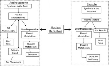
The accumulation of boar taint compounds is determined by the difference between the rates of synthesis and clearance of these compounds in the pig. These metabolic processes are controlled by the expression of a number of different genes that encode enzymes involved in the metabolism of boar taint compounds. Additionally, the intensity of metabolic processes depends on protein expression and enzyme activities, which might vary independently from gene expression. The metabolic pathways of androstenone and skatole are summarised in Figure 1.
Summary of the metabolic pathways of androstenone and skatole and the role of nuclear receptors.

Androstenone biosynthesis
Androstenone is a steroid produced by the Leydig cells of the testis of entire male pigs in parallel with anabolic testicular hormones (Gower, Reference Gower1972; Kwan et al., Reference Kwan, Orengo and Gower1985). Additionally, low levels of androstenone were detected in plasma from female and castrated pigs, suggesting the possible production of androstenone by the adrenal cortex and the ovary (Claus et al., Reference Claus, Hoffmann and Karg1971). Androstenone is derived from pregnenolone through the formation of 5,16-androstadien-3β-ol by the andien-β synthase system. Cytochrome P450C17 (CYP17A1) and cytochrome b5 (CYB5) are particularly important in 5,16-androstadien-3β-ol biosynthesis (Meadus et al., Reference Meadus, Mason and Squires1993; Davis and Squires, Reference Davis and Squires1999). The production of androstenone and other testicular steroids is controlled by the neuroendocrine system, particularly by the lutenizing hormone (LH), which is under stimulatory control of gonadotropin-releasing hormone (GnRH). A transient activation of the hypothalamic–pituitary–gonadal axis during early postnatal life (approximately 2–4 weeks of age) results in an increase in levels of circulating testicular steroids including androstenone (Bonneau, Reference Bonneau1982; Schwarzenberger et al., Reference Schwarzenberger, Toole, Christie and Raeside1993; Sinclair et al., Reference Sinclair, Squires and Raeside2001). Otherwise, the biosynthesis of androstenone is low in young pigs and gradually increases along with other testicular steroids at sexual maturity (Gower, Reference Gower1972; Bonneau, Reference Bonneau1982). Therefore, puberty is a central aspect regulating androstenone biosynthesis in entire male pigs by the maintenance of adult Leydig cell morphology and stimulation of the neuroendocrine system. Genetic background is also an important regulator of the intensity of androstenone biosynthesis. Sexually mature boars that have low levels of fat androstenone but normal levels of plasma testosterone and LH profiles have been identified over 20 years ago (Bonneau et al., Reference Bonneau, Carrié-Lemoine, Prunier, Garnier and Terqui1987).
Androstenone metabolism
The metabolism of androstenone has been mainly studied in testis and liver. The metabolic process is mediated by 3β- and 3α-hydroxysteroid dehydrogenase enzymes (3β-HSD and 3α-HSD) (Dufort et al., Reference Dufort, Soucy, Lacoste and Luu-The2001; Doran et al., Reference Doran, Whittington, Wood and McGivan2004; Sinclair et al., Reference Sinclair, Hancock, Gilmore and Squires2005b), as well as Phase II conjugating enzymes, such as hydroxysteroid sulfotransferases (SULT2A1 and SULT2B1) and UDP-glucuronosyltransferase (UGTs) (Sinclair et al., Reference Sinclair, Gilmore, Lin, Lou and Squires2006; Moe et al., Reference Moe, Grindflek and Doran2007a).
Testis
A large proportion of androstenone that is synthesised in the testis is immediately sulfoconjugated by hydroxysteroid sulfotransferase (SULT2A1, Sinclair et al., Reference Sinclair, Squires, Raeside and Renaud2005a and/or SULT2B1, Moe et al., Reference Moe, Grindflek and Doran2007a). Androstenone is metabolised to 5α-androst-16-en-3α-ol (3α-androstenol) and 5α-androst-16-en-3β-ol (3β-androstenol), as demonstrated by in vitro (Brophy and Gower, Reference Brophy and Gower1972; Sinclair et al., Reference Sinclair, Squires, Raeside and Renaud2005a) and in vivo studies (Saat et al., Reference Saat, Gower, Harrison and Heap1974). The androstenols are subsequently metabolised to produce more polar conjugated steroids (Sinclair and Squires, Reference Sinclair and Squires2005).
Liver
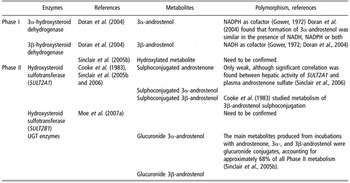
Similar to the process in the testis, androstenone is metabolised in the liver by enzymes 3β-HSD and 3α-HSD (Table 1) with the production of 3β-androstenol and to a lesser extent 3α-androstenol (Doran et al., Reference Doran, Whittington, Wood and McGivan2004; Sinclair et al., Reference Sinclair, Hancock, Gilmore and Squires2005b). Hepatic metabolism of androstenone differs from that in testis in terms of the percentage of produced metabolites. The androstenols then undergo Phase II metabolic reactions to form glucuronide conjugates and sulfoconjugates (Sinclair et al., Reference Sinclair, Hancock, Gilmore and Squires2005b). The mechanism of sulfoconjugation of 3β-androstenol in porcine liver was investigated by Fish et al. (Reference Fish, Cooke and Gower1980) and Cooke et al. (Reference Cooke, Ferguson, Rytina and Gower1983). SULT2A1 is a key Phase II enzyme in the hepatic metabolism of the 16-androstene steroids (Sinclair et al., Reference Sinclair, Hancock, Gilmore and Squires2005b and Reference Sinclair, Gilmore, Lin, Lou and Squires2006). It was recently suggested that SULT2B1 may also be involved (Moe et al., Reference Moe, Grindflek and Doran2007a). Additionally, it is likely that androstenone can be sulfoconjugated through prior enolisation of the 3-keto group by hydroxysteroid sulfotransferase (SULT2A1) (Sinclair et al., Reference Sinclair, Hancock, Gilmore and Squires2005b and Reference Sinclair, Gilmore, Lin, Lou and Squires2006). Sinclair and Squires (Reference Sinclair and Squires2005) reported that a large proportion of androstenone in plasma from some boars is present as sulfoconjugates. However, Zamaratskaia et al. (Reference Zamaratskaia, Lou, Chen, Andresen, Lundström and Squires2007a) found that the range of androstenone levels in the conjugate fraction was much lower than that of the free androstenone. Similarly, Tuomola et al. (Reference Tuomola, Harpio, Knuuttila, Mikola and Lövgren1997) did not detect any androstenone in the conjugate fraction using 47% and 100% methanol to separate conjugated and free androstenone, respectively. More studies to evaluate the significance of androstenone sulfoconjugation to its elimination and accumulation in fat are warranted.
Hepatic metabolism of androstenone

UGT=uridine diphosphate-glucuronosyltransferase.
Other metabolic sites
Part of androstenone is transported from the circulation to the submaxillary glands. Booth (Reference Booth1987) incubated 3H-labelled androstenone, 3α-androstenol and 3β-androstenol with boar saliva and reported that 3α-androstenol was primarily converted to androstenone and androstenone to 3β-androstenol, whereas 3β-androstenol remained unchanged.
Skatole biosynthesis
Skatole is formed from the amino acid l-tryptophan in the large intestine of pigs. Biosynthesis of skatole occurs as a two-step procedure, with tryptophan first converted to 3-indoleacetic acid, which is subsequently converted to skatole (reviewed in Jensen and Jensen, Reference Jensen and Jensen1998). Escherichia coli (E. coli) and Clostridium spp. are responsible for the production of indole-3-acetic acid, and then Lactobacillus and Clostridium convert indole-3-acetic acid to skatole (Jensen et al., Reference Jensen, Cox and Jensen1995). Thus, the amount of skatole produced is primarily regulated by the availability of tryptophan and the composition and activity of intestinal bacteria. A major source of tryptophan for skatole production is from the turnover of the gut-mucosa cells (Claus et al., Reference Claus, Weiler and Herzog1994; Claus and Raab, Reference Claus and Raab1999). A part of skatole that is produced is excreted with faeces and the remaining part is absorbed through the gut wall into the blood. Skatole concentrations in faeces were similar in all genders of pigs (Dehnhard et al., Reference Dehnhard, Bernal-Barragan and Claus1991), although high skatole concentrations in the adipose tissue are mainly (but not only) found in some entire males.
Skatole metabolism
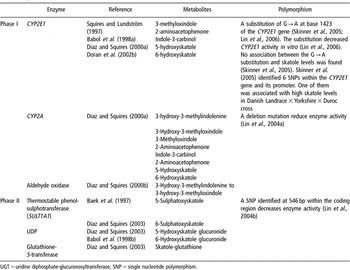
The metabolism of skatole in the liver takes place in two phases (Table 2). Phase I involves the addition of a hydroxyl group that can be used to attach a conjugate in Phase II. The conjugate increases the hydrophilicity (polarity) of the metabolite so that it can be excreted in the urine or bile and the absorption in adipose tissue is reduced. The majority of Phase I skatole metabolism occurs through the cytochrome P450 (CYP) system, a superfamily of heme-containing isoenzymes located within the endoplasmic reticulum in hepatocytes. Currently, CYP isoforms CYP2E1 and CYP2A, and aldehyde oxidase are thought to be primarily responsible for the oxidative metabolism of skatole (Babol et al., Reference Babol, Squires and Lundström1998a and Reference Babol, Squires and Lundström1998b; Diaz and Squires, Reference Diaz and Squires2000a and Reference Diaz and Squires2000b) with CYP2E1 being a major enzyme.
Hepatic metabolism of skatole

UGT=uridine diphosphate-glucuronosyltransferase; SNP=single nucleotide polymorphism.
Diaz et al. (Reference Diaz, Skordos, Yost and Squires1999) have identified seven Phase I metabolites produced using liver microsomes; quantitatively, the major metabolite produced in vitro was 3-hydroxy-3-methylindolenine (45%). The other identified Phase I metabolites are 3-hydroxy-3-methyloxindole, 5-hydroxy-3-methylindole, 6-hydroxy-3-methylindole, 3-methyloxindole, indole-3-carbinol and 2-aminoacetophenone. Some of these metabolites serve then as the substrates for conjugation with either sulphate or glucuronic acid in Phase II (Babol et al., Reference Babol, Squires and Lundström1998b; Diaz and Squires, Reference Diaz and Squires2003). High levels of 6-sulphatoxyskatole in plasma were suggested as indicators of entire male pigs with the ability to rapidly metabolise and excrete skatole (Baek et al., Reference Baek, Hansen-Møller, Friiis, Cornett and Hansen1997), but this needs to be further studied. The enzyme responsible for sulfation was identified as thermostable phenol-sulfotransferase (SULT1A1, Diaz and Squires, Reference Diaz and Squires2003). Modification of the activities and/or expression of skatole-metabolising enzymes may be a useful method for enhancing skatole metabolism and reducing boar taint.
Hepatic metabolism of skatole plays an essential role in its accumulation in fat. High activities of CYP2E1 and CYP2A in mature male pigs are usually associated with low skatole accumulation in fat, whereas low enzyme activities can result in both high and low skatole accumulation (Squires and Lundström, Reference Squires and Lundström1997; Zamaratskaia et al., Reference Zamaratskaia, Squires, Babol, Andersson, Andersson and Lundström2005a).
There is some discrepancy in the literature on the relative importance of CYP2A and CYP2E1 in skatole metabolism, which may be due to the age of animals used in the different studies. Diaz and Squires (Reference Diaz and Squires2000a) investigated the role of different CYP enzymes on skatole metabolism using selective chemical inhibitors. They demonstrated that CYP2A inhibitors reduced the production of all known skatole metabolites, while CYP2E1 inhibitor 4-methylpyrazole reduced the production of five out of seven skatole metabolites. The inhibition of skatole metabolism by 4-methylpyrazole may be, however, due to the combined inhibition of both CYP2E1 and CYP2A, as it was shown that 4-methylpyrazole does not solely inactivate CYP2E1, but may inactivate CYP2A as well (Draper et al., Reference Draper, Madan and Parkinson1997). Terner et al. (Reference Terner, Gilmore, Lou and Squires2006) found that CYP2E1 is more important than CYP2A for skatole metabolism in porcine hepatocytes isolated from pigs at approximately 3 weeks of age. They found that the simultaneous inhibition of CYP2E1 and CYP2A did not reduce the formation of certain skatole metabolites any further than the inhibition of CYP2E1 alone.
Other data also suggest that the overall metabolism of skatole in the young pig differs from that in mature pigs. Lanthier et al. (Reference Lanthier, Lou and Squires2007) found that the accumulation of skatole in the prepubertal pig was not related to the activities of CYP2E1, CYP2A and aldehyde oxidase, although SULT1A1 activity was negatively correlated with skatole levels in plasma and fat. In this paper, skatole levels were measured in intestinal content to distinguish between high and low skatole producers. Stronger correlations between skatole and SULT1A1 were found when only the high producers were included.
Interestingly, Doran et al. (Reference Doran, Whittington, Wood and McGivan2002a) demonstrated that skatole induced the expression of CYP2E1 protein in primary cultured hepatocytes. However, high skatole levels in vivo did not change the rate of formation of skatole metabolites in the liver (Babol and Squires, Reference Babol and Squires1999).
Androstenone, skatole and hormonal status of animals
Androstenone biosynthesis is controlled by the same mechanism as other testicular steroids, namely through the activation of the hypothalamic–pituitary–gonadal axis during puberty. Thus, during puberty androstenone levels drastically increase simultaneously with other testicular steroids (Gower, Reference Gower1972; Bonneau, Reference Bonneau1982). Skatole levels also increase at puberty (Babol et al., Reference Babol, Zamaratskaia, Juneja and Lundström2004), possibly after an increase in testicular steroids (Zamaratskaia et al., Reference Zamaratskaia, Babol, Andersson and Lundström2004a and Reference Zamaratskaia, Babol, Madej, Squires and Lundström2004b). The additional evidence linking increased skatole levels with puberty include the possible regulation of skatole hepatic metabolism by testicular steroids.
Initial attempts to investigate the relationship between skatole and steroids, particularly androstenone in fat, using correlation analysis resulted in inconsistent results (discussed in Walstra et al., Reference Walstra, Claudi-Magnussen, Chevillon, von Seth, Diestre, Matthews, Homer and Bonneau1999). The Pearson correlation coefficients in different studies varied from close to zero (Zamaratskaia et al., Reference Zamaratskaia, Babol, Andersson, Andersson and Lundström2005b) to 0.73 (Bonneau et al., Reference Bonneau, LeDenmat, Vaudelet, VelosoNunes, Mortensen and Mortensen1992). Examination of multiple regression relations showed that the levels of testicular hormones testosterone, estrone sulphate and free estrone in combination with testes and bulbo-urethral gland sizes were the best predictors of skatole in fat (Zamaratskaia et al., Reference Zamaratskaia, Rydhmer, Chen, Madej, Andersson and Lundström2005d). The boar is known for its high amounts of estrogens (Claus and Hoffmann, Reference Claus and Hoffmann1980), which are produced in testicular Leydig cells (Raeside and Renaud, Reference Raeside and Renaud1983; Mutembei et al., Reference Mutembei, Pesch, Schuler and Hoffmann2005) and are positively correlated to skatole levels (Babol et al., Reference Babol, Squires and Lundstrøm1999; Zamaratskaia et al., Reference Zamaratskaia, Madej, Babol, Squires and Lundström2005c).
Useful information on the possible involvement of steroids in skatole metabolism has been produced by in vitro studies. Androstenone was recognised as a potential inhibitor of the expression of CYP2E1 (Doran et al., Reference Doran, Whittington, Wood and McGivan2002b; Tambyrajah et al., Reference Tambyrajah, Doran, Wood and McGivan2004) and CYP2A (Chen et al., Reference Chen, Cue, Lundström, Wood and Doran2008). The possibility of a direct inhibition of CYP2E1 and CYP2A activities by androstenone, 17β-oestradiol and testosterone was also investigated in liver microsomes from male and female pigs (Zamaratskaia et al., Reference Zamaratskaia, Gilmore, Lundström and Squires2007b). In this study, androstenone was identified as a potential competitive inhibitor of CYP2E1 activity in both male and female microsomes, and estradiol inhibited CYP2E1 activity in microsomes from male pigs. It should be noted, however, that concentrations of the steroids used in the studies of Doran et al. (Reference Doran, Whittington, Wood and McGivan2002b) and Zamaratskaia et al. (Reference Zamaratskaia, Gilmore, Lundström and Squires2007b) were higher than physiological levels.
Stimulation of pigs with hCG induces a temporary increase in the levels of testicular steroids (Andresen, Reference Andresen1975; Carlström et al., Reference Carlström, Malmfors, Lundström, Edqvist and Gahne1975; Bonneau et al., Reference Bonneau, Meusy-Dessolle, Léglise and Claus1982; Chen et al., Reference Chen, Zamaratskaia, Madej and Lundström2006), and might, therefore, be a useful model system to study the impact of steroids on skatole metabolism in vivo. However, there were differences in the response of individual pigs to hCG stimulation. A Swedish study did not find any effect of high steroid levels after hCG administration on skatole levels either in plasma and fat (Chen et al., Reference Chen, Zamaratskaia, Madej and Lundström2006), or in the liver (Zamaratskaia et al., Reference Zamaratskaia, Chen and Lundström2006) from the pigs of a crossbreed (Landrace × Swedish Yorkshire). Activities of CYP2E1 and CYP2A in the same pigs were not affected by hCG administration (Zamaratskaia et al., Reference Zamaratskaia, Chen and Lundström2006). However, a decrease in CYP2E1 and CYP2A activities in pigs of Landrace and Duroc breeds following hCG administration was reported in a Norwegian study; this decrease resulted in high skatole accumulation in fat (Zamaratskaia et al., Reference Zamaratskaia, Oskam, Ropstad, Tajet and Andresen2008a). Thus, it is not established whether the transient increase in steroid levels after hCG administration has an effect on skatole levels, or if this effect is related to hepatic skatole metabolism.
On the basis of the results from the different studies described above, it can be suggested that the pubertal increase in skatole levels is likely due to the high androstenone and oestrogen levels in pigs at this stage of development. Testosterone is not involved in the regulation of skatole levels. The role of other testicular-derived compounds needs to be investigated.
Various nuclear receptors and other transcription factors have also been implicated in regulating the expression of genes that encode enzymes involved in skatole and androstenone metabolism (Figure 1). Tambyrajah et al. (Reference Tambyrajah, Doran, Wood and McGivan2004) reported that the CYP2E1 promoter is activated by the transcription factors COUP-TF1 and HNF-1 in the liver. Sinclair et al. (Reference Sinclair, Gilmore, Lin, Lou and Squires2006) demonstrated that ligands that activate the nuclear receptors constituting androstane receptor (CAR), pregnane X receptor (PXR) and farnesoid X receptor (FXR) increase the expression of SULT2A1 in Leydig cells of the testis. Gillberg et al. (Reference Gillberg, Skaanild and Friis2006) found that activation of the CAR receptor in primary porcine hepatocytes increased the activity of CYP2A. Thus, it is likely that the metabolic pathways of androstenone and skatole are co-regulated through these nuclear receptors (Figure 1). In this regard, some of the metabolites of androstenone might bind to these nuclear receptors to affect the expression of skatole-metabolising genes (Dufort et al., Reference Dufort, Soucy, Lacoste and Luu-The2001).
Manipulation of boar taint
Modification of androstenone biosynthesis: immunocastration
Active immunisation against GnRH, so-called immunocastration, can be used to reduce the levels of testicular steroids. Immunocastration reliably reduces the production of androstenone along with the size of reproductive organs, sperm numbers and aggressive behaviour (Bonneau et al., Reference Bonneau, Dufour, Chouvet, Roulet, Meadus and Squires1994; Dunshea et al., Reference Dunshea, Colantoni, Howard, McCauley, Jackson, Long, Lopaticki, Nugent, Simons, Walker and Hennessy2001; Cronin et al., Reference Cronin, Dunshea, Butler, McCauley, Barnett and Hemsworth2003; Oliver et al., Reference Oliver, McCauley, Harrell, Suster, Kerton and Dunshea2003; Jaros et al., Reference Jaros, Bürgi, Stärk, Claus, Hennessy and Thun2005; Zamaratskaia et al., Reference Zamaratskaia, Andersson, Chen, Andersson, Madej and Lundström2008b). Skatole and indole levels are also reduced by immunocastration (Dunshea et al., Reference Dunshea, Colantoni, Howard, McCauley, Jackson, Long, Lopaticki, Nugent, Simons, Walker and Hennessy2001; Metz et al., Reference Metz, Hohl, Waidelich, Drochner and Claus2002; Zamaratskaia et al., Reference Zamaratskaia, Andersson, Chen, Andersson, Madej and Lundström2008b). A potentially promising vaccine, Improvac™ (Pfizer Ltd., formerly CSL Limited, Parkville, Victoria, Australia), has recently been tested in some European countries. Vaccination with Improvac™ is performed twice in the growing/finishing period, 4 weeks apart, with the booster injection given 4 weeks prior to slaughter. Vaccination involves the administration of a modified form of the GnRH to provoke the development of anti-GnRH antibodies, which bind to GnRH to prevent the stimulus for secretion of pituitary LH. Thus, a subsequent reduction of testicular steroid hormone secretion occurs. The reduction of skatole and indole levels to low or undetectable levels in immunocastrated pigs is most likely due to enhanced metabolic clearance by the liver after suppressed steroid production, as occurs in surgically castrated pigs. IGF-1 concentrations in plasma were also reduced in immunocastrated pigs (Claus et al., Reference Claus, Lacorn, Danowski, Pearce and Bauer2007); this might affect skatole production (Claus et al., Reference Claus, Weiler and Herzog1994). Therefore, immunocastration allows the production of heavy male pigs with improved meat quality due to reduced androstenone and skatole levels and reduced aggressive behaviour. However, some improvements would increase the efficiency and practicality of the vaccination procedure (Einarsson, Reference Einarsson2006). For example, the current recommended period between the second injection and slaughter is 4–6 weeks, which means that the second vaccination will be given to rather heavy pigs. This can be quite difficult, especially if pigs are group-penned (Einarsson, Reference Einarsson2006); hence, a longer time between the second injection and slaughter is desirable. Only a few reports on the long-term effect of vaccination in pigs are available (Hilbe et al., Reference Hilbe, Jaros, Ehrensperger, Zlinszky, Janett, Hässig and Thun2006; Zamaratskaia et al., Reference Zamaratskaia, Rydhmer, Andersson, Chen, Lowagie, Andersson and Lundström2008c). Both studies demonstrated that the effect of immunocastration can last much longer than 4–6 weeks (up to 22 weeks according to Zamaratskaia et al., Reference Zamaratskaia, Rydhmer, Andersson, Chen, Lowagie, Andersson and Lundström2008c). The prolonged effect of Improvac™ may make it possible to give the second injection of vaccine earlier than the current directions for use, and thus provide greater flexibility in the application of immunocastration. A negative effect of such an approach would be a lower effect of the endogenous male steroids on growth and leanness. Certainly, the ideal vaccine would require a single application instead of current two; however, this might be a disadvantage from operator safety perspectives.
Modification of skatole production and metabolism: dietary composition
Generally, the amount of skatole stored in adipose tissue depends on the rate of skatole production, intestinal transit time, intestinal absorption and hepatic metabolism. The first three of these factors can be easily manipulated by alterations in dietary composition; however, the mode of action of dietary components on skatole levels is still being studied. Previous studies have shown that including certain carbohydrates in the diet affects the microflora of the gastrointestinal tract and influences intestinal function, which in turn might induce changes in intestinal skatole biosynthesis. The absorption of skatole from the intestine might also be affected by diet. Undigested carbohydrates increase faecal wet and dry weight (Wang et al., Reference Wang, Zhu, Li, Wang and Jensen2004) and decrease intestinal transit time (Drochner, Reference Drochner1993), which reduces the rate of skatole absorption from the large intestine. Little is known about the mechanism of skatole uptake from the intestine. The amount of skatole absorbed through the intestinal wall depends to a great extent on water content and intestinal transit time (Jensen and Jensen, Reference Jensen and Jensen1998).
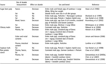
The effect of including different carbohydrates in the diet on skatole levels is well documented (Table 3). Including chicory inulin or raw potato starch (RPS) to the pig’s diet consistently lowered skatole levels in porcine tissue and faeces. Thus, those carbohydrates are fermented to a variable extent by the colonic microflora, with important potential implications for animal health and pork quality.
Effect of dietary carbohydrates on skatole levels in pigs

Limited information is available regarding the effect of diet on the levels of the other tryptophan metabolite, indole. Feeding RPS did not affect indole levels in either fat or plasma (Chen et al., Reference Chen, Zamaratskaia, Andersson and Lundström2007). It was previously suggested that reduction of indolic compounds in the pigs fed RPS is due to the inhibition of cell apoptosis in the colon and thus reduced tryptophan availability (Claus et al., Reference Claus, Losel, Lacorn, Mentschel and Schenkel2003; Mentschel and Claus, Reference Mentschel and Claus2003). The fact that two tryptophan metabolites, skatole and indole, were affected differently by RPS in the same animals is not consistent with this theory and suggests that factors other than apoptotic rate are modified by RPS in the diet. These factors would be specific for skatole formation and would have minor if any effect on indole.
Only limited progress has been made towards determining whether skatole metabolism can be manipulated by diet. Generally, diet can affect the activity of various isoforms of hepatic CYP, which may in turn affect the metabolism of substrates for those enzymes (Sonawane et al., Reference Sonawane, Coates, Yaffe and Koldovsky1983). However, the results on the relationship between diet and skatole-metabolising enzymes are variable. Zamaratskaia et al. (Reference Zamaratskaia, Squires, Babol, Andersson, Andersson and Lundström2005a and Reference Zamaratskaia, Chen and Lundström2006) reported the activities of CYP2E1 and CYP2A were similar in entire male pigs fed with and without RPS. On the other hand, Whittington et al. (Reference Whittington, Nute, Hughes, McGivan, Lean, Wood and Doran2004) reported that including sugar beet in the diet resulted in an increased level of CYP2E1 protein. Furthermore, CYP2A activities in female pigs fed RPS were higher compared to pigs fed control diet (Zamaratskaia et al., Reference Zamaratskaia, Chen and Lundström2006). However, those differences in CYP2E1 protein level and CYP2A activities are not necessarily directly attributed to dietary factors. Differences in hormonal status, e.g. oestrogen levels of the pigs, can cause substantial variations in enzyme properties. Whittington et al. (Reference Whittington, Nute, Hughes, McGivan, Lean, Wood and Doran2004) found similar levels of androstenone and testosterone in pigs fed with and without sugar beet, whereas no oestrogen levels were measured by either Whittington et al. (Reference Whittington, Nute, Hughes, McGivan, Lean, Wood and Doran2004) or Zamaratskaia et al. (Reference Zamaratskaia, Chen and Lundström2006). Genetic differences in CYP2E1 and CYP2A protein expression and activities between individual pigs in those studies cannot be excluded. Finally, it should be noted that both studies were performed on a limited number of animals (four or five pigs in each treatment group in Whittington et al. (Reference Whittington, Nute, Hughes, McGivan, Lean, Wood and Doran2004), and from five to 17 pigs in each treatment group in Zamaratskaia et al. (Reference Zamaratskaia, Chen and Lundström2006)). Statistical analysis on a larger population would probably reveal different results.
Slaughter at lower weight (before puberty)
Slaughter of entire males at a younger age and lower weight (before puberty) might minimise the risk of boar taint. The effect of slaughter weight on the incidence of boar taint has been intensively studied (Bonneau, Reference Bonneau1987; Aldal et al., Reference Aldal, Andresen, Egeli, Haugen, Grodum, Fjetland and Eikaas2005; Zamaratskaia et al., Reference Zamaratskaia, Babol, Andersson, Andersson and Lundström2005b; Jaturasitha et al., Reference Jaturasitha, Pichitpantapong, Leangwunta, Khiaosaard, Suppadit and Kreuzer2006; Chen et al., Reference Chen, Zamaratskaia, Andersson and Lundström2007; Nicolau-Solano et al., Reference Nicolau-Solano, Whittington, Wood and Doran2007). The time of puberty markedly differs between breeds, and between individuals within the same breed, resulting in inconsistent conclusions between studies. Zamaratskaia et al. (Reference Zamaratskaia, Babol, Andersson, Andersson and Lundström2005b) showed that androstenone levels did not differ between pigs slaughtered at 90 and 115 kg live weight, whereas Chen et al. (Reference Chen, Zamaratskaia, Andersson and Lundström2007) found that androstenone levels in fat were higher in heavier pigs. These two studies were performed using the same crossbreed, Landrace × Swedish Yorkshire pigs. Nicolau-Solano et al. (Reference Nicolau-Solano, Whittington, Wood and Doran2007) investigated the effect of carcass weight (70 v. 91 kg) on boar taint compounds androstenone and skatole as well as on 3β-HSD in crossbred pigs (Large White (40%) × Landrace (40%) × Duroc (20%)), and demonstrated that androstenone, but not skatole, levels were significantly higher in heavier pigs. This weight-related increase in androstenone levels in that study was accompanied by a reduction in 3β-HSD protein expression.
Aldal et al. (Reference Aldal, Andresen, Egeli, Haugen, Grodum, Fjetland and Eikaas2005) demonstrated with the Noroc breed (Landrace (50%) × Yorkshire (25%) × Duroc (25%)) that a reduction of slaughter weight to 75 kg (live weight) does not at all guarantee the production of taint-free pork. Further reduction of slaughter weight is not an attractive alternative from the economic point of view.
Overall, these studies have shown that slaughtering at a lower weight does not entirely eliminate boar taint.
Genetic effects on boar taint
The accumulation of both androstenone and skatole in fat is affected by genetic factors, and distinct breed differences in the levels of these compounds have been identified in a number of studies. Between 5% and 8% of purebred Hampshire, Yorkshire and Landrace boars have high concentrations of androstenone in fat, whereas 50% of Duroc intact males have high concentrations; fat skatole levels also differ between breeds (Xue et al., Reference Xue, Dial, Holton, Vickers, Squires, Lou, Godbout and Morel1996; Pedersen, Reference Pedersen1998; Hortos et al., Reference Hortos, Rius, De Vries, Lacoste, Gispert and Diestre2000; Doran et al., Reference Doran, Whittington, Wood and McGivan2002a). Genetic selection for animals with low boar taint should be possible due to the relatively high heritability (range from 0.25 to 0.87) of fat androstenone (Willeke, Reference Willeke1993). Likewise, the heritability of skatole is 0.55 for Landrace and 0.23 for Duroc (Tajet et al., Reference Tajet, Andresen and Meuwissen2006). Tajet et al. (Reference Tajet, Andresen and Meuwissen2006) also reported a positive genetic correlation between skatole and androstenone of 0.36 for Landrace and 0.62 for Duroc. This is likely a consequence of the interactions of androstenone and its metabolites with skatole metabolism (Doran et al., Reference Doran, Whittington, Wood and McGivan2002b; Zamaratskaia et al., Reference Zamaratskaia, Gilmore, Lundström and Squires2007b; Chen et al., Reference Chen, Cue, Lundström, Wood and Doran2008 and discussion on nuclear receptors above). Thus, genetic selection for low levels of one boar taint compound may result in an overall decrease in both boar taint compounds at least in some breeds.
Previous attempts at selection against androstenone resulted in decreased performance and sexual maturation due to the lower production of androgens and estrogens. For example, Willeke et al. (Reference Willeke, Claus, Müller, Pirchner and Karg1987) observed a delayed puberty in the gilts of a ‘low androstenone’ line. Using a selection index associating androstenone and bulbo-urethral gland thickness (Sellier et al., Reference Sellier, Le Roy, Foilloux, Gruand and Bonneau2000) resulted in increased bulbo-urethral gland size and no reduction in androstenone due to inaccuracies in estimated genetic parameters for these traits. It is therefore desirable to identify animals that have a decreased genetic capacity to accumulate androstenone in fat while maintaining the normal levels of testicular steroids that are characteristic of intact males. The development of genetic markers to identify these pigs would allow the selection of pigs that are free of taint from androstenone but otherwise grow as normal boars.
Two common approaches for developing genetic markers are the use of anonymous markers and the candidate gene approach. Quantitative trait loci (QTL) are described by the position of markers that are in linkage disequilibrium and most closely associated with differences in the trait phenotype. Candidate genes can be identified by examining genes located within a QTL region (positional candidate gene approach) or by directly developing markers within genes expected to influence the phenotype of interest (functional candidate gene approach). Individuals can be selected for their superior QTL genotype on the basis of their linked marker genotype; however, the marker should be tightly linked to the QTL in order to reduce the possibility of recombination events disrupting the marker–QTL association. Ultimately the best marker involves identifying the genetic change in the gene that directly affects the trait and using that polymorphism as the marker for marker-assisted selection.
Defining the boar taint phenotype
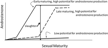
In order to assess genotypic effects on boar taint, it is first important to accurately define the boar taint phenotype of the pigs used in the study. Boars should have mature functional testis so that their true boar taint phenotype and potential for testicular production of 16-androstene steroids can be accurately assessed. Figure 2 illustrates the three possible boar taint phenotypes from androstenone. Boars that have high levels of androstenone do not need to be further characterised for sexual maturity and steroidogenic potential. However, boars with low levels of androstenone should be assessed for their steroidogenic potential. This insures that those boars with low levels of androstenone are not simply immature but have a low potential for the synthesis of androstenone or a high clearance rate of androstenone. Selecting for boars that have low levels of androstenone, but are late maturing, will result in problems with boar taint as the slaughter weight is increased and these boars near sexual maturity. Additionally, such selection may negatively affect the sexual status of related female pigs of a ‘low androstenone’ line (Willeke et al., Reference Willeke, Claus, Müller, Pirchner and Karg1987).
Effect of sexual maturity on boar taint from androstenone (M. Bonneau, personal communication).

Sexual maturity and steroidogenic potential is best assessed by measuring plasma levels of sex steroids, such as estrone sulphate (Schwarzenberger et al., Reference Schwarzenberger, Toole, Christie and Raeside1993; Sinclair et al., Reference Sinclair, Squires and Raeside2001) or free estrone in fat (Zamaratskaia et al., Reference Zamaratskaia, Rydhmer, Chen, Madej, Andersson and Lundström2005d). Sexual maturity may also be assessed by age/weight or size of secondary sex glands of the pigs (e.g. bulbo-urethral gland; Bonneau and Russeil, Reference Bonneau and Russeil1985), but these criteria should be characterised for individual breeds. Unfortunately, the degree of sexual maturity of boars has not been reported in many studies to date, which potentially confounds the results of these studies.
The assessment of sexual maturity also allows the effects of sex steroids and other hormones on the production of skatole in the intestine and metabolism of skatole in the liver to be accurately assessed. Differences in metabolism and clearance of skatole should be assessed in pigs that are producing high levels of skatole and thus have high levels of skatole in faeces (Claus et al., Reference Claus, Dehnhard, Herzhog, Bernal-Barragan and Giménez1993; Lanthier et al., Reference Lanthier, Lou and Squires2007). In this way, we can separate pigs that have low fat skatole due to the efficient metabolism of skatole in the liver from those that have low fat skatole due to decreased production of skatole in the intestinal tract.
Identification of QTLs for boar taint
There have been a few studies designed to identify QTLs for androstenone and skatole accumulation (reviewed in Robic et al., Reference Robic, Larzul and Bonneau2008). Two studies involved an experimental cross between Large White and Meishan pig breeds (Quintanilla et al., Reference Quintanilla, Demeure, Bidanel, Milan, Iannuccelli, Amigues, Gruand, Renard, Chevalet and Bonneau2003; Lee et al., Reference Lee, Archibald, Law, Lloyd, Wood and Haley2005). Quintanilla et al. (Reference Quintanilla, Demeure, Bidanel, Milan, Iannuccelli, Amigues, Gruand, Renard, Chevalet and Bonneau2003) found significant QTLs for androstenone using two different statistical methods on chromosomes (SSC) 3, 7 and 14. The QTL on SSC 7, close to the major histocompatibility complex of the pig, showed the largest effects. Two candidate genes in this region were investigated, CYP21 and CYP11a, but found not to be responsible for the QTL. Lee et al. (Reference Lee, Archibald, Law, Lloyd, Wood and Haley2004) also found a QTL for skatole located on SSC 14. They also found a QTL for androstenone and boar flavour on SSC 6. The potential QTLs identified in these studies explained 4% to 15% of the phenotypic variation in androstenone levels. However, different QTLs were identified by the two groups even within the same pig populations, with only the QTL with the largest effect on SSC 7 consistently found between the two studies. In addition, the slaughter weights were rather light at 80–85 kg live weight. A third study (Varona et al., Reference Varona, Vidal, Quintanilla, Gil, Sanchez, Folch, Hortos, Rius, Amills and Noguera2005), designed to map QTLs in 10 candidate chromosomal regions identified from the previous studies, was carried out with a Landrace outbred population. These authors failed to detect any QTLs for androstenone in any of the candidate chromosome regions and instead detected a QTL for skatole on SSC 6. The variations in the results from the different studies, which may in part be due to differences in live weight (i.e. sexual maturity), number of animals and breed combinations used, severely limit the utility of the available data. More definitive studies utilising different commercial pig breeds are necessary to further map the location of QTLs for androstenone and skatole.
Candidate genes based on metabolic studies
Another approach to developing genetic markers is to investigate polymorphisms, usually single nucleotide polymorphisms (SNP), in candidate genes. Candidate genes can code for key enzymes in the metabolic pathway of boar taint compounds and ideally should not involve other pathways, such as anabolic steroid metabolism. A number of key enzymes involved in the metabolism of both androstenone and skatole have been identified to date.
A number of studies have shown differences in the expression of candidate genes encoding enzymes involved in the metabolism of boar taint compounds between high and low boar taint pigs and between different pig breeds. However, only a few studies have reported SNPs in these genes that are correlated with levels of boar taint. These include an SNP in the CYP2E1 promoter (Skinner et al., Reference Skinner, Doran, McGivan, Haley and Archibald2005), an SNP in the coding region of CYP2E1 (Skinner et al., Reference Skinner, Doran, McGivan, Haley and Archibald2005; Lin et al., Reference Lin, Lou and Squires2006), a single base deletion in the coding region of CYP2A (Lin et al., Reference Lin, Lou and Squires2004a), and an SNP in the coding region of SULT1A1, which may all be related to skatole deposition (Lin et al., Reference Lin, Lou and Squires2004b), and an SNP in the 5′ untranslated region of CYB5 related to decreased androstenone synthesis (Lin et al., Reference Lin, Lou, Peacock and Squires2005). The importance of some of these markers was not supported by other studies (Skinner et al., Reference Skinner, Anderson, Haley and Archibald2006), and this may be due to the small number of animals or differences in the breeds used in these studies. No polymorphisms in transcription factor and nuclear receptor genes have yet been reported. In light of their central role in regulation of gene expression, polymorphisms in these genes are potentially more important than polymorphisms in individual metabolic genes (reviewed in Robic et al., Reference Robic, Larzul and Bonneau2008).
There have been very few large-scale studies that have quantified the effects of polymorphisms in candidate genes for boar taint in a variety of pig breeds. The effects of polymorphisms in CYB5 reported to date are small (Peacock et al., Reference Peacock, Lou, Lundström and Squires2007; Zamaratskaia et al., Reference Zamaratskaia, Lou, Peacock, Rydhmer, Andersson, Juneja, Chen, Lundström and Squires2008d) as are the effects of the detected QTLs for androstenone and skatole (Quintanilla et al., Reference Quintanilla, Demeure, Bidanel, Milan, Iannuccelli, Amigues, Gruand, Renard, Chevalet and Bonneau2003; Lee et al., Reference Lee, Archibald, Law, Lloyd, Wood and Haley2005), suggesting that boar taint is similar to many other traits in that it is the product of small effects in many genes. The data available to date suggest that there is not likely one major metabolic gene controlling boar taint. The effects of different polymorphisms vary among different lines and breeds of pigs as do the levels of androstenone and skatole, likely because some alleles may be fixed in different lines. Further large-scale studies to quantify the effects of SNPs in candidate genes for boar taint in a variety of breeds are warranted.
Gene expression analysis – microarray and proteomics
A major limitation of studying metabolic pathways to identify candidate genes is that you can only find those genes that are directly involved in the particular pathway being studied. A much broader approach is to conduct transcriptional profiling using DNA microarrays, in which the expression of thousands of genes (the ‘transcriptome’) is compared between animals with two different phenotypes. A recent study was published on the differences in gene expression profiles between boars with high and low fat androstenone (Moe et al., Reference Moe, Meuwissen, Lien, Bendixen, Wang, Conley, Berget, Tajet and Grindflek2007b). A wide variety of genes were differentially expressed between the two groups. This included genes previously identified (e.g. CYB5) as well as other genes involved in steroidogenesis and electron transport. However, the degree of sexual maturity and steroidogenic potential of the boars were not reported in this study. Previous studies by Stewart et al. (Reference Stewart, Lou, Squires and Coussens2005) comparing gene expression profiles between boars with high and low levels of plasma estrone sulphate using a smaller (1,700 gene) human microarray identified a number of the same genes as the study by Moe et al. (Reference Moe, Meuwissen, Lien, Bendixen, Wang, Conley, Berget, Tajet and Grindflek2007b) comparing low- and high-androstenone boars. Thus, in order to identify those genes that are specific to boar taint, it is important to differentiate between boars with high and low levels of steroidogenesis and boars with high and low levels of androstenone. This allows the separation of genes for ‘maleness’, which we want to keep, from genes that are important for boar taint, which we want to eliminate.
In addition to transcriptome analysis, proteomic approaches examine the levels of different proteins that are expressed between animals with two extremes of a trait. This involves separation of the proteins by 2-D electrophoresis or chromatographic methods, followed by quantification of the proteins and identification by mass spectrometry. Taken together, these analyses can identify differences in the expression of genes that may be responsible for the trait of interest. Results of proteomic analysis in low and high boar taint pigs have not yet been published in the scientific literature.
It is also important to note that microarrays and proteomics measure differences in gene expression and do not measure the presence of polymorphisms that alter the coding sequence and affect the activity of the gene product. Any polymorphisms that exist in these differentially expressed genes are likely to be found in the promoter region.
Recommendations for future research
Biochemistry of boar taint
Skatole biosynthesis in the large intestine has been a subject of huge research efforts. However, there is still gap in our understanding of microbiological aspects of skatole production. Analyses of the intestinal microbiota in the pigs with low and high potential for skatole production could reveal the mechanism(s) by which tryptophan degradation is regulated in the large intestine. This will help us to further manipulate the microbiotic environment to reduce skatole production.
The importance of CYP2E1 and CYP2A enzymes in skatole metabolism is well established. Additional information regarding the impact of various testicular steroids on hepatic metabolism is needed. Androstenone metabolism, particularly the formation of androstenone sulfoconjugates and its significance to androstenone elimination and accumulation in fat, should be further studied.
Genetics of boar taint
Future research on the genetics of boar taint should consider a number of factors. First of all, it is important to carefully define the genetic potential for boar taint by assessing the sexual maturity of all animals used in genetic studies. Studies that include sufficient numbers of animals from commercially relevant breeds are required to give sufficient statistical power to identify associations between genetic markers (SNPs) and boar taint. It is also important to rationalise the studies to identify QTLs for boar taint so that a clearly established list of QTLs is available for comparison with the chromosomal location of candidate genes for boar taint. Further studies of gene expression analysis should be conducted using pigs from high and low boar taint phenotypes but with similar steroidogenic potentials in order to identify potential candidate genes. Finally, studies to further understand the integration of the metabolism of boar taint compounds are warranted and these studies will also lead to more useful candidate genes.
Conclusions
Many aspects are important in the development of boar taint, of which genetics, age/weight, hormonal status of animals, and the rates of production and metabolic clearance of boar taint compounds are probably the most important. Boar taint level can be manipulated by modifying these factors. For example, immunocastration drastically changes the hormonal status of pigs and reduces the levels of androstenone and skatole. Dietary manipulations can reduce the risk of high skatole levels via the reduction of skatole intestinal synthesis or skatole absorption through the intestinal walls.
Boar taint due to high levels of skatole and androstenone is highly heritable and not all market weight entire males have boar taint; it should thus be possible to select for pigs that do not have boar taint. In these studies, it is critical to assess the steroidogenic potential of the pigs in order to separate late-maturing pigs from those with a low genetic potential for boar taint. A number of candidate genes for boar taint have been identified and work is continuing to identify new candidate genes and to develop genetic markers for low boar taint based on SNPs in these genes.
Acknowledgements
A major part of this review was carried out within the EU FP6 Specific Support Action project no. 043969 ‘PIGCAS’.