Introduction
Why should different parts of the retina be tuned to different parts of the electromagnetic spectrum?
This question, logically, follows discoveries of differences in visual pigments across the retinas of mammals, reptiles, amphibians, and fishes; intraretinal differences in colored oil droplets in birds; and variations in spectral transmission across the cornea and other ocular media in fishes. Attempts by visual ecologists to address possible functions of these intraretinal variations have been hampered, in most cases, by a lack of knowledge about the animal’s visual ecology and behavior. However, in a couple of cases, described herein, there is evidence to support the hypothesis that such intraretinal variability optimizes visual sensitivity for specific visual tasks. This is in contrast to the alternate hypothesis that suggests that intraretinal variations may be an artifact of retinal development or phylogenetic constraint that serves little or no visual function. This review summarizes the literature on intraretinal variability in spectral sensitivity (referred to subsequently as IVSS for short). For a more general description of the visual process, visual ecology, and intraocular filters, the reader is directed to various excellent reviews (Walls & Judd, Reference Walls and Judd1933; Walls, Reference Walls1942; Muntz, Reference Muntz and Ali1975; Knowles & Dartnall, Reference Knowles, Dartnall and Davson1977; Lythgoe, Reference Lythgoe1979; Heinermann, Reference Heinermann1984; Loew & Lythgoe, Reference Loew and Lythgoe1985; Goldsmith, Reference Goldsmith1990; Szél et al., Reference Szél, Rohlich, Caffe and van Veen1996; Bowmaker, Reference Bowmaker1998; Douglas & Marshall, Reference Douglas, Marshall, Archer, Djamgoz, Loew, Partridge and Vallerga1999; Peichl, Reference Peichl2005; Osorio & Vorobyev, Reference Osorio and Vorobyev2008; Jacobs, Reference Jacobs2009).
Variations in neural processing across the retina will not be dealt with here. The diversity of mechanisms mediating IVSS is already so great, and to date, variation in the interactions between filters, receivers, and processing across the retina is a relatively unexplored area. The assumption is, therefore, that the maximum number of dimensions to color vision is constrained by the number of photoreceptors in the retina that possess different spectral sensitivities, either inherently or as a result of intraocular filters. The role of neural processing in understanding the implications of IVSS on visual perception cannot be overstated, and great strides are being made towards an understanding of these processes, but it is not within the scope of this review.
The main take home message presented herein is, that while a great deal is known about the proximate causes, that is, the mechanisms mediating differences in spectral sensitivity across the retina, almost nothing is known about the ultimate causes, that is, the functional significance of differential spectral sensitivity in different regions of the retina, despite the fact that this phenomenon has been found in every major group of vertebrates.
Proximate and ultimate causes
The various mechanisms mediating IVSS can be divided into two categories: filters and receivers. Of the many types of intraocular filters, only those reported to cause differences in spectral sensitivity across the retina will earn mention here. This is not to say that IVSS does not result from other types of filters but that no evidence exists to date.
Filters
Cornea
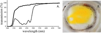
This is the first structure in the path of light as it enters the eye, and all vertebrate corneas filter electromagnetic radiation to some degree; however, variations in spectral filtering across the cornea are prominent mostly among fishes. Spectral transmission curves of vertebrate corneas show sharp decreases at short wavelengths, with relatively high transmission at middle and long wavelengths (Fig. 1). Reducing the relative quantity of short wavelength radiant flux is thought to: decrease damage by ultraviolet (UV) radiation, decrease noise resulting from photons scattering off the optical axis after entering the eye, and increase contrast by reducing the effects of glare and veiling light in the environment. This is the same reason that pilots, drivers, and skiers often use yellow lenses to improve visibility through the atmosphere under high-intensity situations (Lythgoe, Reference Lythgoe1979; Douglas & Marshall, Reference Douglas, Marshall, Archer, Djamgoz, Loew, Partridge and Vallerga1999).
(A) Spectral transmission of a partially pigmented vertebrate cornea (Anampses geographicus; Labridae). Solid line is the transmission through a clear area of the cornea and dotted line is through a yellow-pigmented area of the cornea. (B) Photograph of the pseudopupil pattern of corneal pigmentation in Hemigymnus fasciatus (Labridae). Data set and photo supplied by Ulrike Siebeck.

Corneal spectral transmission curves are quantified by the wavelength at which transmission drops to 50% of the maximum on the short wavelength arm, referred to as the T50 value. While transmission spectra of most corneas have been measured centrally, in some cases where transmission has been measured at different locations, variations in T50 have been observed.
Among fishes, members of the families Balistidae, Cyprinidae, Monacanthidae, and Tetraodontidae have higher carotenoid pigment density in dorsal and/or ventral areas of the cornea resulting in T50 values as high as 516 nm compared to lows of 350 nm centrally (Fig. 1A) (Douglas & McGuigan, Reference Douglas and McGuigan1989; Siebeck & Marshall, Reference Siebeck and Marshall2001). Differences in spectral transmittance across corneas are correlated with differences in spectral content of light impinging on the eye from different directions, with higher intensity and higher short wavelength content typically coming from the upper visual field. More complex patterns of corneal pigmentation are reported among the families Labridae and Scaridae, some with pupil like areas (pseudopupils) of lower pigment density (Fig. 1B) (Kondrashev et al., Reference Kondrashev, Gamburtzeva, Gnjubkina, Orlov and My1986). The precise function of the pseudopupil has yet to be determined, but it is probable that these windows in the short wavelength-filtering cornea are aligned with the optical axis of the area centralis. This would provide a greater intensity and broader spectrum of light to an area of the retina that contains: smaller cones and also possibly a greater number of spectral classes of cones than elsewhere in the retina, while still acting as a shield to the rest of the retina with all of the benefits of short wavelength filters mentioned above (Kondrashev et al., Reference Kondrashev, Gamburtzeva, Gnjubkina, Orlov and My1986; Siebeck et al., Reference Siebeck, Collin, Ghoddusi and Marshall2003). The pseudopupil may be an example of the trade-off between acuity, sensitivity, and hue discrimination, where the benefits of short wavelength filtering endowed upon the rest of the retina are not optimal for increasing acuity or hue discrimination in the region of the retina behind the pseudopupil.
A further specialization, found in some surface and shallow-water bottom-dwelling fishes, are occlusable corneal filters, in which screening pigments (short wavelength absorbing) can be moved across the cornea to varying degrees (Appleby & Muntz, Reference Appleby and Muntz1979; Kondrashev et al., Reference Kondrashev, Gamburtzeva, Gnjubkina, Orlov and My1986; Siebeck et al., Reference Siebeck, Collin, Ghoddusi and Marshall2003). The degree of occlusion is regulated by light intensity with the pigment retracting as intensity decreases, thus providing all of the benefits of a yellow cornea during the bright hours with none of the disadvantages when light levels drop. The dorsal, and sometimes also ventral (Siebeck et al., Reference Siebeck, Collin, Ghoddusi and Marshall2003), short wavelength filtering results in differential spectral filtering across the retina, and offsets greater downwelling, and in shallow waters upwelling light intensities.
Ellipsoids
The most widespread and diverse group of filters are those located in the photoreceptors themselves. Immediately vitread of the photoreceptor outer segment is a structure called the ellipsoid, which may contain one of a variety of pigmented structures (ellipsosomes, paraboloids, oil droplets, or diffuse yellow pigments). Possible functions of these structures include: acting as microlenses to increase photon capture by focusing light into the outer segment and thereby reduce within retina scatter, providing cellular machinery such as mitochondria, acting as an energy storage system, and spectrally filtering incoming light to shift or narrow photoreceptor spectral sensitivity to improve hue discrimination functions (Walls & Judd, Reference Walls and Judd1933; Walls, Reference Walls1942; Douglas & Marshall, Reference Douglas, Marshall, Archer, Djamgoz, Loew, Partridge and Vallerga1999). To date, there are no reports of variability in the spectral properties of ellipsosomes or parabaloids across the retina of any species, but such variations do exist in oil droplets and diffuse yellow pigments.
Oil droplets are found in the ellipsoids of some fishes, amphibians, reptiles, birds, and monotreme and marsupial mammals (reviewed in Walls, Reference Walls1942; Douglas & Marshall, Reference Douglas, Marshall, Archer, Djamgoz, Loew, Partridge and Vallerga1999; Hemmi & Grunert, Reference Hemmi and Grunert1999; Collin & Shand, Reference Collin, Shand, Collin and Marshall2003). Oil droplets come in a diverse array of colors, as viewed by the human eye, which result from the absorbance of, primarily, carotenoid pigments (Goldsmith et al., Reference Goldsmith, Collins and Licht1984). Intraretinal variability of colored oil droplets in birds is well documented and takes two forms: variation in oil droplet types and therefore associated cone types (Partridge, Reference Partridge1989; Peiponen, Reference Peiponen1992; Hart, Reference Hart2001; Rahman et al., Reference Rahman, Aoyama and Sugita2007, Reference Rahman, Yoshida, Maeda, Tanaka and Sugita2010) and variation in spectral transmission of oil droplets (Walls, Reference Walls1942; Hart et al., Reference Hart, Lisney and Collin2006; Knott et al., Reference Knott, Berg, Morgan, Buchanan, Bowmaker and Bennett2010). In the retina of the pigeon, for example, there is a predominance of yellow, orange, and red oil droplets dorsotemporally in an area known as the red field, whereas the rest of the retina possesses predominantly colorless, pale green, and yellow oil droplets (King-Smith, Reference King-Smith1969; Muntz, Reference Muntz and Dartnall1972; Bowmaker, Reference Bowmaker1977; Bowmaker & Knowles, Reference Bowmaker and Knowles1977). Regional variation in spectral transmission of oil droplets across the retinas of pigeons have been correlated with differences in spectral sensitivity and hue discrimination, with peak spectral sensitivity being 575 nm in the yellow field and 600 nm in the red field (Bowmaker, Reference Bowmaker1977; Brenner et al., Reference Brenner, Spaan, Wortel and Nuboer1983; Nuboer & Wortel, Reference Nuboer and Wortel1985a,Reference Nuboer and Wortelb; Wortel et al., Reference Wortel, Rugenbrink and Nuboer1987; Remy & Emmerton, Reference Remy and Emmerton1989). The long wavelength-shifted spectral sensitivity in the dorsotemporally located red field is proposed to improve discrimination of foliage (Lythgoe, Reference Lythgoe1979). In the South American opossum, oil droplets are restricted to the ventral retina, which is in opposition to the tapetum (discussed below) that is restricted to the dorsal retina (Ahnelt et al., Reference Ahnelt, Hokoc and Rohlich1995). To date, there is no evidence to suggest that the function of this ventral only distribution of oil droplets is to alter spectral sensitivity; however, there is also no evidence to suggest that there are differential requirements for microlenses or energy storage/production units across the retina, so the ultimate cause of the ventrally located oil droplets in South American opossum remains enigmatic.
Diffuse yellow pigments (Walls, Reference Walls1942) refers to the macula lutea, which directly translates to yellow spot and was once thought to be exclusive to higher primates but has since been found in chameleons (Hulke, Reference Hulke1867). The yellow color results from xanthophyll carotenoids, lutein, and zeaxanthin (Bone et al., Reference Bone, Landrum and Tarsis1985) deposited in cone photoreceptor axons. The density of these pigments decreases rapidly at about 6–8 degrees eccentricity from the center of the fovea. The effect is to markedly reduce the transmission of short wavelength light that reaches the photoreceptor layer, in the foveal region. These pigments are birefrigent and are also thought to be responsible for the entoptic effect known as Haidinger’s brush, which gives humans the ability to detect the orientation of the electric field vector of polarized light (Bone, Reference Bone1980; Hemenger, Reference Hemenger1982; Misson, Reference Misson2003). While the macula lutea necessarily alters spectral sensitivity, we do not perceive these differences across the eye due to higher order processing. Such phenomena are common in the visual system and remind us that sensitivity and perception are not necessarily equal, thus we must be cautious not to assume that a lack of perception equals a lack of visual function. Thus, while the absorption of short wavelengths by the macula lutea may reduce chromatic aberration and the scatter of short wavelength light thus improving visual acuity, as well as removing dangerous short wavelength radiation and oxidizing agents such as cytoxic-free radicals (Kirschfeld, Reference Kirschfeld1982; Douglas & Marshall, Reference Douglas, Marshall, Archer, Djamgoz, Loew, Partridge and Vallerga1999), any functional significance with respect to spectral filtering and visual ecology is unknown.
Visual pigment self screening
The photolabile visual pigments found in photoreceptors can filter light before it reaches visual pigments further along the outer segment. In long photoreceptors (>100 μm) or when the light travels through the same receptor more than once, as is the case with reflecting tapeta, this self-screening results in a broadening of the spectral sensitivity curve (Warrant & Nilsson, Reference Warrant and Nilsson1998). Dorsal–ventral differences in tapetal structure and photoreceptor length are common among vertebrates, particularly fishes (personal observation), and may result in differences in spectral sensitivity that have not yet been documented. In some deep-sea fishes, a special case of self screening exists where banks of photoreceptors lie in front of one another on the optical axis (Locket, Reference Locket and Crescitelli1977; Denton & Locket, Reference Denton and Locket1989; Fröhlich et al., Reference Fröhlich, Negishi and Wagner1995; Hess et al., Reference Hess, Melzer and Smola1998). These may, if the appropriate neural processing exists, allow for comparisons between receptors in the same visual axis thus providing the potential for hue discrimination even though all the receptors in each bank possess the same visual pigment. Self screening in such banked retinas shifts the peak spectral sensitivity of rods from 480 nm in the most vitread bank to as high as 565 nm in the most sclerad bank, with a decrease in absorption of less than a factor of 2 at 565 nm but by a factor of a million at 480 nm (Denton & Locket, Reference Denton and Locket1989). While the effects of differences in self screening on spectral sensitivity are relatively small, they will be most prominent in animals inhabiting dimly lit environments and possessing long photoreceptors, where fine tuning sensitivity to the available light is critical. It is difficult to predict the advantages in such systems where a few photons may mean the difference between escape and capture, particularly when we know so little about the behavior and visual ecology of species (e.g., in the deep sea) where these factors are relevant.
Postreceptoral filters
Behind the photoreceptors are the retinal-pigmented epithelium (RPE) and in some cases, a tapetum. While RPE is typically described as black and therefore nonreflective, it does in fact reflect light at long wavelengths, even if only slightly (Boulton et al., Reference Boulton, Docchio, Dayhawbarker, Ramponi and Cubeddu1990). In some fishes, for example, pickerel, RPE reflectance is as high as 30% at 650 nm, which is well within the sensitivity spectrum of the rod visual pigments (reviewed in Douglas & Marshall, Reference Douglas, Marshall, Archer, Djamgoz, Loew, Partridge and Vallerga1999), which in the case of pickerel that use the long wavelength-shifted vitamin A2-based visual pigment (porphyropsin). And while there are no reports of variation in RPE reflectance within an eye, it is likely that this has not yet been investigated.
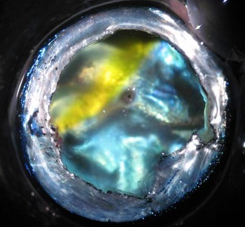
Tapetal reflectance, on the other hand, varies spectrally in different regions of the eye in many vertebrates. The tapetum lucidum is present in nocturnally active, fishes, amphibians, reptiles, and mammals but is notably absent in birds (Walls, Reference Walls1942; Arnott et al., Reference Arnott, Maciolek and Nicol1970; Best & Nicol, Reference Best and Nicol1980; Ollivier et al., Reference Ollivier, Samuelson, Brooks, Lewis, Kallberg and Komaromy2004; Emond et al., Reference Emond, McNeil, Cabana, Guerra and Lachapelle2006). The spectral reflectance of tapeta vary from violet/blue to red depending on the types and densities of reflective and absorptive structures present (Best & Nicol, Reference Best and Nicol1980; Douglas et al., Reference Douglas, Partridge and Marshall1998; Ollivier et al., Reference Ollivier, Samuelson, Brooks, Lewis, Kallberg and Komaromy2004), and dramatic differences in spectral reflectance can occur in different areas of the tapeta (Munk, Reference Munk1980; Douglas et al., Reference Douglas, Partridge and Marshall1998) (Fig. 2). For example, deep-sea big eyes have tapetal areas that reflect blue, green, orange, and red (Wang et al., Reference Wang, Nicol, Thurston and McCants1980). In the case of the myctophid Notoscopelus resplendens, which has a short wavelength-reflecting dorsal tapetum and a mid wavelength-reflecting ventral tapetum (Douglas et al., Reference Douglas, Partridge and Marshall1998), the dorsal retina attains a 5.1% improvement in photon capture of upwelling light compared to the ventral retina, conversely, the ventral retina has a 2.5% improvement over the dorsal retina for downwelling light (for calculations see Appendix 1). Thus, differences in tapetal reflectance can result in IVSS even for species with a single visual pigment. In other species, the tapetum lucidum does not cover the entire retina (e.g., lungfish, South American possum, whale, seal, rodent, horse, sheep, goat, deer; Walls, Reference Tyler and Smith1942; Ahnelt et al., Reference Ahnelt, Hokoc and Rohlich1995; Ollivier et al., Reference Ollivier, Samuelson, Brooks, Lewis, Kallberg and Komaromy2004; Bailes et al., Reference Bailes, Robinson, Trezise and Collin2006; Shinozaki et al., Reference Shinozaki, Hosaka, Imagawa and Uehara2010), necessarily resulting in variations in sensitivity across the retina. In most of these cases, the tapetum is found only in the dorsal retina and can therefore be correlated with the decreased photon flux of light reflected off the ground or upwelling light in the case of aquatic animals. As a comparison to the effect of different colored tapeta (above), the presence of a ventral tapetum increases photon capture by 22%, in the case of the myctophid (for calculations, see Appendix II). To accomplish the same increase in photon catch, a photoreceptor would have to nearly double in length. Doubling photoreceptor length also doubles thermal dark noise (directly proportional to the quantity of visual pigment) and since noise sets the lower limit on vision in dim light (Barlow, Reference Barlow1956), the tapetum presents a significant advantage notwithstanding its potential drawbacks with respect to detectability of eye shine by predators and prey (Ollivier et al., Reference Ollivier, Samuelson, Brooks, Lewis, Kallberg and Komaromy2004). Interestingly, in the aquatic environment, upwelling light is the result of scattering and is dominated by short wavelengths (Tyler & Smith, Reference Tyler and Smith1970; Gaten et al., Reference Gaten, Shelton and Herring1992), while in terrestrial environments, reflected light from the ground and vegetation tends to be long wavelength shifted. A survey of aquatic and terrestrial vertebrates with IVSS (Table 1) shows that the dorsal retinas of terrestrial animals tend to have spectral sensitivities shifted to longer wavelengths (e.g., greater proportion of L/M cones dorsally), while those of aquatic animals are shifted to shorter wavelengths (e.g., higher A1-based chromophore content dorsally). It would be of interest to compare the spectral reflectance of dorsal and ventral tapeta of several aquatic and terrestrial animals to see if this correlation holds there also.
Example of variation in tapetal reflection in Myctophum nitidulum (Myctophidae). Photo supplied by Fanny deBrusserolles.

Examples of intraretinal variability in vertebrate visual pigments.

* Only study to measure the spectral environment in different parts of the visual field to investigate possible correlations with intraretinal differences in spectral sensitivity
Receivers
IVSS due to visual pigments can be divided into three broad categories: 1) variation in chromophore ratio, 2) variation in photoreceptor classes, and 3) variation in opsin expression. However, in some cases, these categories are not mutually exclusive (examples to follow).
Chromophores
In instances where suitable approaches have been used to measure differences in chromophore content across the retina, the ratio of A1:A2-based visual pigments (rhodopsin:porphyropsin) has been found to vary dorsoventrally (reviewed in; Bowmaker, Reference Bowmaker, Cronly-Dillon and Gregory1991). For example, coho salmon (Temple et al., Reference Temple, Plate, Ramsden, Haimberger, Roth and Hawryshyn2006); rudd, rainbow, trout and brown trout (Muntz & Northmore, Reference Muntz and Northmore1971); and archerfish (Temple et al., Reference Temple, Hart, Marshall and Collin2010), all have a higher ratio of A2-based visual pigments ventrally, while bullfrogs (Reuter et al., Reference Reuter, White and Wald1971) and four-eyed fish (Bridges, Reference Bridges1982) have a higher ratio of A2 dorsally. The effect of changes in chromophore ratio on the spectral sensitivity of a photoreceptor varies with the wavelength of peak sensitivity of the visual pigment contained within. A2-based pigments are always long wavelength shifted relative to the same opsin combined with A1, but for visual pigments that absorb at short wavelengths, the shift when changing from A1 to A2 is much less than for visual pigments that absorb at longer wavelengths. For example, an UV visual pigment that absorbs at 350 nm with A1 will absorb at 365 nm with A2, a middle wavelength visual pigment at 500 nm with A1 will absorb at 530 nm with A2, and a long wavelength visual pigment at 570 nm with A1 will absorb at 638 nm with A2 (models predicting effects of chromophore shifts are reviewed in Temple et al., Reference Temple, Ramsden, Haimberger, Veldhoen, Veldhoen, Carter, Roth and Hawryshyn2008a). While dorsal–ventral differences in chromophore ratio are not typically more than 50%, such differences alter peak absorbance by as much as 30–40 nm for long wavelength-sensitive (LWS) cones, thus providing a source of IVSS in species that possess an A1–A2 interchange system. Apart from the advantages bestowed upon the retina by changing spectral sensitivity, there may be advantages with respect to signal to noise ratio since A1-based visual pigments are more thermally stable as a result of the shorter conjugated pi-electron chain in A1 chromophores. This greater thermal stability can result in a nearly 10-fold decrease in thermal dark noise when the same photoreceptor uses A1 instead of A2 (Ala-Laurila et al., Reference Ala-Laurila, Donner, Crouch and Cornwall2007).
Photoreceptor classes
Changing the ratio of different photoreceptor classes is another mechanism that alters spectral sensitivity across the retina. It has been known for nearly a century that spectral sensitivity and hue discrimination differ between the center (fovea) and periphery of our own visual field (Ferree & Rand, Reference Ferree and Rand1919; Willmer & Wright, Reference Willmer and Wright1945; Weale, Reference Weale1953). It has since been demonstrated that such differences are in part mediated by differences in the proportions of the three cone types (Roorda & Williams, Reference Roorda and Williams1999). Primate retinas have no short wavelength-sensitive (SWS), S-cones in the foveola (de Monasterio et al., Reference de Monasterio, McCrane, Newlander and Schein1985; Szél et al., Reference Szél, Diamantstein and Rohlich1988) and the proportions of the three cone types vary greatly towards the periphery (Hagstrom et al., Reference Hagstrom, Neitz and Neitz1998). Such intraretinal variability in the proportions of functional photoreceptor classes have also been reported in many other mammals (Hemmi & Grunert, Reference Hemmi and Grunert1999; Peichl et al., Reference Peichl, Künzle and Vogel2000; Schiviz et al., Reference Schiviz, Ruf, Kuebber-Heiss, Schubert and Ahnelt2008; Shinozaki et al., Reference Shinozaki, Hosaka, Imagawa and Uehara2010) and fishes (Levine et al., Reference Levine, MacNichol, Kraft and Collins1979; Burkhardt et al., Reference Burkhardt, Gottesman, Levine and MacNichol1983) (Table 1). As an example, in salmonids, UV-sensitive cones, which take a corner position in the regular mosaic pattern of photoreceptors, are absent from most of the retina in juvenile salmon but are present in earlier age classes and in mature adults (Beaudet et al., Reference Beaudet, Browman and Hawryshyn1993; Allison et al., Reference Allison, Dann, Veldhoen and Hawryshyn2006). The absence of UV cones in the ventral retina in juvenile salmon has been correlated with a loss of UV sensitivity in the ventral retina (Deutschlander et al., Reference Deutschlander, Greaves, Haimberger and Hawryshyn2001). There is some debate as to whether the UV class of receptor is lost and regained or whether this class of receptor changes opsin expression with ontogeny (Cheng & Novales Flamarique, Reference Cheng and Novales Flamarique2004; Allison et al., Reference Allison, Dann, Veldhoen and Hawryshyn2006). The possibility that photoreceptors change opsin expression is counter to the “one opsin in each receptor” paradigm, and there is now a great deal of evidence to support such changes in other vertebrate photoreceptors (see below). The functional significance, or lack thereof, of variations in photoreceptor classes across the retina and of variations in opsin expression (next section) have been incorporated into a discussion, which follows, of the two competing hypotheses that attempt to explain the existence of IVSS in general.
Opsin expression
Differential opsin expression across the retina is a mechanism of intraretinal variability that was thought to be an exception (Peichl, Reference Peichl2005; Bowmaker, Reference Bowmaker2008); however, when efforts have been made to look for it, it has been found in a growing number of vertebrates from many taxa and may prove to be the rule. Well-studied examples include mouse and zebrafish, but evidence for intraretinal variable opsin expression has been found in many small mammals and fishes (Table 1). In mice, for example, LWS opsin dominates dorsally, while the SWS1 opsin dominates ventrally to the exclusion of the LWS opsin (Szél et al., Reference Szél, Rohlich, Caffe, Juliusson, Aguirre and van Veen1992; Wang et al., Reference Wang, Macke, Merbs, Zack, Klaunberg, Bennett, Gearhart and Nathans1992). At the transition zone, between the dorsal and ventral hemispheres, SWS1 and LWS opsins are coexpressed in most cones (Röhlich et al., Reference Röhlich, van Veen and Szél1994). The exclusive expression of SWS1 ventrally results in an increase in short wavelength sensitivity in the upward-looking field of view (Calderone & Jacobs, Reference Calderone and Jacobs1995; Peichl et al., Reference Peichl, Künzle and Vogel2000).
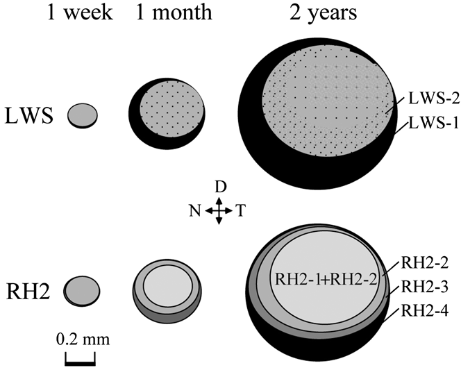
In zebrafish, the situation is more complex; there is not only spatial variability in opsin expression patterns across the retina but also temporal variability. Zebrafish have four cone classes (two single cones and a double cone) and express nine cone opsins: two LWSs, one rod opsin (RH1), four middle-wavelength sensitive (RH2) opsin subtypes, one SWS1, and one SWS2. The pattern of LWS expression does not change considerably from 1 week to 2 years of age, with a large central to dorsotemporal area in which LWS-2 (shorter wavelength absorbing of the two LWS opsins) predominates and a smaller peripheral area in which LWS-1 (longer wavelength absorbing of the two LWS opsins) predominates (Fig. 3). The expression pattern of the four RH2 opsins does change with ontogeny, and these changes can be summarized as a transition from predominantly RH2-1 over most of the retina at 3 days to a series of concentric rings of different opsin expression with the short wavelength-absorbing opsins RH2-1 and RH2-2 in the large central to dorsotemporal area surrounded by a ring of RH2-3 and then RH2-4 at 2 years (Fig. 3; Takechi & Kawamura, Reference Takechi and Kawamura2005). These changes in opsin expression have been referred to as “transdifferentiation” (Ahnelt & Kolb, Reference Ahnelt and Kolb2000), which is not to be confused with “transmutation,” which was the name of a theory put forth by Walls (Reference Walls and Judd1942) to describe an evolutionary change from rods to cones. To date, no obvious function for this circular pattern of opsin expression has been proposed; however, it is worth noting that the two shorter wavelength-absorbing RH2 opsins are expressed in the dorsal hemisphere, which matches the use of shorter wavelength-sensitive A1-based visual pigment in the dorsal retinas of other fishes and may be correlated to the greater relative content of shorter wavelengths in upwelling light (as described above).
Temporal variations in spatial variability in opsin expression patterns of LWS and RH2 opsins in zebrafish (Danio rerio). Opsin subtypes are labeled by gray scale and the key is in the 2-year panel; black dots indicate sparse expression. Figure adapted from Takechi and Kawamura (Reference Takechi and Kawamura2005).

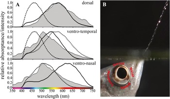
When changes in opsin expression are combined with changes in chromophore ratio (e.g., salmonids, Temple et al., Reference Temple, Ramsden, Haimberger, Veldhoen, Veldhoen, Carter, Roth and Hawryshyn2008a,Reference Temple, Veldhoen, Haimberger, Phelan, Veldhoen and Hawryshynb) photoreceptors in different regions can be finely tuned. In archerfish, such combined changes result in three different sets of visual pigments in three different regions of the retina (Temple et al., Reference Temple, Hart, Marshall and Collin2010). Dorsally, archerfish have predominantly A1-based visual pigments with two cone spectral peaks (maximum absorbance = λ max) at 454 and 570 nm. Ventronasally, they have a greater A2 content with two cone λ max values 515 and 620 nm. While ventrotemporally in the area of the retina that is used by archerfish to look up out of the water at aerial prey at which they will spit jets of water, they have predominantly A1-based visual pigments with three cone λ max values 453, 534, and 564 nm (Fig. 4). These different sets of visual pigments not only change the spectral sensitivity of the archerfish eye for different parts of their field of view, but they also give it the potential to have dichromatic color vision in some areas and trichromatic color vision in others. These differing spectral sensitivities in different directions have been correlated with different visual tasks (Temple et al., Reference Temple, Hart, Marshall and Collin2010) and provide support for the functional hypothesis of IVSS.
Intraretinal variability in visual pigments in archerfish (Toxotes chatareus). Photoreceptor absorptance (corrected for optical media transmission) for single (dashed lines) and double (solid and dotted lines) cones from three regions of the retina compared to environmental light impinging on those areas (gray-filled curves = normalized spectral radiance). Figure adapted from Temple et al. (Reference Temple, Hart, Marshall and Collin2010).

Two hypotheses to explain existence of IVSS
There are two predominant hypotheses to explain intraretinal variability in visual pigments among vertebrates. One suggests that regional differences are functional and provide some visual advantage through optimization of sensitivity, contrast detection, hue discrimination, or signal to noise ratio (Reuter et al., Reference Reuter, White and Wald1971; Szél et al., Reference Szél, Rohlich, Caffe, Juliusson, Aguirre and van Veen1992, Reference Szél, van Veen and Rohlich1994, Reference Szél, Rohlich, Caffe and van Veen1996; Juliusson et al., Reference Juliusson, Bergstrom, Rohlich, Ehinger, Vanveen and Szél1994; Arrese et al., Reference Arrese, Oddy, Runham, Hart, Shand, Hunt and Beazley2005; Peichl et al., Reference Peichl, Chavez, Ocampo, Mena, Bozinovic and Palacios2005; Rocha et al., Reference Rocha, Ahnelt, Peichl, Saito, Silveira and De Lima2009; Temple et al., Reference Temple, Hart, Marshall and Collin2010). The other suggests that regional differences are a product of development that reflects ancestral stages of visual pigment evolution, and that rather than providing a selective advantage, regional differences exist where selective pressures are weak and are due to developmental/phylogenetic constraints (Ahnelt et al., Reference Ahnelt, Hokoc and Rohlich1995; Szél et al., Reference Szél, Rohlich, Caffe and van Veen1996; Gaillard et al., Reference Gaillard, Kuny and Sauve2009).
The functional hypothesis is based on the fact that visual environments, in most habitats, are not uniform with respect to the spectral quality of light, and that different visual tasks may be required in different areas of the field of view. Support for the functional hypothesis comes from correlations between the spectral sensitivity of regions of an animal’s retina, and the spectral content of particular parts of their field of view or the requirements of a particular visual task known to occur in a particular part the animal’s field of view. Three examples follow.
Many small rodents, like the house mouse (Mus musculus; Table 1) have a ventral region of the retina rich in S-cones (Szél et al., Reference Szél, Rohlich, Caffe, Juliusson, Aguirre and van Veen1992), with a λ max at 365 nm that provides increased sensitivity to short wavelengths in the upward direction (Jacobs et al., Reference Jacobs, Williams and Fenwick2004). This ventral S-cone-rich field is proposed to increase contrast of aerial predators against a background of blue sky, referred to as the “skylight hypothesis.” Interestingly, skylight during most of the day has a spectral irradiance centered around 500 nm; it is only at crepuscular hours, a period when mice are most active, that the spectrum shifts, leaving two peaks one around 450 nm and the other close to 700 nm (McFarland & Munz, Reference McFarland, Munz, Evans, Bainbridge and Rackham1975). As yet there is no behavioral evidence supporting this hypothesis, however, given the ability to modify the visual pigments in this model (Jacobs et al., Reference Jacobs, Williams, Cahill and Nathans2007), such experiments are certainly tenable.
The quokka (Setonix brachyurus) and the quenda (Isoodon obesulus) together represent another example of intraretinal variability in visual pigments (Table 1). Both of these marsupials have a distribution of M and L cones that peaks in the dorsotemporal retina just above the midline with concentric rings of decreasing cone density towards the periphery. And both have a more dorsally located dorsotemporal area of increased S-cones. However, in addition, the quenda has a second ventronasally located area of increased S-cone density. This small difference between these two relatively similar species has been attributed to differences in their habitat. Whereas the quokka prefers open grasslands, the quenda is more likely to be found in forested habitats, and the upward-looking S-cone area combined with M- and L-cones may provide hue discrimination for detecting predators among foliage (Arrese et al., Reference Arrese, Oddy, Runham, Hart, Shand, Hunt and Beazley2005).
The third example is a fish and represents the only example where spectral measurements from the natural environment have been compared to the spectral sensitivity of photoreceptors in different regions of the retinal. The archerfish (Toxotes chatareus) has different sets of visual pigments in three retinal regions (described above; Fig. 4). The spectral sensitivity of the two classes of photoreceptors in the dorsal retina provide one matched and one offset detector relative to the spectral background of upwelling light in the murky waters of rivers, lakes, and mangroves where they are found (Fig. 4). Matched and offset detectors optimize sensitivity to bright and dark targets (potential predators) approaching from below (Lythgoe, Reference Lythgoe, Bainbidge, Evans and Rackham1966; Munz & McFarland, Reference Munz and McFarland1975). The spectral sensitivities of the two classes of photoreceptors in the ventronasal retina again provide one matched and one offset detector relative to the spectral background of downwelling skylight (Fig. 4). In this region of the retina, single cones, which are matched, are well suited for detecting dark silhouetted targets (potential aerial predators) approaching from above. While offset twin cones may function in detecting targets reflecting upwelling light that is long wavelength shifted (Temple et al., Reference Temple, Hart, Marshall and Collin2010). Finally, the three pigments in the ventrotemporal retina of the archerfish (453, 534, and 564 nm) are similar to those in primates (e.g., humans 420, 533, and 564 nm), which are thought to be adapted to detecting fruit against a background of foliage (Osorio & Vorobyev, Reference Osorio and Vorobyev1996), a task not unlike the archerfish detecting insects against a background of foliage (Temple et al., Reference Temple, Hart, Marshall and Collin2010).
Support for specific visual functions of IVSS in the above examples has been based on correlations derived from classic visual ecology models applied to rather limited data sets; therefore, conclusions about function are necessarily tenuous. In general, we simply do not possess enough information about the visual ecology and behavior of most species studied. Recognition of the prevalence of IVSS across vertebrate classes will hopefully lead to more research including manipulative experiments designed to address function specifically.
Evidence against a functional hypothesis to explain the presence of IVSS, at least in the case of the ventral S-cone-rich areas in mice and rodents, is based on the existence of two species that share the same general habitat, but one shows regionalization of the retina and one does not. The presence of a ventral S-cone-rich area in M. spiculegus but not in Apodmeus microps, which are both found on the Great Hungarian Plain, has been proposed as evidence that the “skylight hypothesis” is flawed (Szél et al., Reference Szél, Rohlich, Caffe and van Veen1996).
The alternate hypothesis is that intraretinal variability in visual pigments are the result of developmental/phylogenetic constraints and was proposed, in part, because in many cases, no clear function has been attributed to differences in spectral sensitivity across the retina (Ahnelt et al., Reference Ahnelt, Hokoc and Rohlich1995; Szél et al., Reference Szél, Rohlich, Caffe and van Veen1996; Gaillard et al., Reference Gaillard, Kuny and Sauve2009), and furthermore, because an example exists where two species sharing the same habitat do not both possess intraretinal variability (as described above; Szél et al., Reference Szél, Rohlich, Caffe and van Veen1996). The implication of this hypothesis is that areas of the retina where variability occur are under relaxed selective pressure and represent vestiges of an ancestral state of opsin evolution (Szél et al., Reference Szél, Rohlich, Caffe and van Veen1996). The change in opsin expression, “transdifferentiation,” with development in some species, even in the absence of intraretinal variability in the adult retina (e.g. rat Szél et al., Reference Szél, van Veen and Rohlich1994; Ahnelt & Kolb, Reference Ahnelt and Kolb2000), is presented as further support for this argument and follows the tenet that development recapitulates evolution.
Evidence against the hypothesis that intraretinal variability, at least in mammals, represents developmental/phylogenetic constraint is based on two main arguments: firstly, principles of niche partitioning (Christopher & Barrett, Reference Christopher and Barrett2006) suggest that in the case of M. spiculegus and A. microps, it is highly unlikely that these two species are filling the same niche and therefore not liable to be living in the same visual habitat or sharing the same visual tasks, despite being similar morphologically and sympatric in part of their distribution. Just because two species live in the same habitat does not necessitate the same visual sensitivity. Even more closely related species, like the cichlids of Lake Malawi and Lake Victoria, express opsins with slightly different λ max values despite living in the same lake. The differences in opsin expression among these fishes have been correlated with specific visual tasks (Carleton et al., Reference Carleton, Parry, Bowmaker, Hunt and Seehausen2005; Parry et al., Reference Parry, Carleton, Spady, Carboo, Hunt and Bowmaker2005).
The second argument against developmental/phylogenetic constraint is theoretical. Vision is a sensory modality that provides information about the three-dimensional position of objects through the detection of changes in intensity, hue, polarization, and movement. The visual system mediates the collection of this information through an imperfect system that must accommodate a trade-off between sensitivity and spatial resolving power (Land & Nilsson, Reference Land and Nilsson2002), and it cannot be spectrally tuned to maximize contrast of all colors at all times in all directions, due to limitations of space in the retina. The human fovea is a perfect example, in order to maximize acuity; trichromatic color vision and dim-light sensitivity have been compromised by the absence of blue cones and rods in the fovea. It is simply not possible to have more than one photoreceptor in one location in the retina. Therefore, animals have been forced to compromise through the selection of visual pigments that optimize sensitivity and contrast detection for visual tasks that are most likely to improve fitness. These visual tasks may differ at different stages of life history and are often different in different parts of the field of view. One of the strongest selective pressures on most species is predation and since detection is the first step in evasion, vision with its rapid data acquisition, and information about size and distance is critical in predator detection in most vertebrates. Even when the threat of predation only occurs during a brief period of the day or a specific season, the selective pressure may be great enough to drive the visual system to have properties that may not be optimal for the remainder of the animal’s normal behaviors. In this context, it is hard to imagine an area of the retina having photoreceptors that are not under selective pressure to optimize target detection under at least one circumstance. Further evidence, to support this notion, comes from the fact that the intraretinal variations described throughout this review do not represent phenomena that are unique to individuals but rather are a consistent feature for all members of the species, suggesting that unlike iris color patterns in humans or body color patterning in guppies, selective pressures are driving the maintenance of the specific patterns of IVSS that have been reported.
Conclusions
IVSS is a widespread phenomenon among vertebrates and is mediated by several different mechanisms, which in and of itself supports the notion that it provides a functional advantage to the visual system. The phenomenon of different functions being attributed to different parts of the eye is not unique to vertebrates. Many invertebrates have distinct areas that have markedly different spectral sensitivities based on the presence of different cell types expressing different opsins, and some also have areas specific for polarization vision (Stavenga, Reference Stavenga2002). While the mechanisms mediating intraretinal variability in vertebrates are mostly well understood, the functional significance is often unclear. This should come as no surprise since so little is known about the natural behaviors of most vertebrates let alone the specific visual tasks that they must confront. That not enough is known about the visual ecology of animals of interest is a common conclusion of most papers that address intraretinal variability in vertebrates. Douglas and Marshall (Reference Douglas, Marshall, Archer, Djamgoz, Loew, Partridge and Vallerga1999) describe the conundrum facing all visual ecologists: “...correlating an animal’s environment with its visual system is fraught with difficulties primarily because the environmental factors most relevant to an animal are usually not known in enough detail.” The challenge then is to fill gaps in our knowledge. More field studies are needed of animals performing natural behaviors in natural settings to greater our understanding of the specific visual tasks (Muntz, Reference Muntz and Ali1975), and more needs to be known about the selective pressures on animals of interest.
Appendix I
Functional advantage of one region of the tapeta over another, for a given photic environment, is given by:

where X is the sum of the total relative photon capture across all wavelengths for one area of the retina (e.g., dorsal) exposed to a specific light environment (e.g., upwelling irradiance). X′ is the same as X but for the other area of the retina (e.g., ventral) exposed to the same light environment (e.g., upwelling). X and X′ each equal C(λ) + C′(λ) for the respective area of the retina and


where I(λ) is the spectral irradiance of the light environment (upwelling and downwelling spectral irradiance were extracted from fig. 7 of Gaten et al., Reference Gaten, Shelton and Herring1992). I′(λ) is the remaining photon irradiance after the light has passed through the photoreceptor and after reflection off of the tapetum. P(λ) is the absorptance spectrum of the photoreceptor.

where R(λ) is the remaining photon irradiance after light has passed through the photoreceptor the first time and equals I(λ)–C(λ), where I(λ) and C(λ) are as above. r(λ) is the relative reflectance of the tapetum (data extracted from fig. 5f of Douglas et al., Reference Douglas, Partridge and Marshall1998 and normalized to correct for measurement technique as suggested by author J.C.P.)

where A(λ) is the absorbance spectrum of the photoreceptor, which is the normalized absorbance spectrum for a visual pigment (based on templates from Govardovskii et al., Reference Govardovskii, Fyhrquist, Reuter, Kuzmin and Donner2000) with λ max at 486 as found in Notoscopelus resplendens (Douglas & Partridge, Reference Douglas and Partridge1997), multiplied by the length of the photoreceptor (set at 50 μm as per O’Day & Fernandez, Reference O’Day and Fernandez1976) and multiplied by the specific absorbance of the visual pigment (set at 0.013 per μm as per Turner et al., Reference Turner, White, Collins, Partridge and Douglas2009).
Appendix II
Functional advantage of the presence of a tapetum was calculated as above where X is the sum of the total relative photon capture across all wavelengths in the presence of a tapetum, and X′ is the same but in the absence of tapetum. For this example, the calculations were done using the values for the ventral tapetum of N. resplendens as described in Appendix I and the spectral irradiance of downwelling light also described in Appendix I.
Acknowledgments
I wish to express my gratitude to Nathan Hart, Ágoston Szél, two anonymous reviewers, and the editors, for comments and contributions that have improved this manuscript. Thank you also to Ulrike Siebeck, and Fanny deBrusserolles for use of their images. I have been supported by Postdoctoral fellowships from The University of Queensland and the Natural Sciences and Engineering Research Council of Canada.